Atheroma, Thrombosis and Embolism
 Call Kai
 Learn
 Practice Test
 Spaced Repetition
 Match
 Flashcards
 Knowt Play
1/32

There's no tags or description
Looks like no tags are added yet.
33 Terms
atheroma
focal intimal lesion consisting of lipid-rich necrotic core covered by fibrous cap within medium and large arteries
non-modifiable risk factor
increasing age
male gender
family history/ genetics
modifiable risk factors for atheroma
hyperlipidaemia
hypertension
cigarette smoking
diabetes mellitus
obesity
sedentary lifestyle
C-reactive proteins
causes of atheroma
starts with damage or injury to endothelium of artery
damage may be caused by:
high blood pressure
high cholesterol
irritant e.g. nicotine
diseases e.g. diabetes
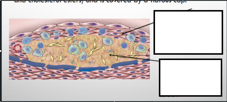
parts of an atheroma
fibrous cap
smooth muscle cells
macrophages
foam cells
lymphocytes
collagen
elastin
necrotic centre
cell debris
cholesterol crystals
foam cells
calcium

stages of atherosclerosis formation
endothelial dysfunction/injury
fatty streak
fibrofatty plaque
complicated plaque
endothelial dysfunction/injury
risk factors cause
incr. endothelial permeability
leukocyte adhesion
lipoprotein entry into vessel wall
results in LDL accumulation within intima
morphology of the fatty streak
earliest visible lesion
intimal accumulation of lipid-laden macrophages
key cells in the fatty streak
monocytes→ macrophages
foam cells
morphology of fibrofatty plaque
smooth muscle migration from media
collagen deposition
formation of fibrous cap
lipid-rich necrotic core
components of fibrofatty plaque
fibrous cap
lipid core
inflammatory cells
complications associated with complicated plaque
plaque rupture
plaque erosion
haemorrhage into plaque
calcification
superimposed thrombosis
clinical consequences of complicated plaque
myocardial infarction
stroke
peripheral artery disease
pathophysiology of atherosclerosis
develops as chronic inflammatory response of arterial wall to endothelial injury
lesion progression occurs through interactions of modified lipoproteins, monocyte-derived macrophages, T lymph. and normal cells
contemporary view of atherosclerosis expressed by response-to-injure hypothesis
vessels commonly affected by atherosclerosis
bifurcations→ sites of turbulent flow
abdo. aorta
coronary arteries
popliteal arteries
carotid vessels
circle of willis
fatty streak
earliest lesion in atherosclerosis
composed of lipid filled foamy macrophages
starts are multiple flat yellow spots that coalesce into streaks
lesions not significantly raised and don’t cause flow disturbance
morphological evolution of atherosclerosis
not all fatty streak are destined to progress to atheromatous plaque
coronary fatty streaks start to form in adolescence at the same anatomical sites that later tend to develop plaques
atherosclerotic plaque
consists of intimal thickening and lipid accumulation
appears white-yellow
superimposed thrombus on plaque appears red
impinges on vessel lumen
sequelae of atherosclerosis
haemorrhage into plaque
atherosclerotic plaques cause turbulence, damages endothelium→ rupture, ulceration, erosion
induces thrombosis
lumen occlusion→ ischaemia/infarction
atheroembolism
aneurysm formation
thrombosis
solid mass of blood constituents formed within vascular system in vivo
arterial thrombosis most commonly superimposed on atheroma
venous thrombosis most commonly due to stasis
virchow’s triad
components leading to thrombosis:
endothelial injury
abnormal blood flow
hypercoagulability
arterial thrombosis
typically from rupture of atheromatous plaque
found in arteries/ left heart chambers
composed mainly of platelets
diseases associated with arterial thrombosis and treatment
diseases
acute coronary syndrome
ischaemic stroke
claudication
treatment→ antiplatelet agents (clopidogrel)
venous thrombosis (VTE)
typically from combination of factors from Virchow’s triad
found in venous sinusoids of muscle and valves of veins
mainly composed of fibrin
diseases associated with VTE and treatment
diseases
deep vein thrombosis (DVT)
pulmonary embolism
treatment
anticoagulants→ heparin, warfarin
risk factors for endothelial damage
endothelial dysfunction
smoking
hypertension
endothelial damage
surgery
catheter/PICC lines
trauma
risk factors for hypercoagulability
hereditary
factor V Leiden
prothrombin G2021OA
protein C and S deficiency
acquired
cancer
chemotherapy
OCR/HRT
pregnancy
obesity
HIT→ heparin-induced thrombocytopenia
risk factors for stasis
immobility
polycythemia
sequelae of thrombosis
dissolution (fibrin broken down)
incorporation into vessel wall
re-canalisation
occlusion of vessel
embolisation
embolism
mass of material in vascular system able to become lodged in vessel and block its lumen
most emboli derived from thrombi
pulmonary embolus
derived from DVT
travels through venous system and right side of heart
lodges in pulm. artery or in smaller arterial branches
types of emboli
thrombus derived
atheromatous plaque material
vegetation on heart valves (infective carditis)
fragments of tumour
amniotic fluid
gas
fat
systemic emboli
generally originate from left side of heart or atheromatous plaque
sequelae of
MI
AF
infective endocarditis
can cause
CVA or TIA
gangrene, bowel necrosis