1.4 NEURONES AND SYNAPSES
 Call Kai
 Learn
 Practice Test
 Spaced Repetition
 Match
 Flashcards
 Knowt Play
1/45

There's no tags or description
Looks like no tags are added yet.
46 Terms
Nerves
these are bundles of neurones (nerve cells).
Centron
the cell body of a neuron
Dendrites
numerous extensions that conduct impulses along Dendron towards cell body.
Synapses
junctions between neurones that provide fine control integration in nerve pathway.
Why nervous control over hormone action
Faster and more precise
stimuli
Changes that bring about responses
receptor (e.g. a sense organ)
stimulus is detected
effector ( Muscle or gland)
brings about a response.
central nervous system (CNS)
brain and spinal cord, coordinator and links effectors and receptors.
Why Internal communication mechanism essential.
Receptors and effectors are often far apart
neurone Def
(nerve cell) is functional unit of nervous system. function to conduct nerve impulses

Sensory neurones
conduct impulses from receptors to CNS.
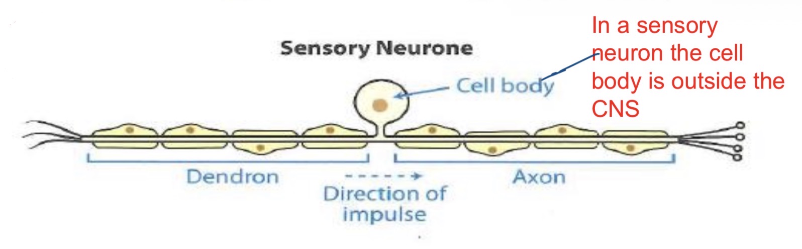
Connector neurones
( relay, association or intermediate) connect sensory and motor neurones within CNS.


Motor neurones
conduct impulses from CNS to effector (muscle or glands).

STRUCTURE OF A NEURONE:
Each neurone contains cell body (centron), which contains nucleus and other organelles as well as Nissl’s granules.
has extensions- dendrons (or dendrites if very small and numerous), which transmits impulses to cell body.
Extensions called axons, which transmit impulses away from cell body and terminate in synaptic bulbs.
In mammals, most of nerve fibres are myelinated. covered by myelin-made by Schwann cell.
There are small spaces between Schwann cells, giving rise to gaps in
sheath -nodes of Ranvier. Where axon exposed and saltatory conduction occurs.( jumping of action potential from node to node. Depolarisation occurs at nodes of Ranvier only.
Myelin
protects neurones and speeds up impulses.


NERVE IMPULSE
1. Resting potential
2. Action potential
3. Refractory period.
Polarised
there is an electrochemical gradient across membrane.
Depolarised
When sodium ions move into axon and cause inside of axon to become positive compared to outside.
Repolarised
When sodium ions move out of axon and cause inside of axon to become negative compared to outside.
Hyperpolarised
change in cells membrane potential that makes it more negative by allowing in negative chlorine ions during refractory period and inhibitory synapses
RESTING POTENTIAL
At rest, axon membrane is polarised and resting potential is -70mV.
Inside of membrane is negative relative to outside
maintained because cell membrane largely impermeable to flow of positive sodium ions.
Stimulation of neurone must be strong enough to reach the threshold intensity.

ACTION POTENTIAL
When neurone stimulated cell membrane becomes permeable to ions- diffuse into neurone down concentration gradient.
Once threshold reached ion gated channels open.
Sodium floods across membrane causing depolarisation at Nodes of Ranvier.
inside of neuron positive relative to outside (+40mV) .
This depolarisation and reversal of potential is an action potential.
REFRACTORY PERIOD
At peak of action potential recovery period starts and Na+ ions diffuse and pumped out of neurone.
rapidly restores resting potential and membrane becomes impermeable to Na+ ions.
During this recovery period a further impulse cannot occur as gated ion channels closed. slight overshoot and membrane becomes slightly morenegative (hyperpolarisation)
Hyper polarisation functions
Ensures action potential propagated in one direction only.
limits number of action potential and ensures each one is a separate entity/on its own
Axon all periods
1. Resting potential with inside of axon -70mV compared to outside.
2. Membrane becomes permeable and positive ions diffuse in – membrane is starting to become depolarised.
3. At -55mV(inside relative to outside) ion gated channels open and positive Ions flood in rapidly. Rapid depolarisation occurs. (Action potential)
4. Positive ions diffuse and are pumped out of axon. refractory period. slight overshoot hyper polarisation.
5. resting potential is restored and axon can conduct another nerve impulse.

Saltatory conduction
depolarisation takes place at nodes of Ranvier only, local circuits pass from node which is depolarised causing depolarisation at the next node
Threshold stimulus
refers to level of stimulus a neuron requires before an action potential is produced – a small amount of depolarisation can occur without an action potential but at a critical value (threshold) an action potential will occur.

All-or-nothing law.
increasing strength of stimulus will not result in a stronger impulse, once threshold is reached. As maximum number of action potentials that can be generated in a particular time due to time for repolarisation;
Sub threshold stimulus
sub threshold stimulus not result in an action potential. Stimulus above threshold will result in an action potential.
An increase in stimulus strength increases action potential frequency.
Further increase in stimulus results in nofurther increase in action potential.
IMPULSE PROPAGATION – LOCAL CIRCUITS
Action potentials move very rapidly along neurones (a wave of depolarisation).
On inside - Positive ions from depolarised zone pass along Inside of membrane toward polarised zone immediately in front.
On outside positive ions move back from polarised zone into depolarised zone.
These processes occur simultaneously that creates a wave of depolarisation that moves rapidly along the neurone
HOW TO INCREASE THE SPEED
Myelination increases conduction speed by saltatory conduction jumping of impulses from node to node because depolarisation can occur there.
Larger diameter. - provides greater surface area for ion exchange. less leakage proportionally or ions which could affect results. Giant axons found in earthworms, prawns and squid. (More ion gated channels)
Increasing temperature affects rate of diffusion of ions and will speed up action potentials. (more kinetic energy)

Synapse diagram

synapses
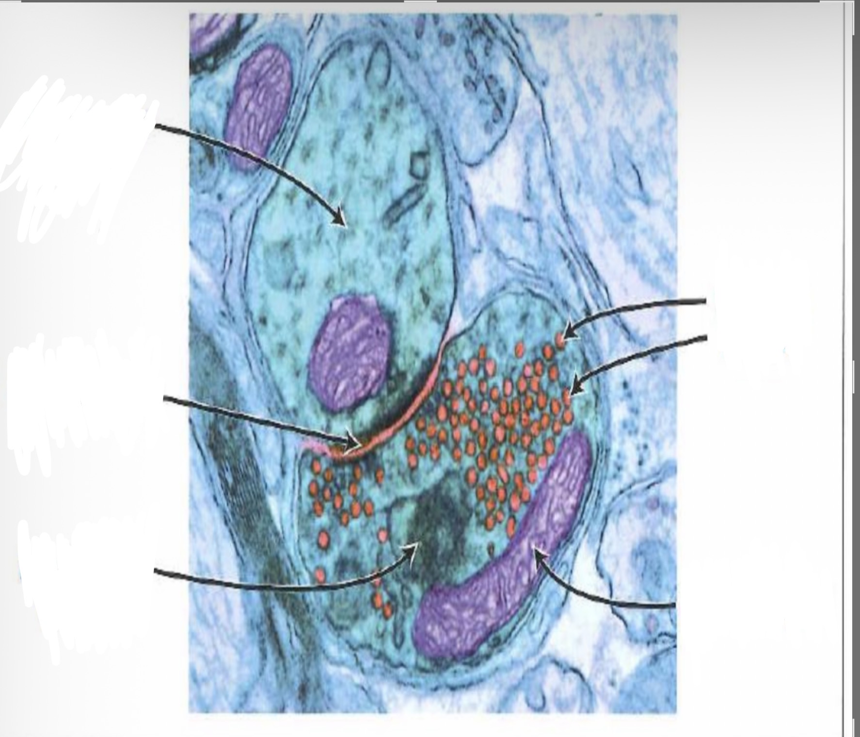
At end of axon, impulse arrives at synaptic bulbs. -contain neurotransmitters e.g. acetylcholine or noradrenaline.
These very close to their target cells, but small gap (20nm wide) between them -synaptic cleft.
membrane of neurone just before cleft- pre-synaptic membrane and one on other side- post-synaptic membrane.
whole structure- synapse.
membrane of post synaptic neuron contains receptors complementary to the neurotransmitter involved.
Impulse arrives at synaptic bulb and calcium ions diffuse into synaptic bulb.
Calcium ions stimulate movement of vesicles towards pre-synaptic membrane.
Vesicles fuse with pre-synaptic membrane and release acetylcholine molecules into cleft (exocytosis – active process requires ATP).
Acetylcholine molecules diffuse across cleft and attach to complementary receptors on post-synaptic membrane.
Causes opening of Na+ ion channels in membrane of post synaptic membrane.
Post-synaptic membrane becomes depolarised as Na+ diffuses in, which generates an excitatory post-synaptic potential (EPSP).
If EPSP reaches a threshold intensity, action potential evoked and impulse propagated in post-synaptic cell.
Acetylcholine hydrolysed by acetylcholinesterase enzyme into choline and Ethanoic acid ( prevents continued stimulation and action potentials in post synaptic neuron.).
Breakdown products diffuse across cleft; reabsorbed into synaptic bulb and resynthesised into acetylcholine (requires ATP produced by mitochondria)
FUNCTIONS OF SYNAPSES
Synapses slow down transmission slightly,
Synapses enable nerve impulses to pass from neuron to neuron – allows nervous communication to continue throughout the body.
ensure transmission occurs unidirectionality –vesicles found in presynaptic neuron only and receptors are found on post synaptic neuron only.Synaptic knob only at one end of neurone
protect effectors from over-stimulation,
integrate activity of different neurones which synapse with a single postsynaptic neurone.
Synapses provide flexibility – if no synapse each stimulus would provide an automatic and never changing response . Each receptor join with coordinator or effector and response would always be same
Summation important in providing flexibility that synapses demonstrate.
infrequent action potential reaching a synapse
may not be sufficient to cause an action potential in post synaptic membrane, however a series of impulses travelling along same neuron or a number of pre-synaptic neurones operating together may be enough to trigger an EPSP.
Junction of two neurones

INHIBITORY SYNAPSE function
(Function – Make it more difficult for synaptic transmission to take place
INHIBITORY SYNAPSE
Neurotransmitters from inhibitory synapse combine with receptors on post synaptic membrane.
It may allow negative chloride ions to move in.
It stops threshold being reached and an EPSP.
It prevents depolarisation of the post synaptic neuron.
If hyperpolarisation in inhibitory synapse
makes it difficult to reach threshold level and produce an EPSP
GABA
Inhibitory synapses reduce input of background stimuli which may affect reflex actions.
GABA a neurotransmitter released at inhibitory synapse in mammals. causes negative ions to flow into post synaptic neurones causing Hyperpolarisation (like refractory period) and it inhibits EPSP. functions: reducing anxiety and panic attacks by “damping down” nerve pathways In brain.
Nicotine Effect
Stimulates release of acetylcholine.
Causes depolarisation of the post synaptic membrane
Positive ions move in and an EPSP and threshold occurs.
Curare effect
Blocks receptors at neuromuscular junctions prevents synaptic transmission. No neurotransmitters can join – no ions move in. No transmission at neuromuscular junctions – loss of muscle function/paralysis.
Opioids Effect
Blocks calcium channels in presynaptic junction. Less exocytosis. Less neurotransmitter released and less EPSP’s. Used for pain relief