cns pathology labs
 Call Kai
 Learn
 Practice Test
 Spaced Repetition
 Match
 Flashcards
 Knowt Play
1/31

There's no tags or description
Looks like no tags are added yet.
32 Terms
g: purulent patches
m: focally dense inflammatory cellular infiltrate
tissue edema
acute pyogenic meningitis

acute pyogenic meningitis

acute pyogenic meningitis

brain abscess
swelling liquefied center filled with pus
brain abscess
in children and cerebellum
g: circumscribed
cystic mass
solid mural nodules
m: bipolar astrocytes
grade 1: pilocytic astrocytoma
g: ill defined
infiltrative
white growth
m: moderate cellularity
moderate atypia
fibrillary/ protoplasmic astrocytes
grade 2: low grade diffuse atrocytoma
g: infiltrative growth
m: high cellularity
mitotic activity
no necrosis
pleomorphic astrocytes
grade 3: anaplastic astrocytoma
g: infiltrative growth
large
hemorrhage
necrosis
cyst
m: high cellularity
marked cell anaplasia
palisading necrosis
vascular endothelial proliferation
grade 4: glioblastoma multiforme astrocytoma

grade 1: pilocytic astrocytoma

grade 1: pilocytic astrocytoma

grade 2: low grade diffuse atrocytoma
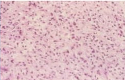
grade 2: low grade diffuse atrocytoma

grade 3: anaplastic astrocytoma

grade 3: anaplastic astrocytoma

grade 4: glioblastoma multiforme astrocytoma

grade 4: glioblastoma multiforme astrocytoma
intra axial
slow growing
dystrophic calcification
closely packed cells
fried agg appearance
oligodendroglioma

oligodendroglioma
intra axial
in 4th ventricle
childhood and adolescence
leads to hydrocephalus
m: grade 2 and 3
perivascular pseudorosettes
ependymal rosette
ependymoma

ependymoma

ependymoma

medulloblastoma
cerebellum and children
grade 4
highly malignant rapidly growing
subtle round cell
moderate pleomorphism
high mitotic count
homer wright rosettes
medulloblastoma
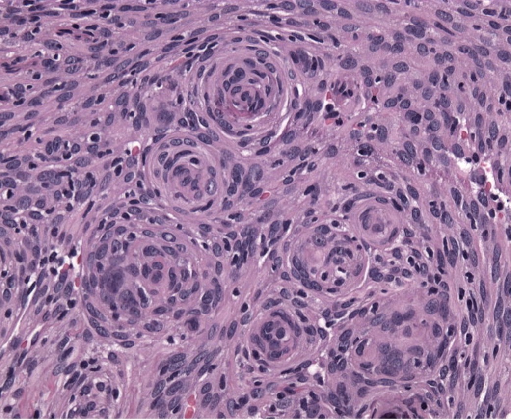
meningioma

psamomma bodies in meningioma

meningioma
concentric cell whorls
proliferated meningothelilal cells
oval cells indistinct cell borders
pale cytoplasm regular round nuclei
psamomma bodies
dystrophic calcification
meningioma

brain metastasis
tubule formation
well circumscribed and demarcated
hyperchromatasia
variation in nuclear size and shape
mitosis
brain metastasis

shwannoma
shwann cells
antoni A: spindle shaped elongated nuclei parallel bundles nuclei has palisade pattern
antoni B: larger ovoid haphazardly areanged microcytic stroma and myxomatous.
shwannoma