Chapter 2: How We See the Invisible World
 Call Kai
 Learn
 Practice Test
 Spaced Repetition
 Match
 Flashcards
 Knowt Play
1/63

There's no tags or description
Looks like no tags are added yet.
64 Terms
Visible Light
Electromagnetic Waves/Radiation the human eye can pick up

Wavelength
Distance between one peak of a wave and the next peak

Amplitude
The height of each peak

Frequency
Rate of vibration | # of wavelengths in a certain time period

Reflection
The bouncing back of a ray of light when the ray hits a surface that it does not go through

Absorption
Material captures the energy of a ray of light

Transmission
Lightwave travels through material

Refraction
Direction change in lightwave in a new medium (Bending of light)

Refractive Index
A measure of the light-bending ability of a medium
Lens
Collects light then refracts it to converge into one point (image or focal point)

Convex Lens
Light refracts towards focal point on the other side of lens | light focuses into one point after passing through lens

Concave lens
Light refracts away from focal point after passing through lens | light spreads/scatters after passing through lens

Focal length
Distance to the focal point

Photon
A fundamental particle of light and electromagnetic radiation

Electromagnetic Spectrum
The different types of electromagnetic radiation

Fluorescent dyes
-Absorb UV or blue light
-Use the energy to emit photons of different colors (Visible)
Magnification
Lens ability to enlarge image
Resolution
Ability to view the separation of two different points
Ex., microscope resolving power: 0.6 nm | microscope can distinguish two points 0.6 nm or more apart
How does smaller resolving power affect resolution?
- Smaller resolving power means higher resolution, and smaller details are more visible

Contrast
Visible difference between parts of specimen
Galileo Galilei
Used a compound microscope (two lenses) to examine insect parts

Robert Hooke (1665)
Observed cork cells (dead wood cells) described as "Honeycomb" or "Small Boxes of Air" Micrographia (1665)

Light Microscopes
Use light beams (up to 1000x)

Electron Microscopes
Use short-wavelegth electron beams (100,000x)

Scanning Probe Microscopy
Uses sharp probes over surface of specimen (no light or electrons)

Brightfield Microscope
Usually, a compound microscope (2 or more lenses)" ocular (10X) and objective (4X to 100X)

Total Magnifcation
Ocular lens X Objective Lens

What is the purpose of immersion oil in a Brightfield Microscope?
To keep light from bending in high-magnification lens

Brightfield Illumination
Dark objects are visible against a bright background
- Light reflected off the specimen does not enter the objective lens

Darkfield Illumination
Light objects are visible against a dark background
- Only light reflected off the specimen enters the objective lens when opaque disk placed in condenser

Phase-Contrast Microscopy
Brings together direct rays and diffracted rays to form an image without staining
- Used for examination of internal living cell structures

Differential Interference Contrast Microscopy (DIC)
Similar to phase contrast: two light beams and prisms split light beams to give more contrast and color

Fluorescence Microscopy
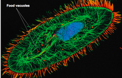
Uses UV Light - Cells stained with fluorescent dyes (Fluorochromes) absorb UV and emit visible light
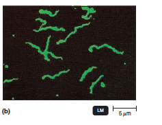
Confocal Microscopy
Cells are stained with fluorochrome dyes, then short wavelength (blue) light used to excite specimen --> Illumination and 3D image from computer

Two-Photon Microscopy
Cells are stained with fluorochrome dyes, then long-wavelength (red, two-photon) excite the dyes
- Used to study living cells up to 1mm deep (biofilms)
Electron Microscopy
Uses electrons instead of light
- Shorter wavelength of electrons gives greater resolution
- Used on nonliving material, subcellular structures and some molecular structures (single strands of DNA)

Transmission Electron Microscopy (TEM)
A beam of electrons passes through ultrathin sections of specimens, an electromagnetic lens, then focuses on a projector lens
- Heavy-metal salt stains

What is the magnification range and resolution of Transmission Electron Microscopy?
10,000x to 100,000x; 2.5 nm resolution
Scanning Electron Microscopy (SEM)
Beam of electrons scan surface of specimen; secondary electrons bounce off surface of the cell to provide a 3D study of the cell surface
What is the magnification range and resolution of Scanning Electron Microscopy?
1000x to 10,000x; resolution of 10 nm
Scanning Probe Microscopy
A very sharp probe passes over the specimen --> produces information to create images with magnifications up to 100,000,000X | ex. Scanning tunneling probe, Atomic force probe
- Used for research to observe individual atoms on surfaces

Scanning Tunneling Microscopy (STM)
Tungsten probe used to scan specimen and reveal details of surface

Atomic Force Microscopy
Uses a metal-and-diamond probe placed onto a specimen and produces three-dimensional images

Wet Mount
Live Specimen placed on a slide in a drop of liquid and a cover slip to study cell behavior

Fixed Specimen
Heat fixing or chemically treating the specimen to kill microorganisms

Smear
Thin film of microorganisms spread in a drop of water on the slide

Stain
Coloring of microbe with a dye to emphasize certain structures

Chromophore
The colored ion in the stain
Counterion
The uncolored ion in a stain
What is the charge of a chromophore in a basic dye?
Cation (Positive)
What is the charge of a chromophore in an acidic dye?
Anion (Negative)
Simple Stain
Single dye highlights entire microorganism making cell structures and shape visible in the same color

Mordant
The chemical substance that fixes or intensifies stain to enlargen specimen

Basic Stain
Stains negatively charged molecules and structures such as nucleic acids and proteins

Acidic Stain
Stains positively charged molecules and structures

Negative stain
Stains backround, not the specimen

Differential Stains
Used to distinguish organisms based on their interactions with two or more stains; organisms appear in different colors

Gram Staining (Hans Christian Gram 1884)
Distinguishes bacteria with different types of cell walls
- Gram-positive bacteria: thick peptidoglycan cell walls
-Gram-negative bacteria: thin peptidoglycan cell walls + layer of lipopolysaccharides

Acid-Fast Stain
Only binds to bacteria containing waxy material in their cell walls (not decolorized by alcohol
- Identifies Mycobacterium and Nocardia

Capsules Staining
Capsules - gelatinous covering that does not accept most dyes
- Presence indicates the microbe's virulence (ability to cause disease)
- Suspension of India ink/nigrosin contrasts with backround causing a halo effect around the cell

Endospore Staining
Spores appear green within red/pink cells
Primary stain: malachite green (with heat)
Decolorize cells: water
Counterstain: safranin

What endospore-producing bacteria are Endospore-Staining techniques used for?
Bacillus, Clostridium, and Clostridioides
Flagella Staining
Staining that thickens the flagella by applying mordant (tannic acid or potassium acid), then staining (pararosaniline or basic fuschin)

How are specimens prepared for Electron Microscopy?
Cells are embedded in plastic resin, dehydrated in multiple ethanol solution soaks, cut into thin sections (ultramicrotome), and then fixed to fine copper wire or carbon-fire grids and stained
