KN 232 - Week 9 Structure List
 Call Kai
 Learn
 Practice Test
 Spaced Repetition
 Match
 Flashcards
 Knowt Play
1/65

There's no tags or description
Looks like no tags are added yet.
66 Terms
Cranial Nerves
One of 12 pairs of nerves that are part of the PNS and originate from the brain

CN I — Olfactory nerve
Sensory function: smell

CN II — Optic nerve
Sensory function: vision
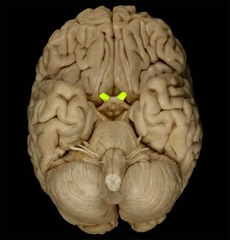
CN III — Oculomotor nerve
- Somatic motor: moves the eyes and elevates the eyelid
- Parasympathetic motor: constricts the pupil and changes lens shape for near vision

CN IV — Trochlear nerve
Somatic motor: controls the superior oblique extrinsic eye muscle

CN V — Trigeminal nerve
- Sensory: sensation from the face, anterior scalp, nasal cavity, most of the oral cavity, teeth, anterior two-thirds of the tongue, part of the ear, and meninges
- Motor: muscles of mastication and other listed muscles

CN VI — Abducens nerve
Somatic motor: controls the lateral rectus extrinsic eye muscle

CN VII — Facial nerve
- Sensory: taste from the anterior two-thirds of the tongue. Somatic motor: facial expression and other listed muscles
- Parasympathetic motor: increases secretion from the lacrimal, submandibular, and sublingual glands

CN VIII — Vestibulocochlear nerve
Sensory: hearing (cochlear branch) and equilibrium (vestibular branch)

CN IX — Glossopharyngeal nerve
- Sensory: taste and general sensation from the posterior one-third of the tongue, part of the pharynx, and sensory information from carotid bodies
- Motor: stylopharyngeus muscle. Parasympathetic motor: increases parotid salivary gland secretion

CN X — Vagus nerve
- Sensory: information from the heart, lungs, most abdominal organs, and parts of the ear, pharynx, and larynx. Motor: most pharyngeal muscles and all laryngeal muscles
- Parasympathetic motor: controls smooth muscle and glands in the heart, lungs, larynx, trachea, and most abdominal organs
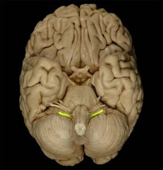
CN XI — Accessory nerve
Somatic motor: controls the trapezius and sternocleidomastoid muscles

CN XII — Hypoglossal nerve
Somatic motor: controls the intrinsic and extrinsic tongue muscles

Spinal Cord
Structural and functional link between the brain and the rest of the body; carries sensory and motor information

Conus medullaris
Tapered inferior end of the spinal cord

Cauda equina
Bundle of spinal nerve roots extending inferiorly from the conus medullaris

Meninges
Protective membranes surrounding the spinal cord and other CNS structures

Dura mater
Tough, outermost meningeal layer that helps stabilize the spinal cord

Arachnoid mater
Web-like meningeal layer located between the dura mater and pia mater

Pia mater
Delicate meningeal layer that adheres to the spinal cord

Epidural space
Space between the dura mater and vertebra; contains fat, areolar connective tissue, and blood vessels

Subarachnoid space
Space deep to the arachnoid mater through which cerebrospinal fluid (CSF) flows

Dorsal root
Carries sensory information into the spinal cord

Ventral root
Carries motor fibers away from the spinal cord

Dorsal root ganglion
Contains the cell bodies of sensory neurons

Spinal nerves
Mixed nerves containing sensory and motor fibers; formed where the dorsal and ventral roots join

Cervical spinal nerves
8 pairs of spinal nerves associated with the cervical region

Thoracic spinal nerves
12 pairs of spinal nerves associated with the thoracic region

Lumbar spinal nerves
5 pairs of spinal nerves associated with the lumbar region

Sacral spinal nerves
5 pairs of spinal nerves associated with the sacral region

Coccygeal spinal nerves
1 pair of spinal nerves associated with the coccygeal region

Posterior ramus
Branch of a spinal nerve that supplies deep back muscles and skin of the back

Anterior ramus
Branch of a spinal nerve that supplies the anterolateral trunk and limbs; also forms nerve plexuses

Gray matter
Contains neuron cell bodies, dendrites, unmyelinated axons, and glial cells

White matter
Contains myelinated axons that carry signals to and from the brain

Anterior horn
Contains cell bodies of somatic motor neurons

Lateral horn
Contains cell bodies of autonomic motor neurons; present in spinal cord segments T1-L2

Posterior horn
Contains sensory neuron axons and interneuron cell bodies

Gray commissure
Band of gray matter surrounding the central canal; contains unmyelinated axons connecting the left and right gray matter

Vertebral artery
Artery that supplies blood to the brain and upper spinal cord

Anterior median fissure
Deep longitudinal groove on the anterior surface of the spinal cord

Posterior median sulcus
Shallow longitudinal groove on the posterior surface of the spinal cord

Ligamentum flavum
Ligament connecting adjacent vertebral laminae and helping support the vertebral column

Internal vertebral venous plexus
Network of veins within the vertebral canal that drains blood from the vertebral region

Central canal
Small channel through the spinal cord that contains cerebrospinal fluid

Denticulate ligament
Lateral extension of the pia mater that helps suspend and stabilize the spinal cord

Brain
Part of the CNS; involved in sensory processing, movement, thought, memory, and other functions

Diencephalon
Brain region containing the thalamus, hypothalamus, and epithalamus

Thalamus
Gateway to the cerebral cortex; relays information to the cortex
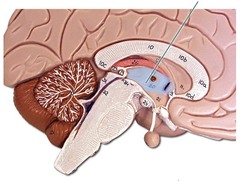
Hypothalamus
Controls the autonomic nervous system and endocrine system

Pituitary gland
Endocrine gland that releases hormones regulating other glands and body functions; its activity is controlled by the hypothalamus
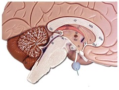
Epithalamus
Region of the diencephalon that contains the pineal gland

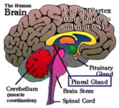
Pineal gland/body
Secretes melatonin
Brainstem
Connects the cerebrum, diencephalon, and cerebellum to the spinal cord; consists of the midbrain, pons, and medulla oblongata

Midbrain
Upper part of the brainstem

Superior colliculi
Midbrain structures involved in visual reflexes and orienting the eyes and head toward visual stimuli

Inferior colliculi
Midbrain structures involved in auditory processing and reflexes

Pons
Part of the brainstem located between the midbrain and medulla oblongata

Medulla oblongata
Inferior part of the brainstem that connects with the spinal cord

Corpus callosum
Largest white-matter tract connecting the left and right cerebral hemispheres

Ventricles
Cavities within the brain lined with ependymal cells that contain cerebrospinal fluid

Lateral ventricles
Two large cavities located within the cerebrum

Third ventricle
Narrow cavity in the middle of the diencephalon

Cerebral aqueduct
Channel connecting the third ventricle to the fourth ventricle

Fourth ventricle
Cavity between the pons and cerebellum; connects with the subarachnoid space and the spinal cord's central canal

Subdural space
Space between the dura mater and arachnoid mater
