Block 2: Circulatory System Slides
 Call Kai
 Learn
Learn Practice Test
 Spaced Repetition
 Match
Match Flashcards
 Knowt Play
1/157

There's no tags or description
Looks like no tags are added yet.
Name | Mastery | Learn | Test | Matching | Spaced | Call with Kai | Chat |
|---|
No analytics yet
Send a link to your students to track their progress
158 Terms

Green: endocardium
Yellow: myocardium
Identify the structures green and yellow boxes (2 structures)
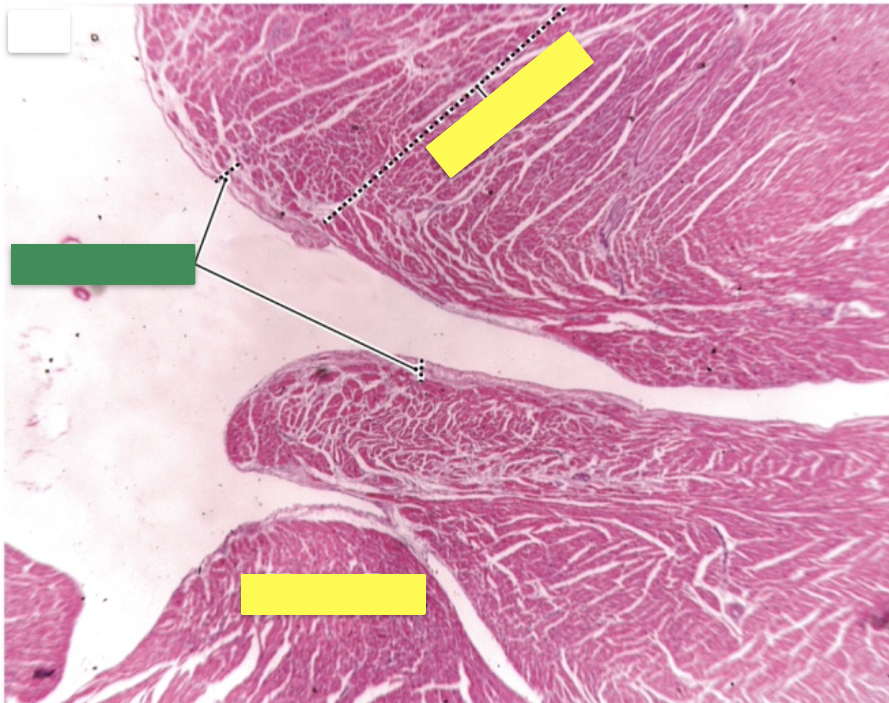

heart (heart wall)
Identify the organ

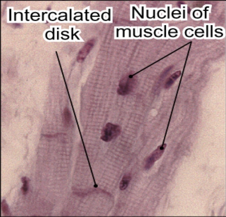
Pink: Intercalated disk
Green: Nuclei of muscle cells
Identify the structures

heart
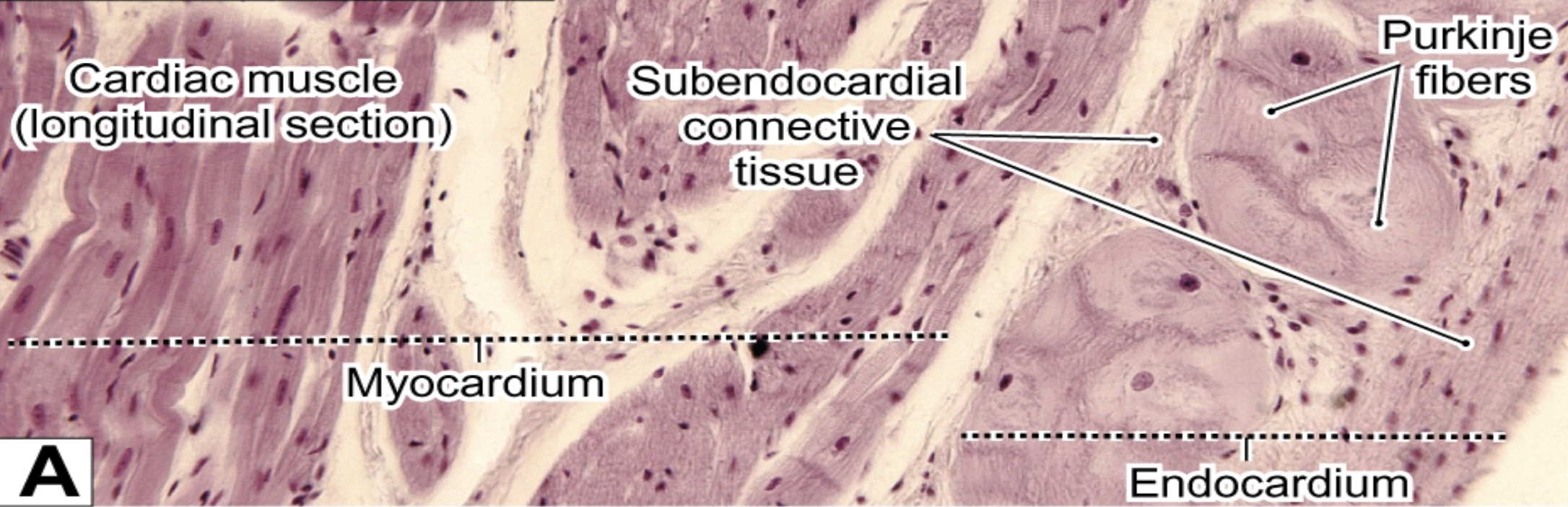
Identify the organ


Purkinje fibers
Identify the structure(s)
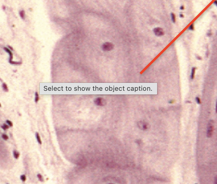

subendocardial connective tissue
Identify the green structure

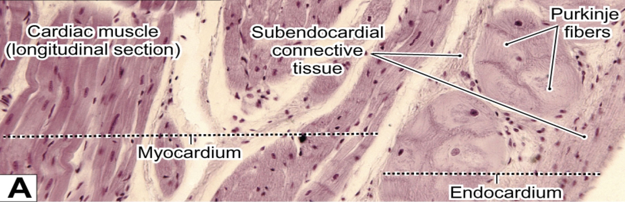
Purkinje fibers
Identify the pink structure


Endocardium
Identify the purple structure

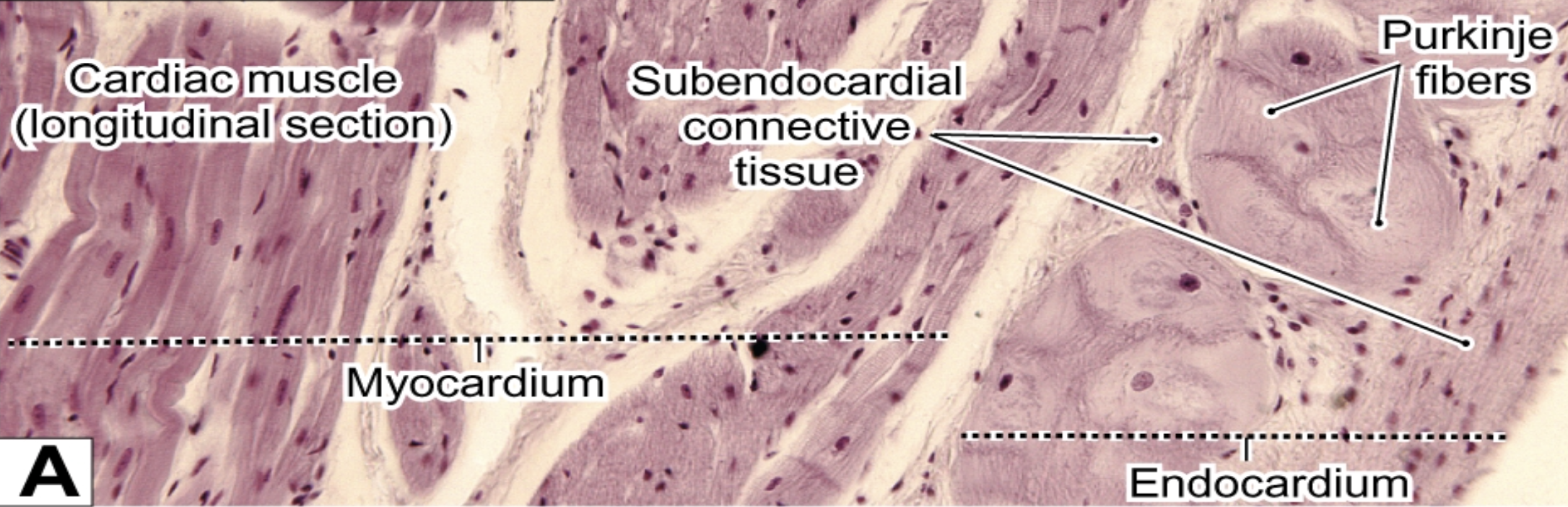
myocardium
Identify the orange structure


Cardiac muscle
Identify the blue structure


heart
Identify the organ


Heart
Identify the organ


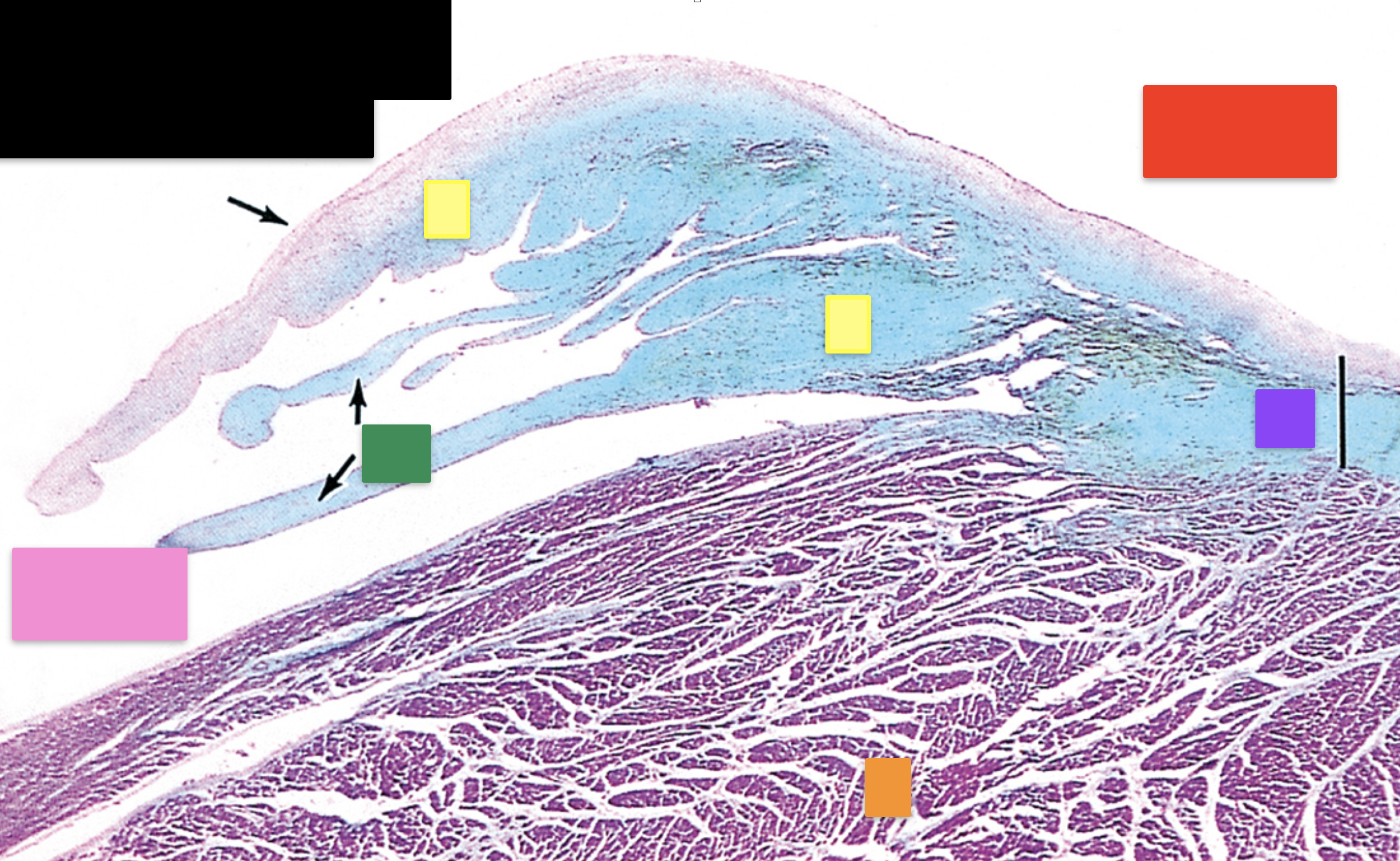
atrium
Identify the red structure

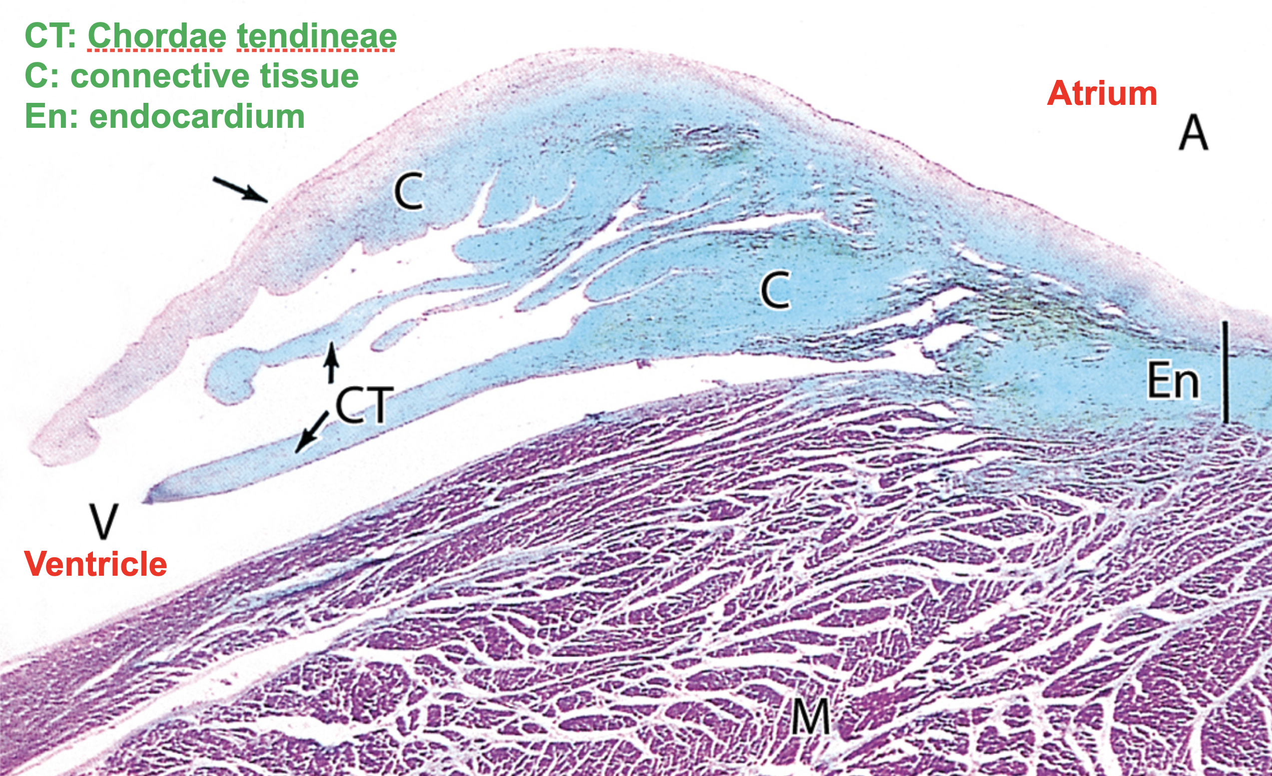
endocardium
Identify the purple structure


myocardium
Identify the orange structure


connective tissue
Identify the yellow structure


chordae tendineae
Identify the green structure


ventricle
Identify the pink structure

heart
identify the organ


intercalated disk
Identify the structure


heart
Identify the organ


branched cardiac muscle
Identify the structure


heart
Identify the organ


nuclei of cardiac muscle cells
Identify the structure


heart
Identify the organ


Red: Lumen of the blood vessels
Orange: Epicardium
Green: Myocardium
Identify the structures

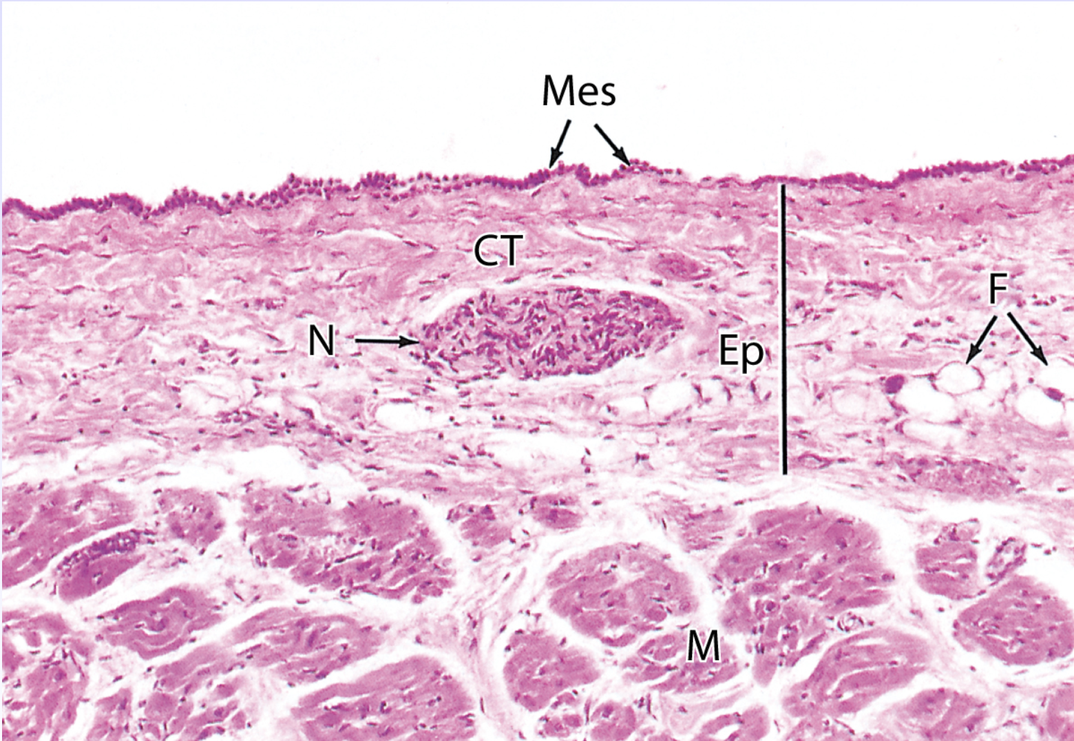
heart
Identify the organ

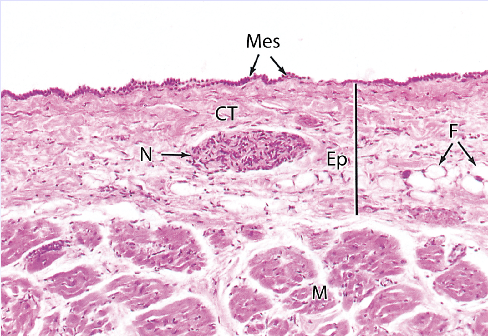
Fat (F)
Identify the structure indicated by the yellow box


Autonomic Nerves (N)
Identify the structure indicated by the blue box

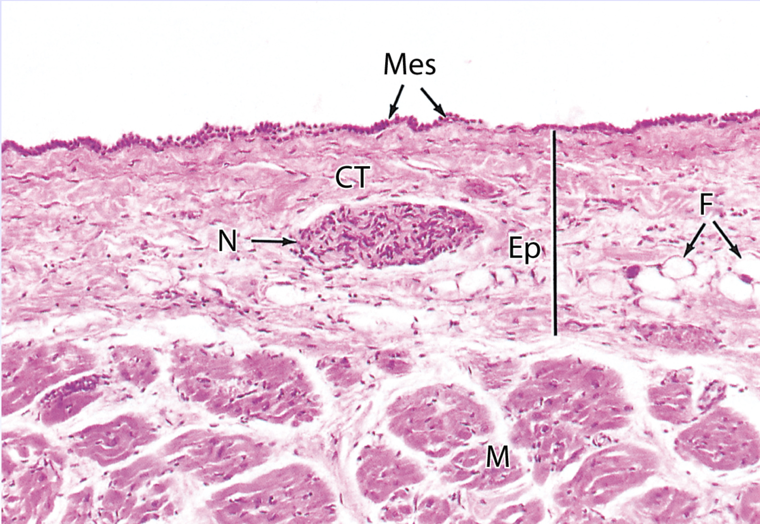
Epicardium (Ep)
Identify the structure indicated by the red box


Myocardium (M)
Identify the structure indicated by the dark green box


Mesothelium (Mes)
Identify the structure indicated by the purple box

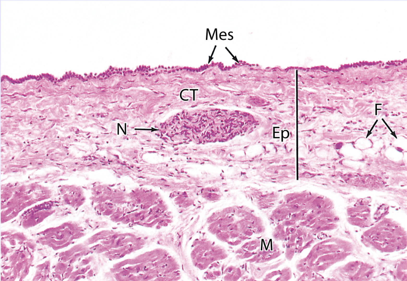
Loose Connective Tissue (CT)
Identify the structure indicated by the light green box (area surrounding it)
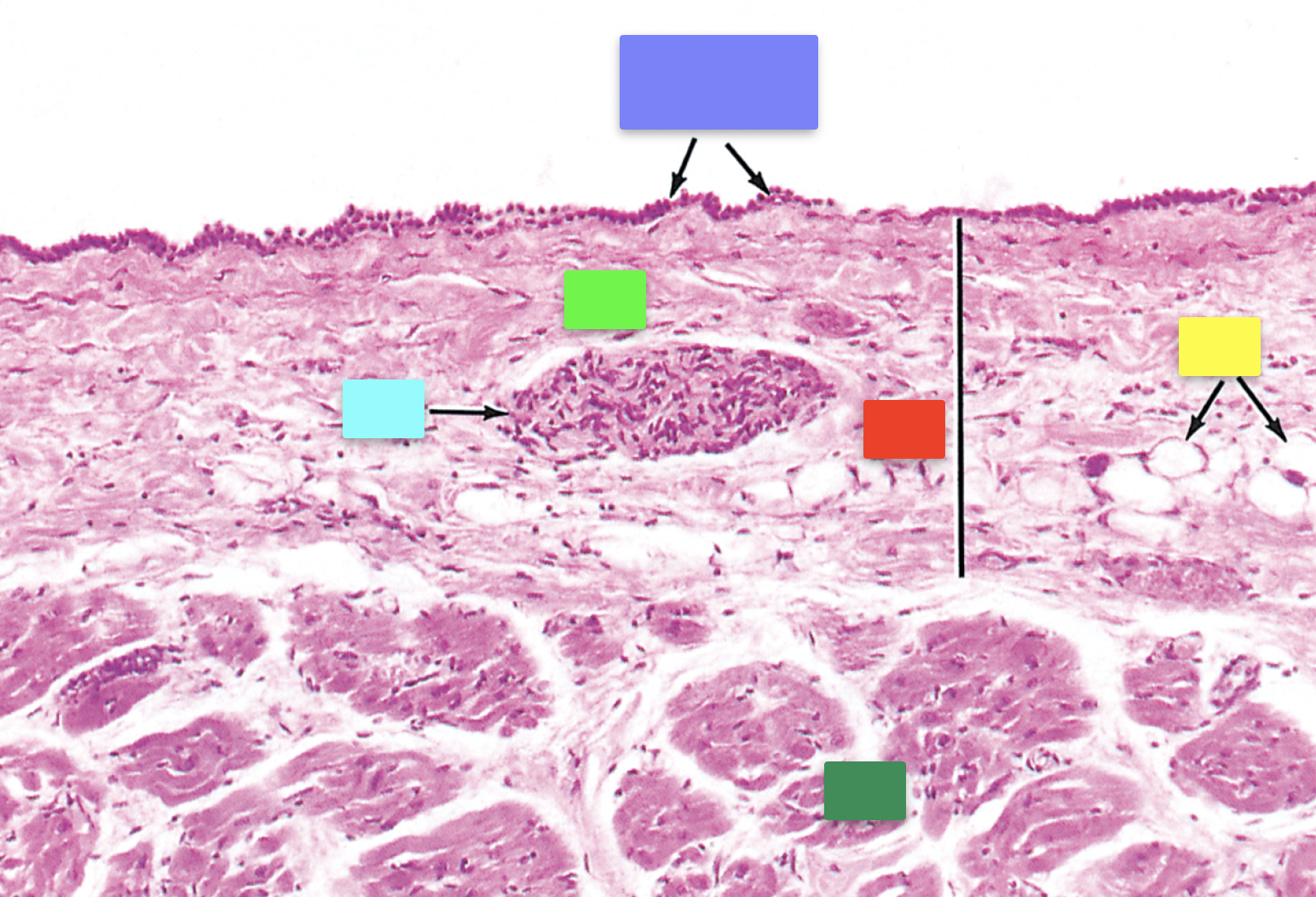
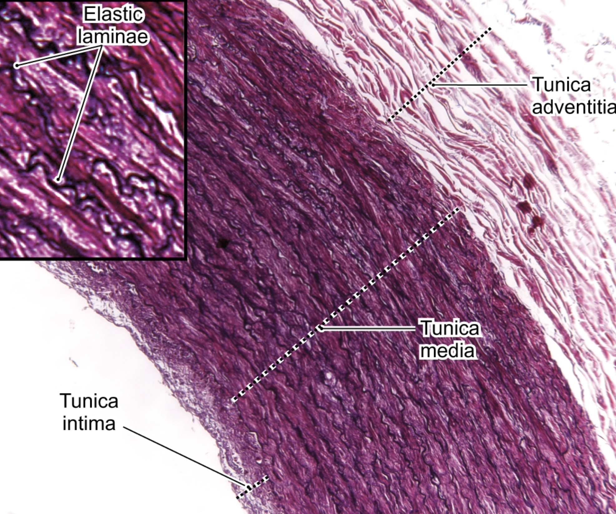
large artery (elastic artery)
Classify the organ


Red: Tunica adventitia
Yellow: Tunica media
Green: Tunica intima
Identify the structures indicated by the red, yellow and green boxes


elastic laminae
Identify this structure

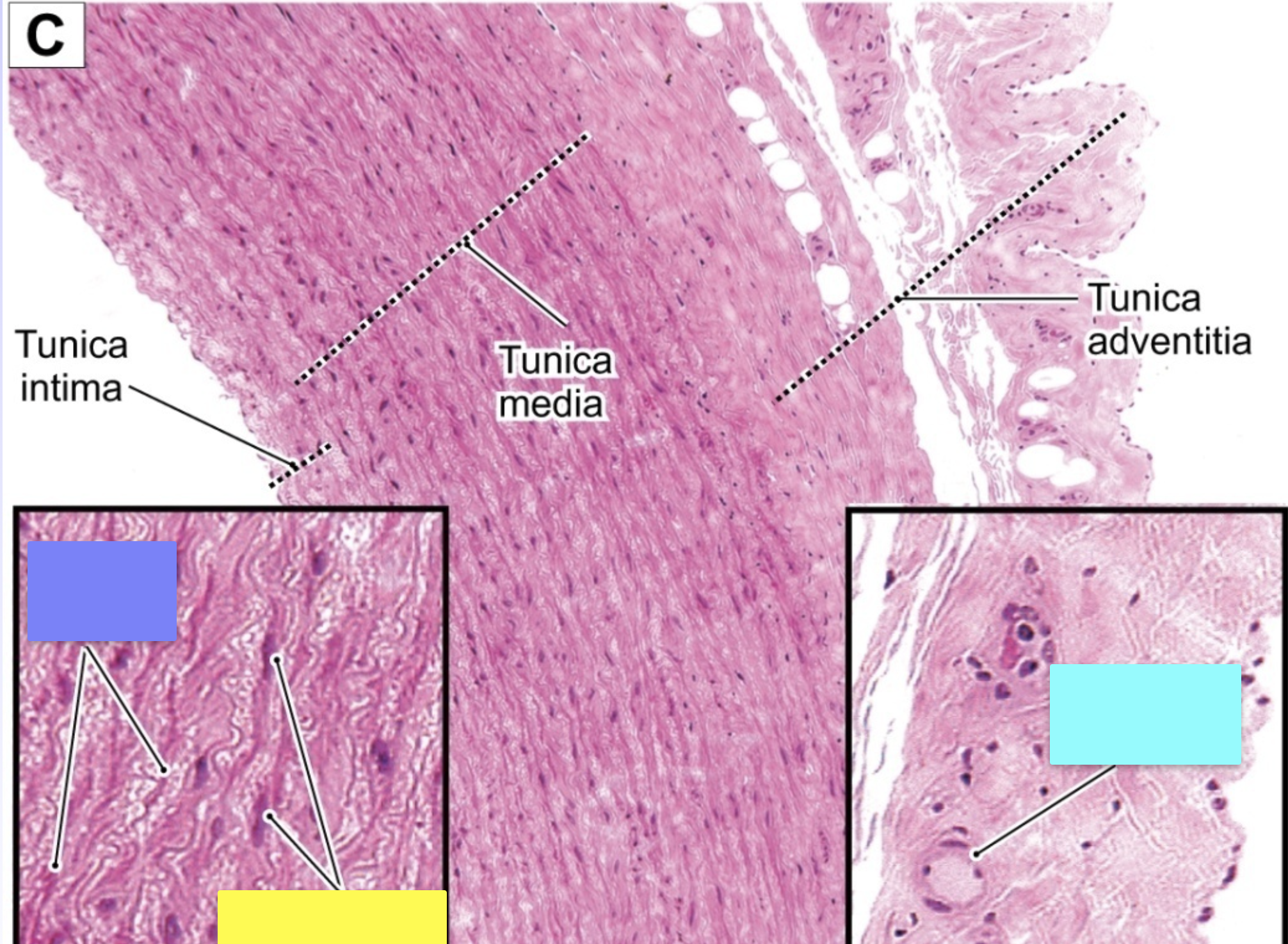
large artery
Classify the organ


Purple: tunica intima
Green: tunica media
Blue: tunica adventitia
Identify the structures indicated by the purple, green and blue boxes


vasa vasorum (in tunica adventitia)
Identify the structure

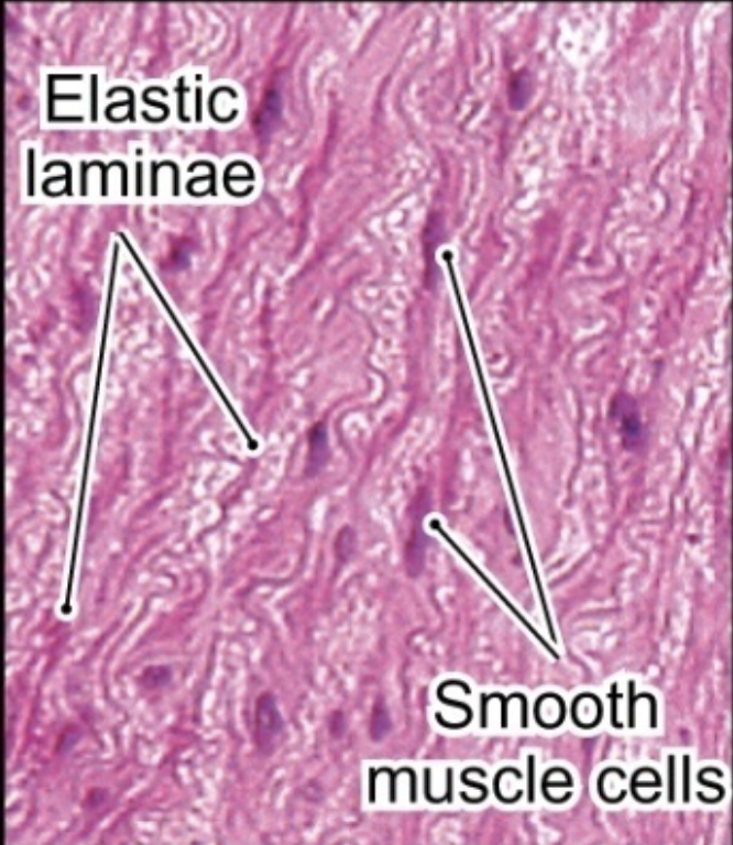
Elastic laminae
Identify the structure indicated by the purple box


Smooth muscle cells
Identify the cells indicated by the yellow box
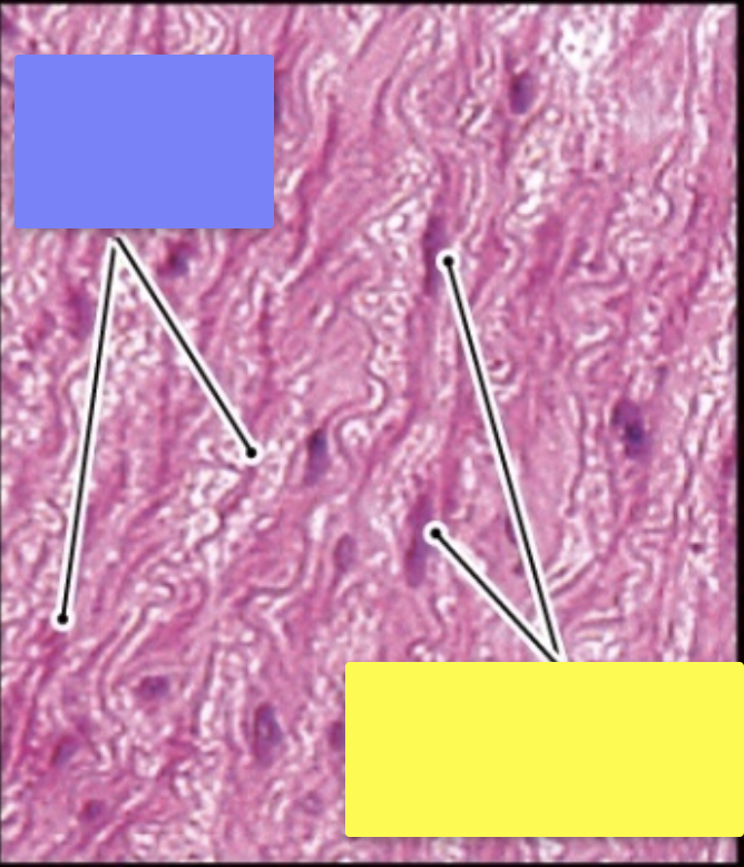

Medium artery (muscular artery)
Classify the organ
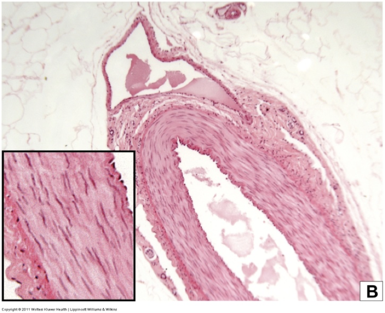
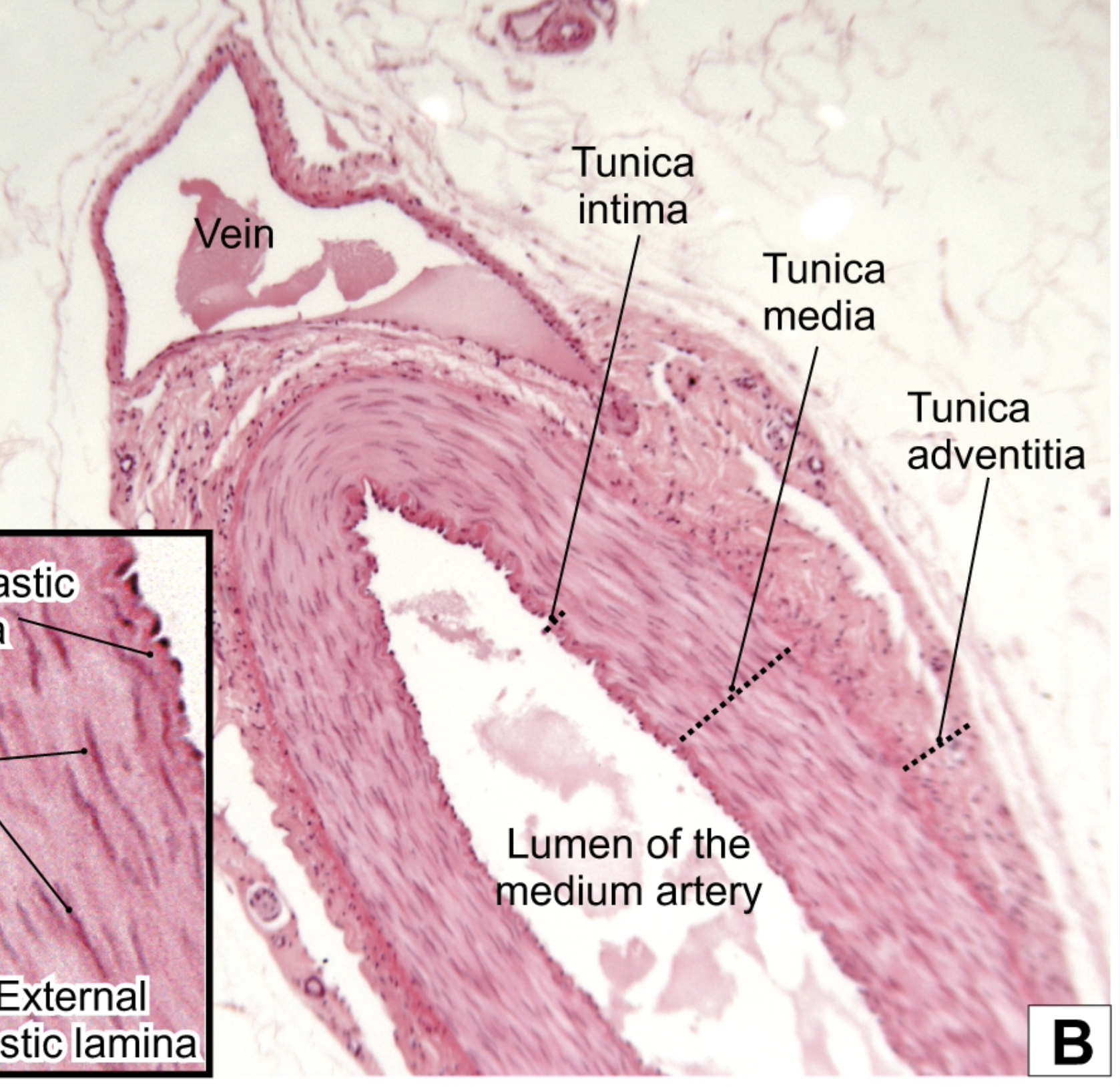
Red: Tunica intima
Dark green: Tunica media
Light green: Tunica adventitia
Yellow: Lumen of the medium artery
Blue: Vein/Lumen of vein
Identify the structures (purple not a structure)


Internal elastic lamina
Identify the structure indicated by the yellow box


Smooth muscle cells
Identify the cells indicated by the red box

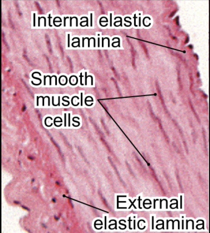
External elastic lamina
Identify the structure indicated by the blue box

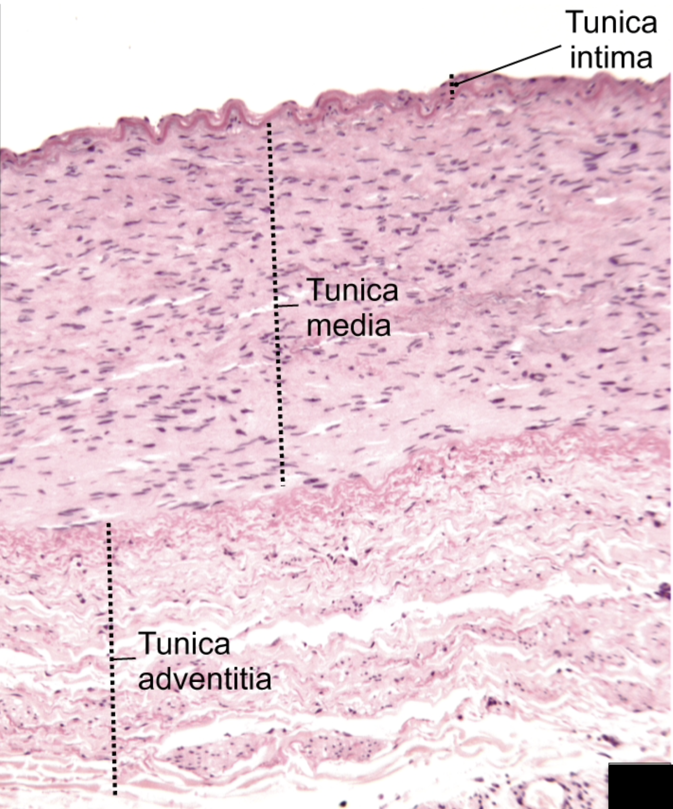
medium artery
Classify the organ


Purple: tunica adventitia
Green: tunica media
Blue: tunica intima
Identify the structures


Subendothelium
Identify the structure indicated by the blue box


Internal elastic lamina
Identify the structure indicated by the red box


endothelium
(if said to classify instead of identify, simple squamous epithelium i think)
Identify the structure indicated by the green box
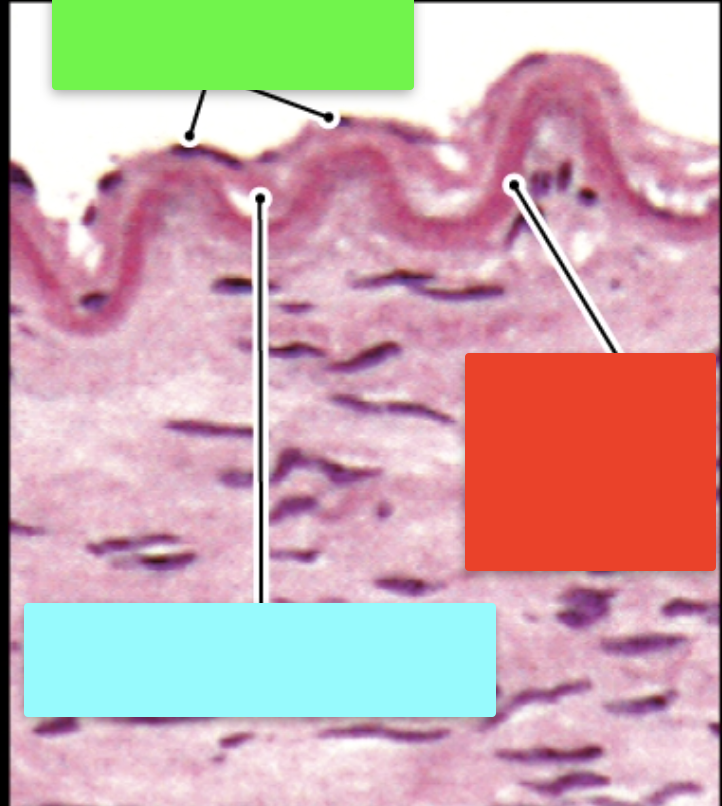

medium artery
Classify the organ


External elastic lamina
Identify the structure indicated by the green box


tunica media
Identify the structure indicated by the red box


internal elastic lamina
Identify the structure indicated by the blue box
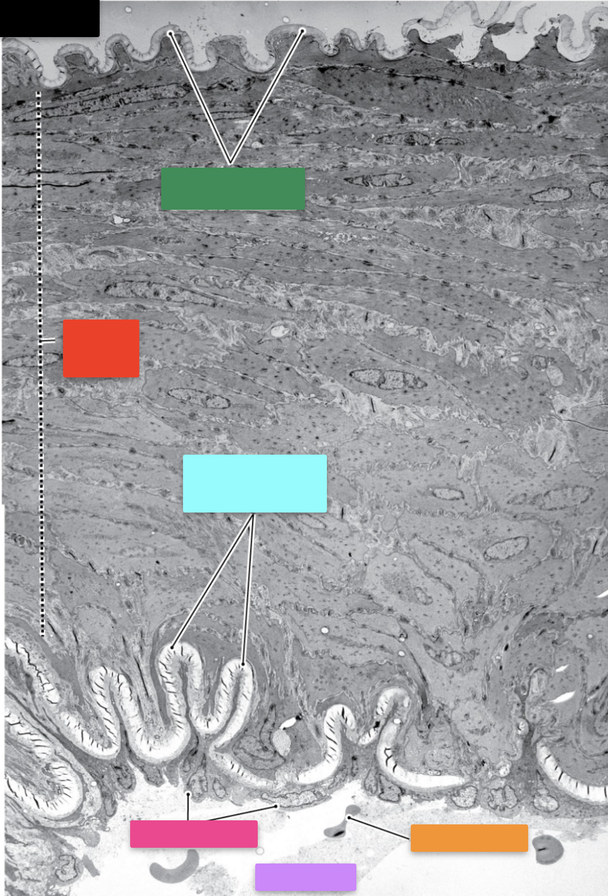

Endothelium
Identify the structure indicated by the pink box


erythrocytes
Identify the structure indicated by the orange box


lumen
Identify the structure indicated by the purple box


Medium artery
Classify the organ


endothelial cell junction
Identify the structure indicated by the purple box


internal elastic lamina
Identify the structure indicated by the red box


tunica media
Identify the structure indicated by the orange box


dense bodies
Identify the structure indicated by the blue box


basal lamina
Identify the structure indicated by the pink box


collagen fibers
Identify the structure indicated by the green box


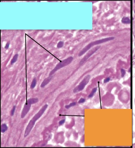
Blue: nuclei of smooth muscle cells
Orange: Internal elastic lamina
Identify these structures

Green: small arteries
Orange: arteriole
Blue: lumen of small vein
Classify the structures


small artery
Classify the organ


Green: Smooth muscle
Orange: erythrocytes
Purple: endothelial cells
Blue: internal elastic lamina
Pink: collagen fiber bundle
Identify the cells/structures indicated by the boxes


arteriole
Classify the structure indicated by the orange box
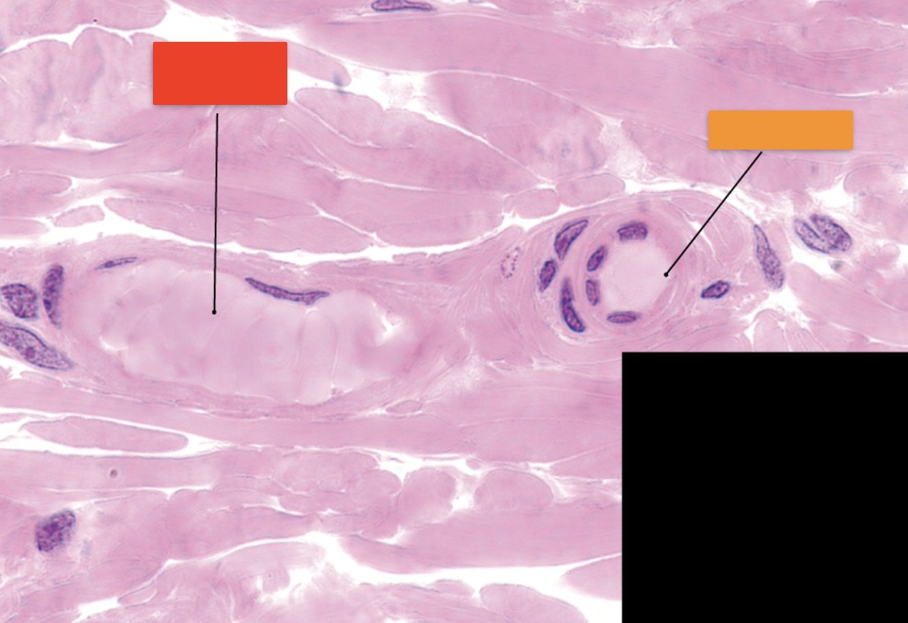

lumen of venule
Identify the structure indicated by the red box


Pink: endothelial cell
Blue: smooth muscle cell
Identify the cells


arteriole
Classify the organ


Pink: nucleus of schwann cell
Orange: internal elastic lamina
Purple: endothelium-smooth muscle cell junctions
Blue: nuclei of endothelial cells
Red: nucleus of smooth muscle cell
Identify the structures

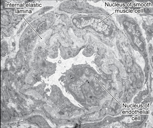
arteriole
Classify the organ


Blue: internal elastic lamina
Pink: nucleus of smooth muscle cell
Orange: nucleus of endothelial cell
Identify the structures


continuous capillaries
Classify the organ


fenestrated capillaries
Classify the organ


discontinuous/sinusoidal capillaries
Classify the organ


continuous capillary
Classify the organ


Pink/Green: intercellular junction
Yellow: erythrocyte
Orange: Marginal fold
Blue: caveolae
Red: Basal lamina of pericyte
Purple: pericyte
Identify the structures


Fenestrated capillary
Classify the organ


Blue: secretory granules containing hormones
Red: endothelial cell cytoplasm
Yellow: diaphragm of fenestration
Green: lumen of fenestrated capillary
Purple: collagen fibers
Identify the structures


discontinuous (sinusoidal) capillary
Classify the organ
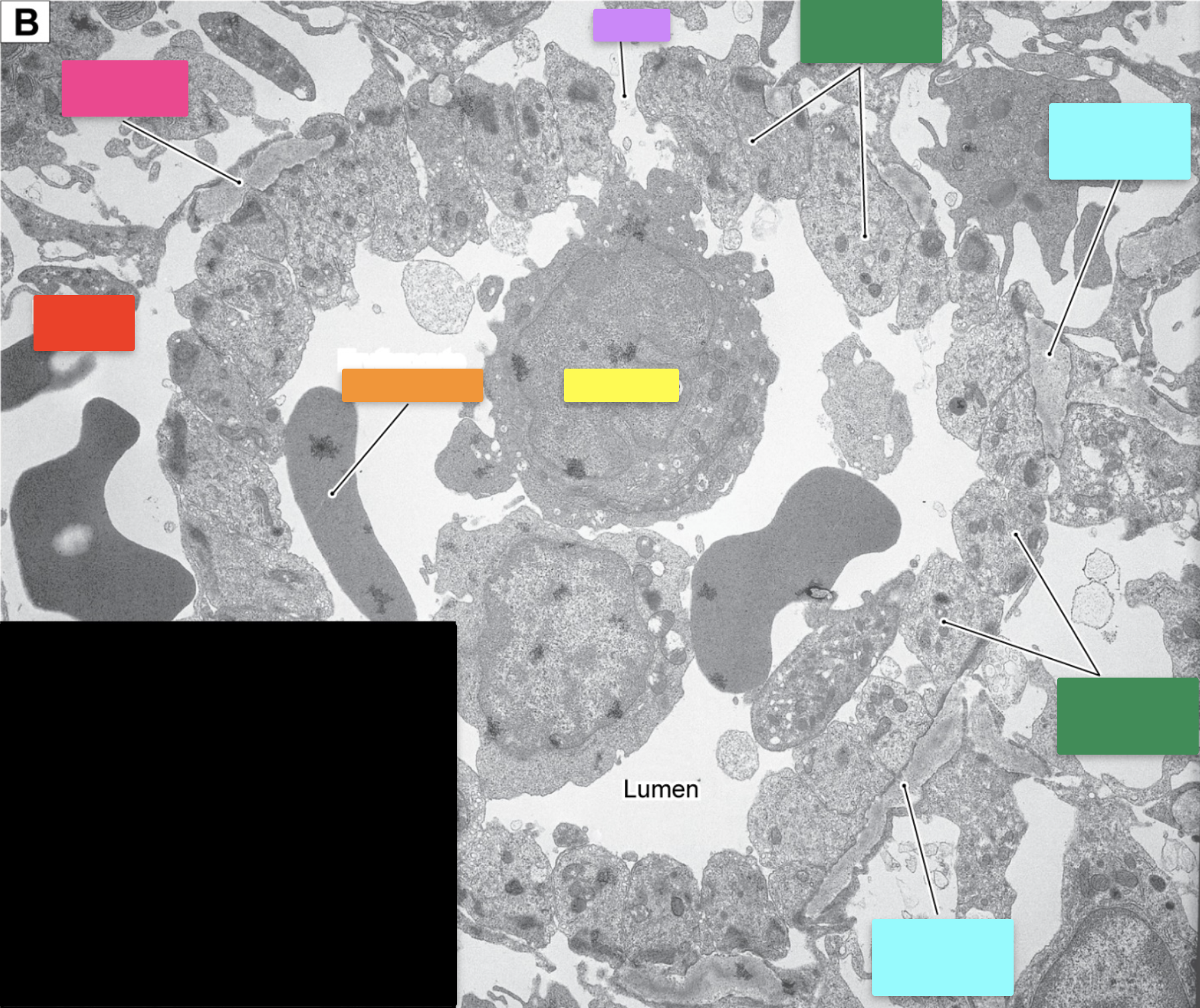

Red: red pulp cord
Green: endothelial cells
Purple: gap
Blue/Pink: basement membrane
Orange: erythrocyte
Yellow: monocyte
Identify the structures


venule
Classify the organ


Pink: Venule
Orange: blood cells
Blue: smooth muscle cells
Green: endothelial cell
Yellow: arteriole
Identify the structures/cells


arteriole
Classify the organ (not cells)


Blue: lumen of small vein
Yellow: smooth muscle nuclei
Green: arteriole
Pink: endothelial cell
Identify the structures (orange not indicating anything)


small vein
Classify the organ


arteriole
Classify the organ indicated by the yellow


Yellow: lumen of small vein
Green: smooth muscle nucleus
Identify the structurees indicated


Purple: internal elastic lamina
Green: lumen of medium vein
Yellow: arteriole
Blue: venule
Orange: nerve fibers
Pink: lumen of medium artery
Identify the structures indicated by the boxes

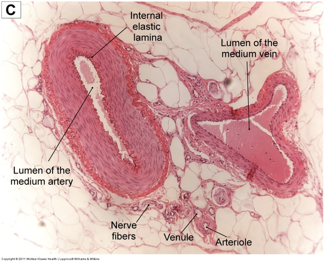
pink: medium artery
green: medium vein
Classify the organs indicated by green and pink


Medium vein
Classify the organ


Red: circular smooth muscle layer
Pink: tunica intima
Green: tunica media
Blue: tunica adventitia
Orange: longitudinal smooth muscle bundles
Identify the indicated structures

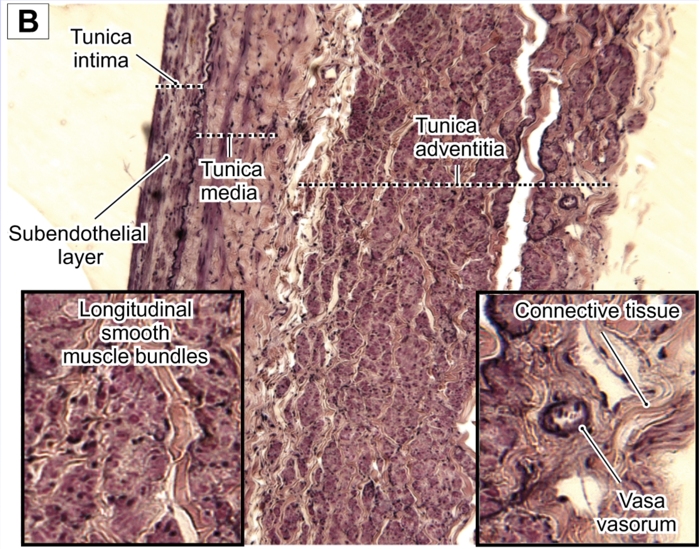
large vein
Classify the organ


Green: tunica intima
Blue: tunica adventitia
Pink: tunica media
Purple: subendothelial layer
Identify the structures indicated


Orange: connective tissue
Blue: vasa vasorum
Identify the structures


large vein
Classify the organ
