39. distal humerus fx
 Call Kai
 Learn
 Practice Test
 Spaced Repetition
 Match
 Flashcards
 Knowt Play
1/43

There's no tags or description
Looks like no tags are added yet.
44 Terms
انتهای دیستال و قدامی Lateral column
قوس مفصلی حدود 180°
Posterior آن فاقد غضروف است؛ بنابراین امکان فیکساسیون خلفی وجود دارد.
طول پیچ باید دقیق باشد تا وارد Radiocapitellar joint نشود.
Trochlea
بین دو ستون قرار دارد.
با Greater sigmoid notch اولنا مفصل میشود.
شکل Spool یا قرقره دارد.
قوس غضروفی تقریباً 270 درجه است.
اصل طلایی AO:
Anatomic articular reduction
Rigid internal fixation
Early range of motion
نکته مهم: اگر فیکساسیون به اندازه کافی پایدار نیست، نباید به قیمت جابهجایی شکستگی، حرکت زودرس را شروع کرد. ابتدا حفظ Reductionشود
نکته: شایعترین مکانیسم کلی، Simple fall از ارتفاع ایستاده بود.
حدود 70% بیماران مبتلا به شکستگی آرنج مستقیماً روی آرنج سقوط میکنند؛ چون فرصت محافظت با دست باز را ندارند.
چرا Capitellum مستعد است؟
مرکز چرخش آن نسبت به شفت هومروس قدامیتر است و در معرض نیروهای Shear قرار می گیرد
سالمندان حتماً علت سقوط را بررسی کن:
Cardiac arrhythmia
Cerebrovascular disease
Polypharmacy
Alcohol dependence
nerve: حدود 26% بیماران ممکن است در زمان مراجعه Ulnnar داشته باشند
Contraindication مطلق TEA
Active infection
Inadequate soft tissue coverage

A distal humerus fracture is defined as a fracture with an epicenter that is located within a square whose base is the distance between the epicondyles on an anteroposterior radiograph
تعریف Distal humerus fracture طبق Muller:
مرکز شکستگی (Epicenter) باید درون مربعی قرار گیرد که قاعده آن برابر فاصله بین دو Epicondyle در نمای AP است.

تعریف Distal humerus fracture طبق Muller:
مرکز شکستگی (Epicenter) باید درون مربعی قرار گیرد که قاعده آن برابر فاصله بین دو Epicondyle در نمای AP است.

طبقهبندی شکستگیهای Capitellum و Trochlea
Bryan–Morrey classificat
نکته حفظی:
Type I = Big bony fragment
Type II = Thin shell
Type III = Comminuted
Type IV = Capitellum + lateral trochl


Lateral collateral ligament complex (LCL)
از سه قسمت تشکیل شده:
Radial collateral ligament (RCL)
Lateral ulnar collateral ligament (LUCL)
Annular ligament
عملکرد:
مقاومت در برابر Varus instability
مقاومت در برابر Posterolateral rotatory instability (PLRI)

سه قسمت:
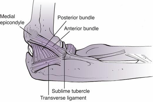
Anterior bundle
Posterior bundle
Transverse ligament
Anterior bundle مهمترین بخش در پایداری آرنج است.
Origin: قسمت Anteroinferior medial epicondyle
Insertion: Sublime tubercle of coronoid
عملکرد:
مقاومت در برابر Valgus instability
مقاومت در برابر Posteromedial rotatory instability

The Hahn–Steinthal (type I) fracture of the capitellum involves the articular surface and a large portion of the subchondral bone. B: The Kocher–Lorenz (type II) fracture involves the articular surface of the capitellum with a thin layer of subchondral bone.
اصل کلی:
اول مفصل را بازسازی کن، سپس آن را به Shaft متصل کن.
نکته طلایی: Central comminution
در صورت وجود Central comminution در Trochlea:
چرا؟
Lag screw میتواند باعث Compression و کوتاهشدن Trochlea در جهت Medial–lateral شود.
نتیجه:
کاهش عرض Trochlea
اختلال Congruity مفصل
احتمال اختلال عملکرد آرنج
بنابراین از Fully threaded position screw برای حفظ Reduction استفاده کن.


classification of distal humerus articular fractures has five patterns.

The distal humerus articular surface is aligned in 4 degrees to 8 degrees of valgus relative to the shaft and is angulated 35 to 40 degrees anteriorly in the sagittal plane. The medial epicondyle is the termination of the medial column and remains on the axis of the shaft in the sagittal view (B), while the lateral epicondyle follows the capitellum into flexion (C). D: Axially, the entire distal humerus articular surface is internally rotated 3 to 8 degrees.

The lateral collateral ligament complex is an important restraint to varus and posterolateral rotatory instability and consists of the radial collateral ligament, the lateral ulnar collateral ligament, and the annular ligament.
The medial collateral ligament functions as an important restraint to valgus and posteromedial rotatory instability.
It consists of an anterior bundle, posterior bundle, and transverse ligament. The anterior bundle is of prime importance in elbow stability and it originates from the anteroinferior aspect of the medial epicondyle, and inserts on the sublime tubercle of the coronoid.

Three peripheral nerves, the median, ulnar, and radial, cross the elbow joint along with a robust collateral blood supply
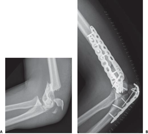
A distal humerus fracture (A) associated with a comminuted olecranon fracture that was used for visualization and ORIF of the articular segment (B).
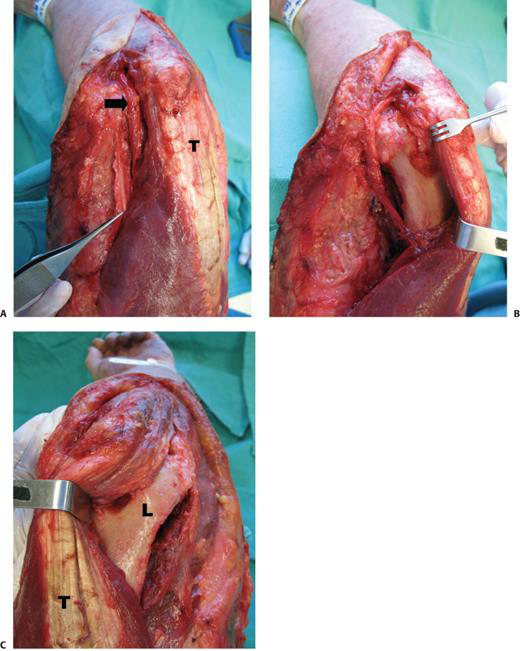
The paratricipital approach is done through a longitudinal posterior skin incision.
A: Medially, the ulnar nerve is identified (arrow). B: The medial intermuscular septum (forceps) is excised, and the triceps muscle is elevated off the posterior aspect of the distal humerus. C: Laterally, the triceps muscle (T) is elevated off the posterolateral aspect of the distal humerus allowing exposure of the lateral column (L), olecranon fossa, and posterior aspect of the trochlea.
عبور Screw از Olecranon fossa ممنوع؛ موجب Impingement و محدودیت Extension میشود.
Screw نباید وارد Articular surface شود؛ باعث Joint penetration میشود.
در انتهای عمل:
بررسی فلوروسکوپیک تمام پیچها
بررسی مستقیم Extra-articular بودن Implantها
بررسی کامل ROM برای رد Impingement و Instability


۱۷. Lateral approaches


The lateral paraolecranon approach described by Studer et al.[249] involves a posterior midline skin incision centered just medial to the tip of the olecranon.
A: Medial and lateral soft tissue flaps are raised and the ulnar nerve is exposed and prepared for transposition. B: The intermuscular septum is excised and a medial arthrotomy is conducted similar to the paratricipital approach. The fascia of anconeus is split longitudinally about 3 to 5 mm lateral to the ulna, which maintains a ribbon of anconeus fascia attached to the ulna that facilitates fascia-to-fascia closure. C: The anconeus is then elevated off the ulna and distal humerus, exposing the posterior aspect of the radiocapitellar joint. At the level of the olecranon tip, the triceps expanse is then split proximally between the lateral cubital retinaculum (lateral) and the triceps tendon proper (medial), which attaches on to the olecranon tip.

Campbell involves a midline split through the triceps tendon and medial head. B, C:The approach can be extended distally by splitting the triceps insertion on the olecranon and raising medial and lateral full-thickness fasciotendinous flaps. To gain further exposure of the posterior trochlea, the elbow is flexed and the olecranon tip may be excised. For ORIF, the medial and lateral collateral ligaments are preserved (asterisk). However, to obtain further exposure for TEA, they may be released (D). O, olecranon; U, ulnar nerve; T, triceps.
(Reprinted with permission from Studer A, et al. The lateral para-olecranon approach for total elbow arthroplasty

The triceps-reflecting anconeus pedicle (TRAP) approach is done through a longitudinal posterior skin incision after identification of the ulnar nerve

Kocher approach to the anterolateral elbow joint uses the interval between extensor carpi ulnaris (ECU) and anconeus.

The extensor digitorum communis (EDC) split approach.
The EDC tendon is split anterior to the mid-equator of the radiocapitellar joint to avoid injury to the lateral ulnar collateral ligament

Potential Pitfalls & Prevention


Post op care

بقهبندی Hastings & Graham
Ho

B: A bicolumn (OTA/AO type C1) fracture treated with ORIF via an olecranon osteotomy. C, D: The distal humerus articular segment is fixated with three medial and three lateral screws placed through parallel plates

Fracture of the capitellum and the lateral ridge of the trochlea.
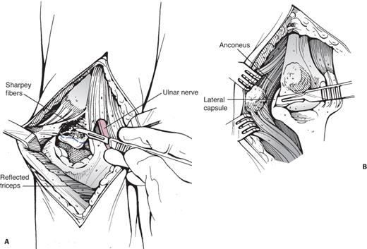
The Bryan–Morrey approach is commonly used for total elbow arthroplasty
Distal humerus fracture (A) treated with hemiarthroplasty and plate fixation of the medial column and suture fixation of the lateral epicondyle (B).


Authors’ preferred treatment algorithm for distal humerus fractures

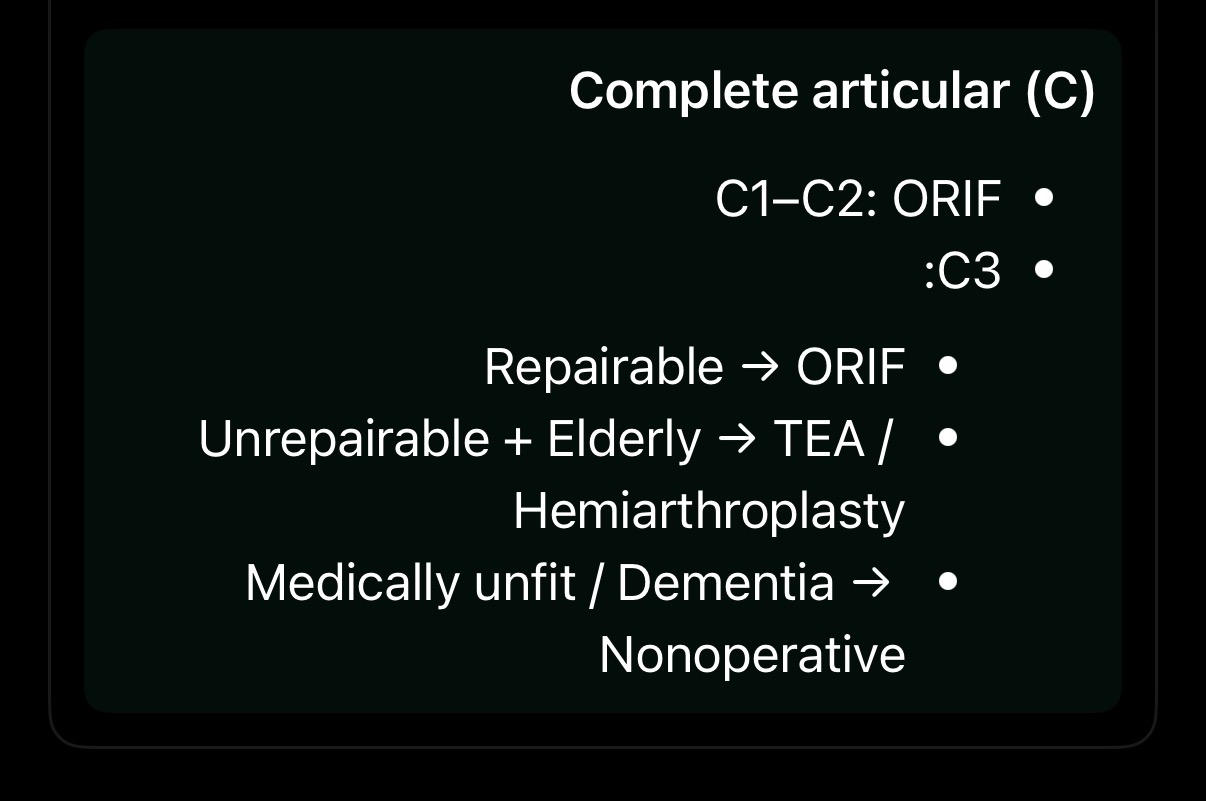
شکستگی بدون جابهجایی (Undisplaced)
درمان غیرجراحی (Nonoperative)
Close monitoring و پیگیری بسیار نزدیک از نظر جابهجایی ثانویه
. شکستگی با جابهجایی (Displaced)
درمان اصولاً جراحی (Operative) است.
الف) Extra-articular – نوع A (A2, A3)
ORIF
رویکرد:
Paratricipital approach
یا Lateral para-olecranon approach
فیکساسیون با Double-column plating
ب) Partial articular – نوع B


– نوع C
coplete articular
