UGA Medical Parasitology - Unit 2: Protozoans
 Call Kai
 Learn
 Practice Test
 Spaced Repetition
 Match
 Flashcards
 Knowt Play
1/54

There's no tags or description
Looks like no tags are added yet.
55 Terms
Robert Hooke
English scientist that discovered cells in all living thingsand pioneered the use of microscopes to observe microscopic structures.
Anton van Leeuwenhoek
Dutch scientist known as the "father of microbiology" who first observed single-celled organisms and bacteria using a microscope he designed.
Protozoan
a protist with animal like characteristics, also means ‘first animals’
Basic Characteristics of Protozoans
1. Heterotrophic: i.e. has to eat other things.
2. Are unicellular.
3. Most are mobile.
Protozoan Subgroups
SAR
ARCHAEPLASTIDA
EXCAVATA
AMOEBAZOA
OPIS
Protozoan Parasite Focus
SAR: Entamoeba histolytica
EXCAVATA: Giardia/Trichomonas/Trypanosomatids
AMOEBAZOA: Entamoeba histolytica
Entamoeba histolytica
no defined shape (constantly changing form)
not free living
feed via trogocytosis
Rather than eat cells whole, E. histolytica
trophozoites will ingest small bites of host
cell material.
• The ‘nibbling’ causes apoptotic death and
leads to an immune response and much of
the pathology of infection.
uses dynamic pseudopodia that extend out to move
Generally they expand asexually through binary fission but sexual
recombination is thought to occur
causes amoebiasis (amoebic dysentery)
Humans are the only reservoir excreting amoebic cysts. The cysts
are environmentally stable (~1 month).
• Transmission occurs through a fecal-oral route (consuming
contaminated food/water)
How does Entamoeba histolytica move?
actin polymerization at the membrane
*also how your immune cells travel around*
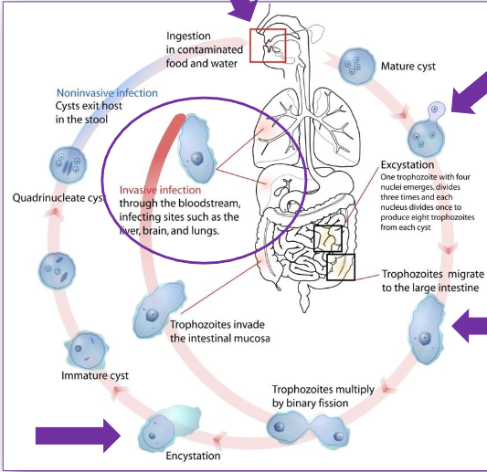
Entamoeba histolytica Life Cycle
Infection begins with cysts being ingested in
contaminated food or water.
• Cysts pass through the stomach and upon reaching
the small intestine the amoeba leaves or “excysts” as
a 4 nuclei containing cell.
• This form, called the ‘trophozoite’, copies its
genomes 1x to give 8 nuclei.
• The parasite then undergoes cytokinesis 3x to make
8 trophozoites from a single cyst.
• The trophozoites migrate to the large intestine, eats
bacteria and begin dividing.
• If they become invasive they can migrate to the
brain, liver and lungs where they cause tissue
destruction and disease.
• Normally, however, they re-encyst and are excreted
by the host to continue the cycle.
Entamoeba histolytica Pathology of Infection
Gradual onset of abdominal pain, watery
stools containing mucus and blood.
• Some patients have only intermittent
diarrhea alternating with constipation.
• Fever is uncommon.
• Parasites begin invading the intestinal
mucosa.
• Infiltration of immune cells and tissue
destruction leads to flask shape ulcers
that eventually allow E. histolytica to
enter into the bloodstream.
• As parasites begin eating and destroying
the colonic epithelium ulcers begin to
form.
• Parasites now have access to
disseminate through the body.
Systemic Phase of Infection:
• Parasites invade the tissues:
• Liver
• Lung
• Brain
• Inflammation and tissue
destruction due to parasite
mediated killing of host cells
can least to systemic
abscesses.
Entamoeba histolytica Treatment and Prevention
Several drugs are available to clear symptomatic
and asymptomatic enteric (luminal) infection:
• Diodohydroxyquin
• Paromomycin
• Diloxanide furoate
• Metronidazole (Flagyl) is the drug of choice for
invasive amoebiasis (90% cure rate).
• Metronidazole should be combined with a luminal
acting drug as it is not fully effective on non-
invasive stages.
• Improved sanitation will help to reduce the
likelihood of transmission.
• Travelers to endemic areas can reduce the risk
of infection by drinking bottled water, not using
ice cubes in drinks, and washing fruits and
vegetables with clean water (or by peeling them
yourself).
Acanthamoeba spp. Life Cycle
Normally free living, these opportunistic pathogens
sometimes infect people through contact with soil,
fresh/sea water, pools, contact lens equipment,
medical devices etc.
• Parasites replicate by mitosis (normal style).
• Acanthamoeba enter the eyes, nose, or broken
skin to gain entry.
• Infection can remain localized at the site of entry or
can spread to other systems of the body and be
deadly.
• Since this is not its normal life cycle of
Acanthamoeba, humans are considered a dead
end host for this organism.
• No known human to human infection.

Acanthamoeba spp. Pathology
If Acanthamoeba enters the eyes it can result in
severe keratitis that can make someone blind.
• This is a serious issue with contact lens users and if
severe will require a corneal graft.
• Systemic infection presents as inflammation of the
lungs or sinuses.
• Skin infections caused by Acanthamoeba can appear
as reddish nodules, skin ulcers, or abscesses in the
skin.
• If the parasite reaches the central nervous system
(CNS) it can cause Granulomatous Amoebic
Encephalitis (GAE).
• GAE starts slowly, with symptoms like headache,
nausea, dizziness, irritability and a low-grade fever.
• GAE is a rare, usually fatal, subacute-to-chronic
central nervous system disease
Acanthamoeba spp. Treatment and Prevention
Miltefosine (Impaido) is a standard prescribed drug
for free-living amoeba infections.
• Several eye medications are available with a
prescription (PHMB-Desomedine, hexamidine and
chlorhexidine)
• To prevent eye infections contact lens users are
advised to carefully follow use and cleaning
directions from their eye care providers.
• It is unknown what can prevent GAE or
disseminated infections as both are extremely rare
anyway.
Balamuthia mandrillaris
Life Cycle/Pathology/Treatment
Can enter the nose or broken skin to gain entry.
• Infection can spread to other systems of the body
and be deadly.
• If the parasite reaches the central nervous system
(CNS) it can cause Granulomatous Amoebic
Encephalitis (GAE).
• Few patients survive Balamuthia infection even
with drug treatment using a combination of
flucytosine, pentamidine, fluconazole, sulfadiazine
and either azithromycin, clarithromycin or even
miltefosine.
• Not known why some people become infected
while others do not. Only ~200 known cases
worldwide.

Naegleria fowleri
“The Brain Eater” Life Cycle
Normally free living these opportunistic pathogens
sometimes come into contact with people who
have fresh water enter deep into the nasal
passages.
• Unlike the other ameobozoons, this is an excavate
and has 3 stages in its life cycle: cyst (1),
trophozoite (2), and flagellate (3). The only
infectious stage of the amoeba is the trophozoite.
• Once penetrating the nasal mucosa the amoeba
migrate to the brain via the olfactory nerves.
• Infection can lead to primary amoebic
meningoencephalitis (PAM).
• No known human to human spread

Naegleria fowleri
Pathology
Enters through nasal cavity, usually when there’s a
sudden rush of water in the cavity while
diving/swimming
• Travels through the cribriform plate and olfactory
bulb where it encounters the brain and begins
feeding.
• Cell death and inflammation causes damage to the
frontal lobe.
• Infection can lead to primary amoebic
meningoencephalitis (PAM).
Stage 1 (2 days)
• Severe frontal headache
• Fever
• Nausea
• Vomiting
• Stage 2 (3 days)
• Stiff neck
• Seizures
• Altered mental status
• Hallucinations
• Coma
Naegleria fowleri
Treatment and Prevention
Only 5 documented cases of people
surviving PAM exist.
• Very few drugs available and only
miltefosine has shown promise as a
potential treatment.
• More research is needed to find
promising therapeutics for these rare
but deadly infections.
• Recommended not to swim in
stagnant or very still lakes and to
avoid aspirating lake water into your
nostrils.
The Kinetoplast
kinet=move plast=organelle
Basic Biology of Trypanosoma cruzi Forms
Normal Eukaryotic Organelles: nucleus, Golgi, ER, mitochondria
Unusual organelles:
• Kinetoplast: Genome of the single giant
mitochondrion containing densly packed DNA
(stains brighter than nuclear DNA).
• Glycosomes: unlike us, trypanosomatids carry out
glycolysis in membrane bound vesicles.
• Reservosomes: pre-lysosomal compartment
present in high amounts in epimastigotes.
• Cytostome/Cytopharynx (SPC): entrance to the
endocytic pathway. Mechanism of large molecule
uptake, i.e. ‘how it eats’.
Acidocalcisome: calcium storage vesicles.
• Contractile vacuole: similar to the CV in amoebas.
• Flagellum and flagellar pocket: how the parasite
moves and the site of exocytosis.
• Subpellicular microtubules: microtubule skeleton
underlying the entire body of the parasite
Salivarian
transmitted via mouth parts “saliva”
Although salivarians expand in the midgut they then
make their way either out of the intestine and to the
salivary glands or up to the foregut and mouthparts in
order to gain direct access to the host bloodstream
during a bloodmeal
Stercorarian
Transmitted via feces
T. Cruzi Life Cycle
stercorarian
Infection begins when a reduviid insect host (kissing
bug) takes a blood meal from a warm blooded animal.
• During the bloodmeal the insect vector excretes parasites
in its feces onto the mammalian host.
• These infectious metacyclic trypomastigotes are are
morphologically distinct from epimastigotes and are
primed and ready to enter their new host using the bite
wound or a nearby mucous membrane.
• The parasites then make their way to the bloodstream
and invade any nucleated host cell, thus initiating a new
phase of the infection process.
• Although they enter via a phagocytic-like mechanism,
parasites soon exit from these lysosome-like structures
and begin replicating as flagellar-less amastigotes.
• Once host cell resources are depleted they reconvert to
their flagellated form and lyse the host cell.
• These released parasites are known as trypomastigotes
and are able to perpetuate subsequent rounds of host cell
infection, replication and lysis
Chagas disease Pathology - acute stage
Acute form: 7-10 days incubation period. High
numbers of parasites in the blood or lymphatic
vessels. Parasites may invade almost all nucleated
cell types, preferentially muscle fiber
Depending on the location and manner of the
initial infection (e.g. mucous membranes or bite
wounds) you have inflammation resulting from
the initial spread of infection.
• During this period, intracellular amastigotes
replicate and begin the process of lysing
infected cells and rapidly spreading.
• This results in initially localized infection and
inflammation resulting in the pathology noted in
Chagoma’s and Romana Sign.
Chagas disease Pathology - indeterminate stage
During this phase of infection parasites
disappear from the blood.
• Nests of amastigotes are generally found in
very low numbers in muscle tissue.
• Antibodies to T. cruzi antigens can be detected.
• Patients are generally asymptomatic.
• Most patients enter chronic phase within 5 to
15 years.
Chagas disease Pathology - chronic stage
• The chronic stage which appears after a silent
period that may last several years.
• The lesions of the chronic phase irreversibly
affect internal organs namely the heart,
oesophagus and colon and the peripheral
nervous system.
• A slow/steady destruction of smooth and
cardiac muscle and parasympathetic ganglia,
due to the combined effects of cellular
destruction and inflammation, results in a
weakening of the ability to carry out normal
functions.
• Results in Myocardiopathy and Megaviscerae
of the smooth muscle organs of the body
• These autopsy images demonstrate T. cruzi
induced megacolon showing massive dilatation
and elongation of the rectosigmoid and distal
descending colon.
• Colonic involvement leads to prolonged
constipation, abdominal pain and fecaloma.
• Increased risk of volvulus (intestinal knotting)
and consequent bowel ischemia (like a heart
attack for the intestine – cut off of blood flow
• These Barium swallow examinations show
widening of the esophagus (top arrows).
• Dilated tortuous esophagus (top arrowheads).
• Widening of the esophagus with tertiary wave
patterns (bottom left arrowheads) due to
aberrant and non-propulsive peristalsis.
• Typical ‘rat-tail’ sign in the distal portion of the
esophagus (bottom right arrows) connecting to
the stomach (s).
Chagas disease - other facts
• Individuals with HIV and chronic T.
cruzi infection may experience a
reactivation.
• This is most commonly manifested by
meningoencephalitis.
• A reactivation myocarditis is the
second most common manifestation
Chagas Disease Diagnoses
Direct Detection:
o Microscopic detection of parasites in blood.
o Hemoculture (growing parasites) from blood.
o Xenodiagnosis (no longer used) where reduviid
insects feed directly on a person followed by
microscopic evaluation of parasites in the insect
mid/hindgut.
• Serodiagnostics:
o Multiple testing methods exist in a variety of
formats (hemaglutination, immunofluorescence
microscopy, ELISA, Western Blot etc.)
o These tests are based on the presence of
antibodies (made by B-cells) that recognize
antigens (typically parasite proteins).
• Molecular Diagnosis:
o By extracting DNA from the blood and performing
PCR (polymerase chain reaction) you can identify
the presence of extremely rare parasites
(extremely sensitive but expensive)
Chagas Disease treatment - Benznidazole (Radanil)
Orally administered frontline drug in the treatment of
Chagas Disease in use since 1971.
• Is part of the nitroimidazole drug family and works by
the production of free radicals.
• Has a dramatic effect on the intracellular amastigote
stage causing parasite lysis.
• Frequent toxicity (allergic skin reactions, peripheral
neuropathy, leucopaenia, convulsions).
• Partially effective in acute stage (strain variability) but
inactive in chronic stage of the disease.
• Prolonged treatment recommended (up to 60 days)
(5-7 mg/kg per day) but side effects make
compliance difficult.
Chagas Disease treatment - Nifurtimox (Lampit)
Orally administered nitro-anion radical metabolite
that acts through the production of toxic free
radicals. Affects DNA stability.
• Used if Benznidazole fails.
• The parasite is more sensitive due to deficiency in
their antioxidant enzyme repertoire.
• Toxicity is a problem (anorexia, nausea, vomiting,
peripheral neuropathy, skin reactions, CNS effects).
• Only partially effective in acute stage (strain
variability) and inactive in the chronic stage of
disease.
• Requires prolonged treatment (up to 90 days) 98-
10 mg/Kg per day).
• Approved for use in the US in 2020.
• Not available in Canada.
Chagas Disease:
Control and Prevention
insecticide spraying
education
blood supply monitoring
Trypanosoma brucei
hemoflagellate: lives in host circulatory system
transmitted by blood feeding arthropod
has many of the same DNA (kDNA)
Causes African sleeping sickness / human african trypanosomiasis
salivarian
T. b. gambiense
Reservoir is primarily humans (Human African Trypanosomiasis (HAT)
Causes a chronic disease that
can remain asymptomatic or mild for months or even
years before reaching the central nervous system
T. b. rhodesiense
Zoonotic disease- wild game and cattle reservoir (Nagana)
Causes an acute disease that
rapidly attacks the body, advancing from the blood
stage to the central nervous system within week
Robert Michael Forde (1861–1948)
had the first observation of trypanosomes
T. Brucei Life Cycle
The female tse tse fly produces larvae, one
at a time. The egg hatches within the female
and the larva develops in the female. During
her life-span a female can only produce a
maximum of 8-10 offspring.
• The larva is laid onto the ground
(larviposition). The female finds a suitable
place to lay the larva. In damp areas females
tend not to concentrate their larviposition in
particular areas. In dry areas larviposition
takes place mainly in well-shaded spots.
• The freshly-laid free-living larva, does not
need to feed and burrows into the soil where
its skin hardens and blackens into a
puparium and within the puparium, pupation
and metamorphosis take place.
T. Brucei Pathology Stage 1
Trypanosomes multiply in the tissue around the
initial bite site.
• This often results in a characteristic local
inflammation known as the trypanosomal “chancre”.
• Usually this is not painful.
• From there they enter the blood and lymphatic
system and begin to disseminate throughout the
circulatory system.
Enlargement of the lymphatic glands along the
posterior cervical lymph node chain (especially in
the posterior triangle of the neck) can be an early
sign of the disease.
• This is known as Winterbottom sign, but is not as
common in T. rhodesiense infection.
• Aspiration of swollen gland often reveals parasites
T. Brucei Pathology Stage 2
Symptoms at this stage are rather mild in
gambiense but can be severe in rhodesiense
with an often fatal outcome.
• After 1-2 week period of asymptomatic
incubation, parasites invade blood leading to
fever and headache.
• Once parasites enter blood stream fever sets
in (low and irregular in gambiense and high
and periodic in rhodesiense.
• General toxic symptoms include headache,
facial edema, nausea and vomiting, back and
bone pain.
• The second stage of trypanosomiasis is
characterized by progressive anemia and cachexia.
• Both features are primarily due to extremely high
serum levels of TNFalpha (pro-inflammatory cytokine).
• TNFalpha (tumor necrosis factor also known as
cachexin) was isolated as a factor inducing wasting in
Nagana.
T. Brucei Pathology advanced stages
• In later stages of infection parasites pass the blood
brain barrier and infect the CNS.
• Presence of parasites leads to meningo-
encephalitis with progressive neurological
involvement, which ultimately ends in coma
(sleeping sickness).
• Untreated trypanosomiasis is always fatal
• The progressive encephalitis can cause severe
dementia with sometimes aggressive behavior.
• Disease progression especially CNS invasion is
much faster in rhodesiense.
• Gambiense can take a year or two while
rhodesiense usually passes the blood brain barrier
within a month.
T. Brucei Diagnosis
• At later stages of the disease there might be no
detectable parasites in the blood.
• Lumbar puncture and microscopic identification of
parasites and detection of elevated protein levels
in the CSF are needed for diagnosis.
• Establishment of CNS infection is important for
therapy decisions
OLD T. b. rhodesiense Treatment
Suramin (I.V. administration); only works in the early stage; multiple doses over about 3 weeks
OLD T. b. gambiense treatment
Pentamidine (intramural injection); only works in early stages
OLD T. Brucei Treatment
Melarsoprol(i.v. infusion); works on late stages; works on both t.b. gambiense and t.b. rhodesiense; Severe toxicity (death 5-10%; encephalopathy 10%; pruritus, cardiac failure
T. Brucei Diagnoses
• Trypansosomiasis is best diagnosed by
demonstration of parasites in blood smears,
lymph node exudates or spinal fluid.
• Parasitemia can be very low at times
resulting in false negative results. It is more
likely to find parasites during symptomatic
periods.
• Several techniques can be used to enrich
rare parasites in blood sample
(centrifugation, chromatography).
• The Card Agglutination Test for
Trypanosomiasis (CATT). Sero-diagnostic
test to identify infected individuals
NEW T. Brucei Treatments - Fexinidazole
• The first treatment for sleeping sickness that relies on pills alone
was approved in 2018.
• Simplified treatment: Oral treatment that can be taken at home. The
new drug, fexinidazole, cures all stages of the disease within 10
days.
• Previously, everyone with the parasites found in a blood test also
had to undergo a spinal tap to see if the parasites had reached their
brains.
• The drug was created in the 1980s by Hoechst, a German drug
company, but later abandoned.
NEW T. Brucei Treatments - Acoziborole
the first single-dose oral treatment for both stages of T.b.
gambiense sleeping sickness, the most common form of the diseas
targets CPSF so mRNA processing will not
occur leading to loss of protein production
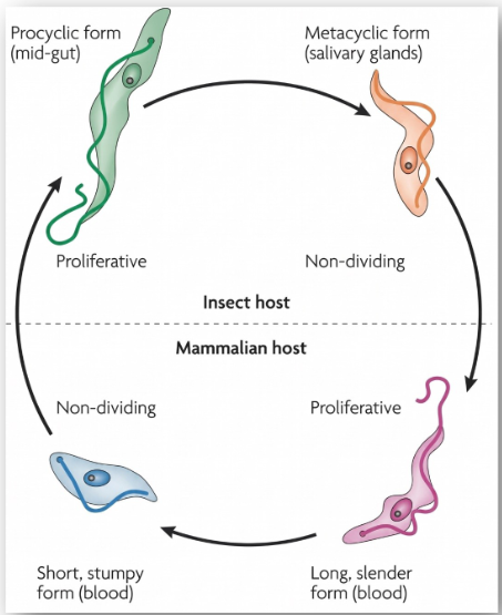
T. Brucei Life Cycle
Trypanosoma brucei
• T. brucei is a salivarian kinetoplastid transmitted by the Tsetse fly.
• Two major strains infect humans: T. b. gambiense and T. b. rhodesiense
• If left untreated it is almost always fatal.
• Surveillance and new drug treatments have made this less of an issue for humans but the economic costs for
cattle production due to Nagana is still a major problem.
• The Tsetse fly is viviparous and gives birth to live young and transmits T. brucei via the salivary glands during a
bloodmeal.
• New oral treatment with fexinidazole, cures all stages of the disease within 10 days.
• T. brucei (like all kinetoplastids) have unique mechanisms regulating gene expression.
• They express genes as polycistronic transcripts that are each individually trans-spliced and polyadenylated.
• Stability, and thus likelihood of translation, is dictated by the presence of life-cycle specific RNA binding proteins.
• Parasites exhibit interesting behaviors like social motility the function and mechanistic basis for which remain to be
worked out.
• The social behavior known as “Quorum Sensing” was recently worked out and is the mechanisms parasites uses
to regulate their numbers in a host.
• As part of their immune evasion strategy T. brucei continually changes its surface coat through a mechanism
known as “Antigenic Variation”.
• Antigenic variation involved the selective expression of different coat proteins that change in order to escape
antibody mediated clearance in the host.
What are the replicative stages of T. brucei called?
Long slender bloodstream form, Procyclic form
What are the major strains of T. brucei that infect people?
T. b. gambiense, T. b. rhodesiense
Who discovered T. brucei and demonstrated transmission?
Sir David Bruce
How is T. brucei gene expression distinct?
lacks obvious promoters and can even use RNA polymerase I (Pol I) to express certain protein-coding genes such as VSG (variant surface glycoprotein) and procyclin, which is highly unusual for eukaryotes
What is trans-splicing?
the process by which a Spliced Leader (SL) RNA, a 39-nucleotide mini-exon, is added to the 5′ end of every mRNA in T. brucei. This is necessary because the parasite transcribes genes as long polycistronic RNAs and must convert them into individual mature mRNAs.
Why are RNA binding proteins so important in T. brucei?
RNA-binding proteins are crucial in Trypanosoma brucei because the parasite lacks extensive transcriptional regulation of individual genes and therefore relies primarily on post-transcriptional regulation.