Mid 2 BIBC 100
 Call Kai
 Learn
 Practice Test
 Spaced Repetition
 Match
 Flashcards
 Knowt Play
1/102

There's no tags or description
Looks like no tags are added yet.
103 Terms
– Monosaccharides (monomer)
• A single polyhydroxy aldehyde or ketone unit
– 5 carbons:
• Riboses (Nucleic acid backbone)
– 6 carbons:
• D-glucose (most abundant 6-carbon sugar, also
called dextrose)
• Fructose (fruit sugar)
• Galactose (milk sugar)
– Oligosaccharides (2-20 monomer)
Disaccharides: 2 monomers
– Sucrose (abundant): D-Glucose + D-Fructose
– Lactose (milk): D-Glucose + Galactose
– Maltose: D-Glucose + D-Glucose
Aldoses
contain an aldehyde
group: the carbonyl is at the end
of the carbon chain.
Ketoses
contain a ketone
group: the carbonyl is at an
internal carbon.
Structual isomer
differ in connectivity:
the atoms are connected in a different order.
• Example: glucose and fructose
• both are hexoses
• glucose is an aldohexose
• fructose is a ketohexose
Stereoisomer
have the same formula and same
connectivity, but differ in 3D arrangement
Chiral carbon
-it is bonded to four different groups.
-Most monosaccharides have these.
-cannot be superimposed on their mirror image (hands)
D/L configuration is determined by the chiral carbon farthest from the carbonyl. If OH is on the …
If on the RIGHT —> D isomer
if on the LEFT —> L isomer
Van Hoff rule
-the way we can determine the number of possible stereoisomers
-2^n rule
-the number of chiral centers (n) determines the max stereoisomers
Aldopentoses have _ sterioisomers
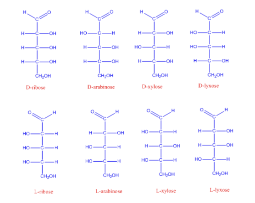
8 stereoisomers
Enantiomers are nonsuperimposable mirror images
-Enantiomers are mirror images of each other
-they differ in configuration at every chiral center
Diastereomers are stereoisomers that are not mirror images
-diastereomers have the same chemical formula and atom to atom connectivity
-they are not mirror images
they differ at some but not all chiral centers
Epimers differ at only one chiral center

-epimers are a subtype of diastereomers
-epimers differ at exactly one chiral center
Monosaccharides cyclize in aqueous solution
Monosaccharides with four or
more carbons can form cyclic
structures.
• Carbonyl C reacts with a hydroxyl
group of the same molecule (often
the second last OH).
• This intramolecular reaction
creates a ring
Pyranoses
sugars with a 6 membered ring (5C + 1 O)
Furanoses
sugars with a 5 membered ring (4C + 1O)
Cyclized sugars are hemiacetals or hemiketals with a new chiral center
aldehyde + alcohol —> hemiacetal
Ketone + alcohol —> hemiketal
if the alcohol is in the same molecules, the rxn forms a ring
this rxn is the reason why many sugars cyclize in h2o
the carbonyl carbon becomes a new chiral center for the cyclic sugar
Sugar cyclization creates alpha and Beta anomers

cyclization creates a new chiral center at the former carbonyl C
this new chiral center is called anomeric carbon
the former carbonyl oxygen becomes a hydroxyl group
if the anomeric OH is opposite to CH2PH (bottom) —> alpha anomer
if the anomeric OH is on the same side as the CH2PH (top) —> Beta anomer
the OH oxygen that attacks the carbonyl becomes the ring oxygen
*know how to draw fischer to haworth and vise versa

Polysaccharide linkages determine structure and function
polysaccharides are long carbohydrate polymers
unlike proteins and nucleic acids, polysac. are not made from a direct template
polysacs. have variable length and branching
storage polysaccharides: commonly have alpha linkages (starch, glycogen)
structural polysaccharides: commonly have Beta linkages (cellulose, chitin, agarose)
Polysacc.s can be homo or heteropolymers
homo-polysaccharides: have ONE type of repeating sugar type
hetero-polysaccharides: have MORE THAN ONE sugar type
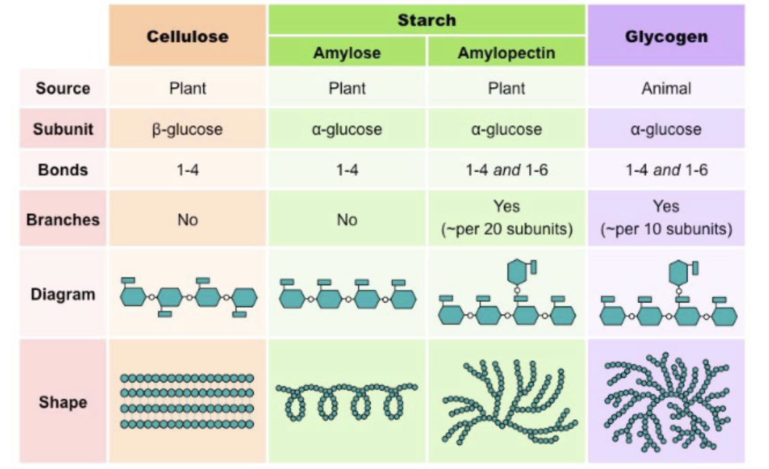
Starch contains linear amylose and branches amylopectin
amylose is a linear a1 to 4 linked glucose polymer
amylopectin is branches . has a1-4 linked chains with a1-6 branch points
glycogen is a highly branches glucose polymer
glycogen is the major glucose storage polysaccharide in animals
it has a1-4 linked glucose chains
has a1-6 branch points
glycogen is more branched than amylopectin
branch points occur roughly every 8-12 residues
Branchin allows rapid glucose release
• Glycogen breakdown enzymes work from chain ends.
• Branching creates many ends.
• More ends allow faster glucose release.
• More branches → more ends → faster mobilization.
storing glucose as glycogen reduces osmotic stress
keeping free glucose molecules would make big pressure towards blood cells which is bad bc they could rupture
to prevent this, glucose is turned into glycogen bc it reduces the number of free particles
alpha 1-4 linked glucose chains favor helical conformations
• α(1→4) linkages bend glucose chains
rather than keeping them fully extended.
• Amylose can form a continuous helix with
~6 glucose residues per turn.
• Unbranched segments of amylopectin and
glycogen can form shorter helices; branch
points interrupt them.
• This helical conformation helps pack
glucose compactly for storage.
beta linkages favor STRAIGHT, tightly packed structural chains
B linked chains are more straight and extended than a linked chains
extended chains can form many hydrogen bonds, especially inter chain H bonds
straight chains pack tightly side by side —> strong structural materials
this contrast with a 1-4 linked storage polymers, which bend into helices
Cellulose is a B 1-4 linked glucose polymer with straight chains
• Cellulose is made of β(1→4)-linked D-glucose units.
• β(1→4) linkages favor extended, relatively straight chains.
• Straight chains can pack side-by-side through many inter-chain hydrogen bonds.
• This tight packing gives cellulose high structural strength.
Humans digest starch but not cellulose because linkage geometry matters
starch and glycogen contain a 1-4 linkages
human amylases and glycosidases can hydrolize a-linkages
and cellulose contains B1-4 linkages
humans lack enzymes that hydrolyze cellulose B1-4 linkages
termites and cows rely on symbiotic microbes that produce cellulases
Glucose polymers differ by linkage geometry and branching
chart
Flow of genetic information:
DNA
Short for deoxiribonucleic acid
Stores genetic info in cells and many viruses
Double stranded
RNA
Short for ribonucleic acid
Transfers and interrupts genetic info
Other functions
Single strand (can fold into tertiary structure)
Flow is: DNA → ← RNA → Protein
Nucleotides and Deoxy nucleotides both have 3 structural elements:
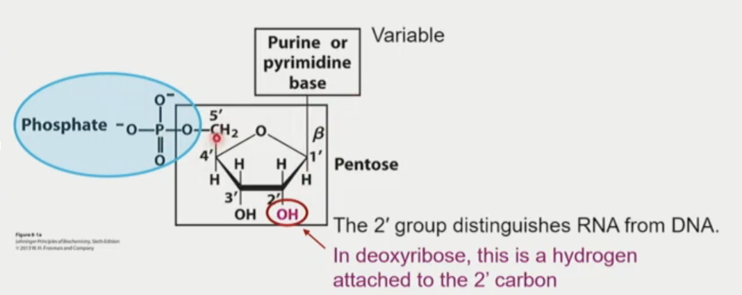
Pentose sugar
Ribose or deoxiribose
Nitrogenous base
Is attached to 1’ C
Phosphate group
1 or more, attached to the 5’
Which is nucleoTIDE and nucleoSIDE

Nucleoside = base + pentose
Nucleotide = base + pentose + phosphate(s)
Are building block of DNA and RNA
Pentose Component:
The pentose is ribose in RNA and deoxiribose in DNA
In nucleic acids, they are in the furanose (5) ring form
Ribose has a 2’ - OH
Deoxyr. Has a 2’-H
The chemical change affects stability and structure
Nitrogenous Base component:

Purines have 2 rings
Pyrimidines have 1 ring
Bases are planar and aromatic
Being planar helps the stack in DNA and RNA
Pyrimidines
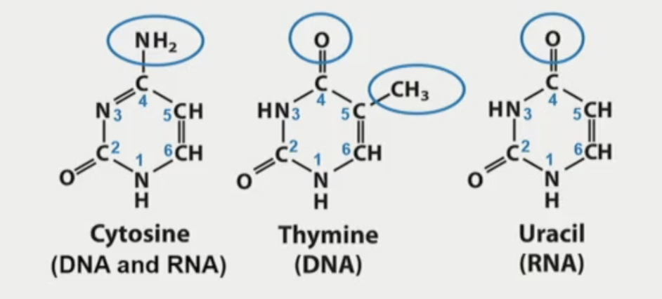
Cytosine (C), Thymine(T), and Uracil(U) are pyrimidines
All have 6-mem-ring
C2: all have carbonyl
C4:
C: amino
T and U: carbonyl
C5:
T: has methyl
Purine
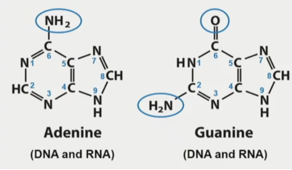
Adenine (A), Guanine(G)
Have 2 rings
C2:
G: amino (NH2)
C6:
G: carbonyl
A: amino
N9 forms sugar C1
Ribonucleotides: RNA building blocks

Purines attached to C1’ of the sugar through N9
Pyrimidines attach to C1’ of the sugar through N1
C2’ of the ribose has a hydroxyl group attached
The phosphate is esterified to C5’ of ribose
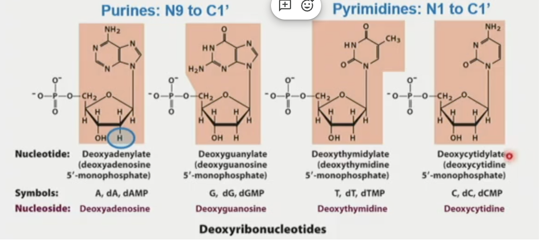
Deoxiribonucleotides: DNA building blocks
Purine N9: to C1’ of deoxyribose
Pyrimidine N1: to C1’ of deoxyribose
C2’ of deoxyribose has a hydrogen
The phosphate is esterified to C5’ of deoxyribose
UV light
nucleic acids absorb UV light at 260nm bc of their nitrogenous bases
DNA can have modified bases (minor)
They are more rare than the standard (major) bases.
There are 4 types
Minor DNA bases can affect gene expression
Examples:
5 methylcytosine
There is methylation in the 5’ C
DNA methylation is associated with gene regulation
RNA often times has the modified MINOR bases
Modified based are common in tRNAs
Can support noncanonical base pairing (this means they can form a bunch of diverse structures that can support different functions
Modified bases expand RNA structural and functional diversity
Nucleotides perform:
Genetic information
Energy carriers
ATP, GTP, etc
Signaling molecules
cyclic forms (AMP, GMP)
Cofactors
NAD, FAD
Nucleotides are linked by phosphodiester bonds

The 3’ -OH of one nucleotide forms a phosphodiester bond with the 5’ phosphate group of the next nucleotide
Nucleic acid strands have a 5’ to 3’ directionality
Repeating phosphodiester bonds create a nucleotide chain
One end has a free 5’ phosphate group
The other ends has a free 3’ hydroxyl OH group
DNA and RNA sequences are written 5’ → 3’
The sugar - phosphate backbone is NEGATIVELY charged
DNA is more stable than RNA
Reason: 2’ OH of RNA
The 2’ -OH makes RNA less chemically stable than DNA
RNA has -OH on the ribose sugar
The 2’OH can attack nearby phosphodiester bond
This can cleave the RNA backbone
DNA lacks the 2’OH, making DNA more chemically stable
double helical structure of DNA
discovered by watson and crick
2 antiparallel DNA stacks
Grooves
helical twist creates major and minor grooves
grooves expose base-pair edges for protein recognition
chemical architecture
hydrophilic exterior : sugar phosphate backbones face OUT
base stacking interior: base point inward and stack along the helix axis
Double helical structure of DNA is mainly stabilized by 2 forces
Base bair H bonds
Base stacking interactions
Base pairing H bonds help hold the 2 DNA strands togther
A pairs with T via 2 H bonds
WEAKER
G pairs with C via 3 H bonds
STRONGER
These standard DNA base pairings are also called Watson Crick pairing
Base pairing is between a purine and a pyrimidine
H bonds form between DONOR and ACCEPTOR groups on the base edges
Pairing is between
Nitrogen from main ring —to—- H of Amino group (N-H)
Oxygen of Ketone =O —-to—- H of amino group
Base stacking also stabilizes the DNA double helix
DNA bases mostly planar
Adjacent bases stack 3.4 A apart
Stacking involves pi interactions, van der waals, and hydrophobic effects
Stacking acts along the helix axis, perpendicular to base pairing H bonds
DNA duplex stability depends on both base pairing and base stacking
DNA melting
heat or extreme pH can denature DNA by separating the two strands
DNA and protein helices use different structural principles
SIMILARITIES:
Both are right handed helical structures
Both have regular H bond patterns
Both directional
DIFFERENCES:
Dna double helix has 2 complementary strands : alpha helix has one polypeptide chain
DNA H bonds form between bases on opposite strands; protein alpha helix h bonds form within the same chain
DNA bases point inward and stack; protein side chains point outward
DNA can form structures beyond the standard double helix
Palindromic sequences
Hairpin structure
Cruciform
Hoogsteen base pairing
Triple helix
Quadruplex
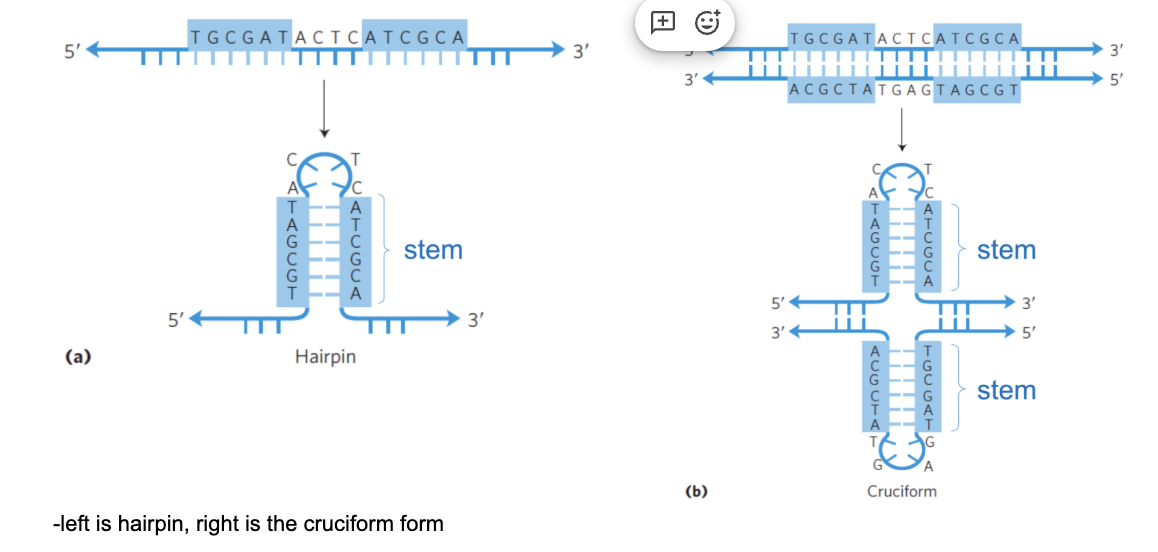
Palindromic DNA sequences
Palindromic DNA sequences can form hairpins and cruciforms
When strands separate locally, each strand can pair with itself
This creates intra-strand base pairing → stem
The intervening sequence forms the loop
One strand can form a hairpin
Both strands forming hairpins can create a cruciform
Deamination changes base identity
Deamination is when it removes an amino group from a base
C deaminates to U
If left unrepaired, the altered base pairing can cause mutation
DNA repair removes inappropriate uracil from DNA
Which can occur in DNA ?
Spontaneous loss of an amino group from the nitrogenous base
Spontaneous loss of a purine base
Crosslinking of two adjacent T bases in the presence of UV light
Depurination removes a base from DNA
Hydrolysis can remove a purine base from DNA
This creates an abasic site
At an abasic site, genetic info is lost
Such sites can destabilize or disrupt the dna strand
Which can occur in DNA ?
Spontaneous loss of an amino group from the nitrogenous base
Spontaneous loss of a purine base
Crosslinking of two adjacent T bases in the presence of UV light
G rich DNA can form G-quadruplexes
4 Guanines (G) can form a planar G-quartet
G bases interact through Hoogsteen type H bonds
Stacked G-quartets form a G -quadruplex
This structure forms in g rich sequences
Including telomeres and regulatory regions
Strands can be arranged parallel or antiparallel
Thymine dimerization induced by uv light
Uv light can covalently link adjacent T bases
This forms thymine dimer
Thymine dimers distort the dna helix
If not repaired, they can lead to mutations
Thymine dimerization induced by uv light
Uv light can covalently link adjacent T bases
This forms thymine dimer
Thymine dimers distort the dna helix
If not repaired, they can lead to mutations
Depurination removes a base from DNA
Hydrolysis can remove a purine base from DNA
This creates an abasic site
At an abasic site, genetic info is lost
Such sites can destabilize or disrupt the dna strand
Depurination removes a base from DNA
Hydrolysis can remove a purine base from DNA
This creates an abasic site
At an abasic site, genetic info is lost
Such sites can destabilize or disrupt the dna strand
Deamination changes base identity
Deamination is when it removes an amino group from a base
C deaminates to U
If left unrepaired, the altered base pairing can cause mutation
DNA repair removes inappropriate uracil from DNA
G rich DNA can form G-quadruplexes
4 Guanines (G) can form a planar G-quartet
G bases interact through Hoogsteen type H bonds
Stacked G-quartets form a G -quadruplex
This structure forms in g rich sequences
Including telomeres and regulatory regions
Strands can be arranged parallel or antiparallel
Hoogsteen pairing uses a difference edge of the base
Watson crick pairing uses the standard base pairing edge
Hoogsteen pairing uses an alternative base edge, often accessible in the major groove
A 3rd strand can interact with an existing base pair
This can support higher order dna base structures
Major classes of RNA perform different cellular functions
Messenger RNA (mRNA): carries genetic info from DNA to the protein synthesis machinery 5%
Transfer RNA (tRNA): delivers AA’s to the ribosome during protein synthesis 15%
Ribosomal RNA (rRNA): forms the structural and catalytic core of ribosomes: 80%
Regulatory and catalytic RNA’s: include riboswithces, small rna’s, microRNAs’ , interfering RNA, guide RNA, and ribozymes
*RNA’s have movre diverse functions than DNA bc they can fold into more diverse structures
Base stacking can give single stranded RNA local helical structure
Adjacent bases interact with one another via base stacking interactions
The helical structure would maximize such interactions
RNA’s form secondary structures by intramolecular base pairing
RNA secondary structure is driven by intramolecular base pairing
Base paired regions are called stems
Stems form right handed RNA double helices
Unpaired regions form loops and bulges
RNA 2* structure is stabilized by H bonding and base stacking
2ndary structure example: tRNA. tRNA folds into a cloverleaf 2ndary structure
Base pared regions form 4 stems: acceptor stem, D stem, anticodon stem, and TpsiC stem
Unpaired regions form 4 loops: D loop, anticodon loop, TpsiC loop, and variable loop
The 3’ CCA end carries the amino acid though the ester bond
The anticodon loop base pairs with a codon in mRNA
RNA can form standard (watson-crick) base pairs
G with C (3 pairs)
A with U (2 pairs)
Paring with between
Nitrogen front the main ring - H of amino groups
Oxygen of ketones - H of amino groups
RNA nucleotides can also engage in noncanonical interactions (non standard)
Can occur:
Non WatsonCrick (WC) base paring between standard bases: A,U,G,C
Non WC base pairing between a standard base and a modified base
2’OH mediated H bonds to a base
RNA’s can fold into compact tertiary structures
Many RNA’s fold into compact 3D structures under physiological conditions
RNA tertiary structure organizes stems, loops, and junctions in space
Folding is supported by base pairing, base stacking, noncanonical interactions, and cations
Like proteins, many RNA’s have unique 3D structures that are critical for function
RNA backbone flexibility supports diverse conformations
Rna has multiple backbone dihedral angles, often labeled a-C (see slide)
This flexibility allows RNA stems and loops to pack in different ways
Nucleotides can adopt nonhelical conformations and noncanonical interactions
This flexibility helps RNA form complex tertiary structures
Cations can help RNA fold into compact tertiary structures
RNA phosphate backbones are - charged and repel each other
Cations shield phosphate repulsion and allow helices to pack closely
Mg2+ can also bind specific sites and stabilize tertiary contacts
tRNA
positions amino acids and anticodon during translation
riboswitches
bind metabolites and regulate gene expression
ribozymes
catalyze biochemical rns
the ribosome uses rRNA that regulate gene expression
Riboswitches are RNA sensors that regulate gene expression
They are RNA elements, often in the 5’UTR of bacterial mRNA’s
They bind small molecules such as metabolites, ions, cofactors, or AA’s
Ligand binding changes RNA structure
The structural change alters transcription or translation of a downstream gene
Ribozymes are folded RNAs with catalytic activity
Ribozymes are RNAs that catalyze chem rxns
Their discovery showed that enzymes are nto always proteins
Folded RNA can form active sites that bind substrates
RNA active sites can position substrates and catalytic groups to accelerate rxns
Ribozymes catalyze 2 major types of rxns
RNa processing rxns
Breaking and joining RNA phosphodiester bonds
Peptide bond formation
Linking amino acids during protein synthesis
Ribosome is a ribozyme
The ribsome is a large rna protein machine that synthesizes proteins
Its a RNA based catalysis, the rRNA in the ribosome is not a protein component
Ribozymes are RNA’s that can catalyze biochemical rxns