BPD: Caries 1 & 3 - Demineralisation, caries types and Stephan curve
 Call Kai
 Learn
Learn Practice Test
 Spaced Repetition
 Match
Match Flashcards
 Knowt Play
1/13

There's no tags or description
Looks like no tags are added yet.
Name | Mastery | Learn | Test | Matching | Spaced | Call with Kai | Chat |
|---|
No analytics yet
Send a link to your students to track their progress
14 Terms
Development of incipient caries overview
Caries is a dynamic process involving repeated periods of demineralisation and remineralisation.
Cariogenic bacteria are naturally present in the mouth and can form dental plaque
Sucrose plays the essential role in the cause of caries: When the bacteria in plaque frequently get access to fermentable carbohydrates (mono- and disaccharides, escpecially sucrose) and breaks it down during bacterial metabolism, they produce organic acids that lower the pH in the plaque, leading to the loss of minerals in the tooth. This is called demineralisation.
Caries bacteria can also produce proteolytic enzymes which leads to proteolysis; breaking down proteins in the organig part of the tooth, particularly dentin.
Demineralisation together with proteolysis, can contribute to the development of caries
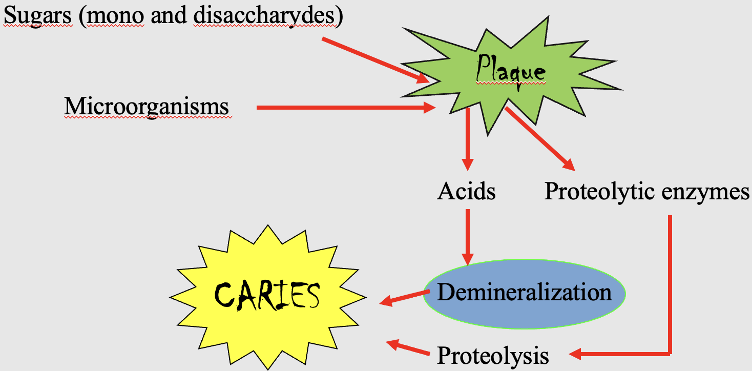
Demineralisation
The underlying process driving caries progression through the different stages.
The tooth repeatedly switches between demineralisation and remineralisation. Which process dominates depends om the pH in the plaque:
Low pH - demineralisation dominates → mineral release/loss
Higher/normal pH - remineralisation can occur → mineral return
If these acid attacks happen frequently enough, the tooth loses more mineral than it can regain and plaque accumulates → the carious lesion progresses to cavitated caries
Incipient/initial caries: demineralisation of enamel
Enamel mainly consists of:
Mainly the unorganic mineral hydroxyapatite (HAP); which contains calcium (Ca²+), phosphate (PO4³-) and hydroxyl (OH-)
A very small organic component, which includes enamel proteins, such as amelogenin og enamelin.
Enamel consists of hydroxyapatite (HAP) crystals, which are organised into enamel rods (prisms). The organic acid from the carogenic bacteria dissolve these minerals, causing calcium and phosphate ions to be released. During remineralisation, these minerals can be returned to the enamel (saliva and fluoride)
During early demineralisation:
Mineral loss begin mainly in the inter-rod spaces, and becomes wider
The enamel rods become thinner
With further mineral loss, the regular arrangement of the rods and HAP crystals become disrupted
The enamel surface may initially remain relatively intact, even though substantial mineral loss is occurring underneath it - this is because minerals from saliva can continuously remineralise/reprecipitate the superficial layer.
The incipient caries (white spot lesion) is partially reversible because the enamel surcae is still intact, and via remineralisation, the minerals return to the enamel and the lesion can be repaired.

Definition of dental caries/caries disease (all of them must be mentioned)
A progressive disease
Affects only dental hard tissues - enamel, dentin and cementum (the pulp does not “get caries”, only inflamed)
Shift in the dynamic balance between demineralisation and remineralisation, in favour of demineralisation
Cariogenic bacteria are present and their metabolic activity
Driven by an acidic local environment (low pH) produced by bacterial metabolism of fermentable carbohydrates
Stages of caries
Incipient caries/initial caries (white spot)
Superficial caries (rough surface)
Caries media (enamel → dentin)
Caries profunda (enamel → approaching pulp)
Caries penetrans (enamel → pulp)

Incipient caries: The white spot lesion
The first clinically visible sign of incipient caries is usually a white spot lesion (incipient caries/intitial caries)
A white spot lesion represents mineral loss beneath a relatively intact enamel surface.
The appear in predictive locations:
Permanent incisors:
Approximal surfaces between teeth
Gingival part of the vestibular/buccal surfaces
Primary/deciduous incisors: neck (ECC)
Decidiuous and permanent molars:
Occlusal surfaces (occlusal fissures of molars)
Approximal surfaces between teeth
Gingival part of vestibular/buccal surfaces
At this stage, there is clinically:
A white spot
Relatively intact surface (subsurface demineralisation)
No roughness
No cavitation
No pain (no complaints from the patient)

Histological zones under a white spot lesion
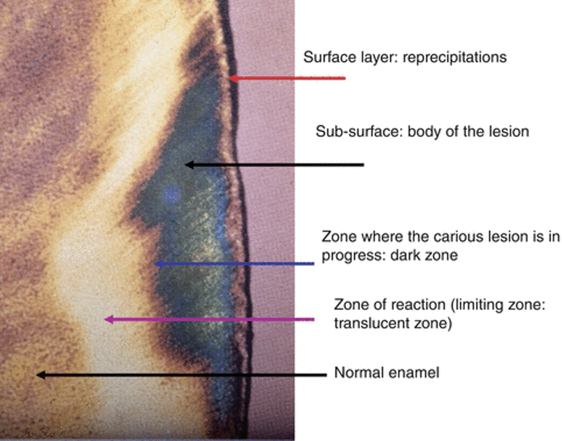
A white spot lesion contains different histological zones
From the enamel surface inward:
Surface layer: Relatively intact (saliva remineralising/reprecipitating)
Sub-surface/body of the lesion: Main demineralised mass
Dark zone: Zone where the carious lesion is in progress
Translucent zone: The deepest advancing part of the lesion, representing the early fron of demineralisation.
Therefore, the visible white spot is only the clinical sign. There is actually a larger subsurface demineralised lesion underneath.
Superficial caries: Crystal/structural collapse
Demineralisation has progressed enough after incipient caries that the enamel structure becomes increasingly porous and weak, and leads to structural collapse.
The enamel surface breaking down leads to surficial microcavitation - tiny cavities that form in the enamel.
The surface becomes clinically rough - can be identified clinically by a dental probe/explorer
It may later become yellowish or brown discoloration
There is no pain
Not reversible: Once the physical enamel has collapsed, that lost structure cannot be restored simply by remineralisation.

Caries media: Visible dark cavity
The caries has progressed through and destroyed the enamel completely, and penetrated into the dentin
A visible dark cavity can be clinically seen
The deeper part of the cavity may have a soft base because the dentin has been demineralised and damaged (fast progression - dentin is more acid-sensitive and easily demineralised)
Pain following irritation, for example from cold, sweet substances or mechanical stimulation

Caries profunda: Deep caries
The caries has progressed through the dentin and approaches the pulp
“Trap on the occlusal surface”: The destruction underneath can be much larger than what is visible from the occlusal surface - the surface opening may look relatively small while caries has spread extensively through the underlying dentin.
The remaining tooth structure may become undermined and weakened.
Spontaneous pain: Pain that occurs even without an obvious external stimulus.
Urgent treatment is needed: The lesion is getting close to the pulp!
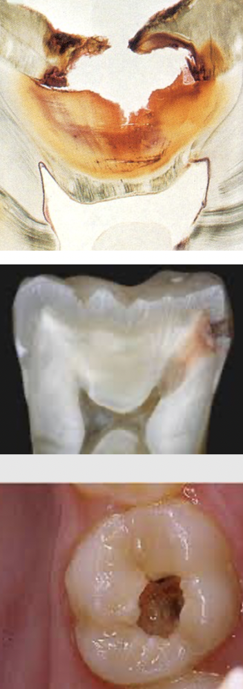
Caries penetrans: Barely visible coronal tooth
The caries has progressed to the pulpal region, and bacteria and their products can irritate/infect the pulp, causing pulpal inflammation (pulpitis) - increased pressure and pain
If the infection then spreads through the root canal and out through the root apex, it can infect the surrounding tissues and cause consequential diseases, like abscess and fever.
The remaining tooth structure can become severely weakened and may eventually fracture.

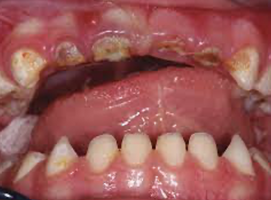
Early Childhood caries (ECC)
Caries occuring in primary (deciduous) teeth in young children
Different circumstances but the same histological procedure (caries lesion progression)
The order of typical locations of caries: Starts on the maxillary deciduous incisors (52, 51, 61, 62) on the gingival part of the vestibular/buccal surfaces. Later the deciduous first molars (54, 64, 74, 84) and maxillary deciduous canines (53, 63)
Affects the mandibular deciduous incisors less often, partly because they are protected by the tongue and salivary flow.
Can progress quickly, because deciduous teeth have thinner enamel and dentin
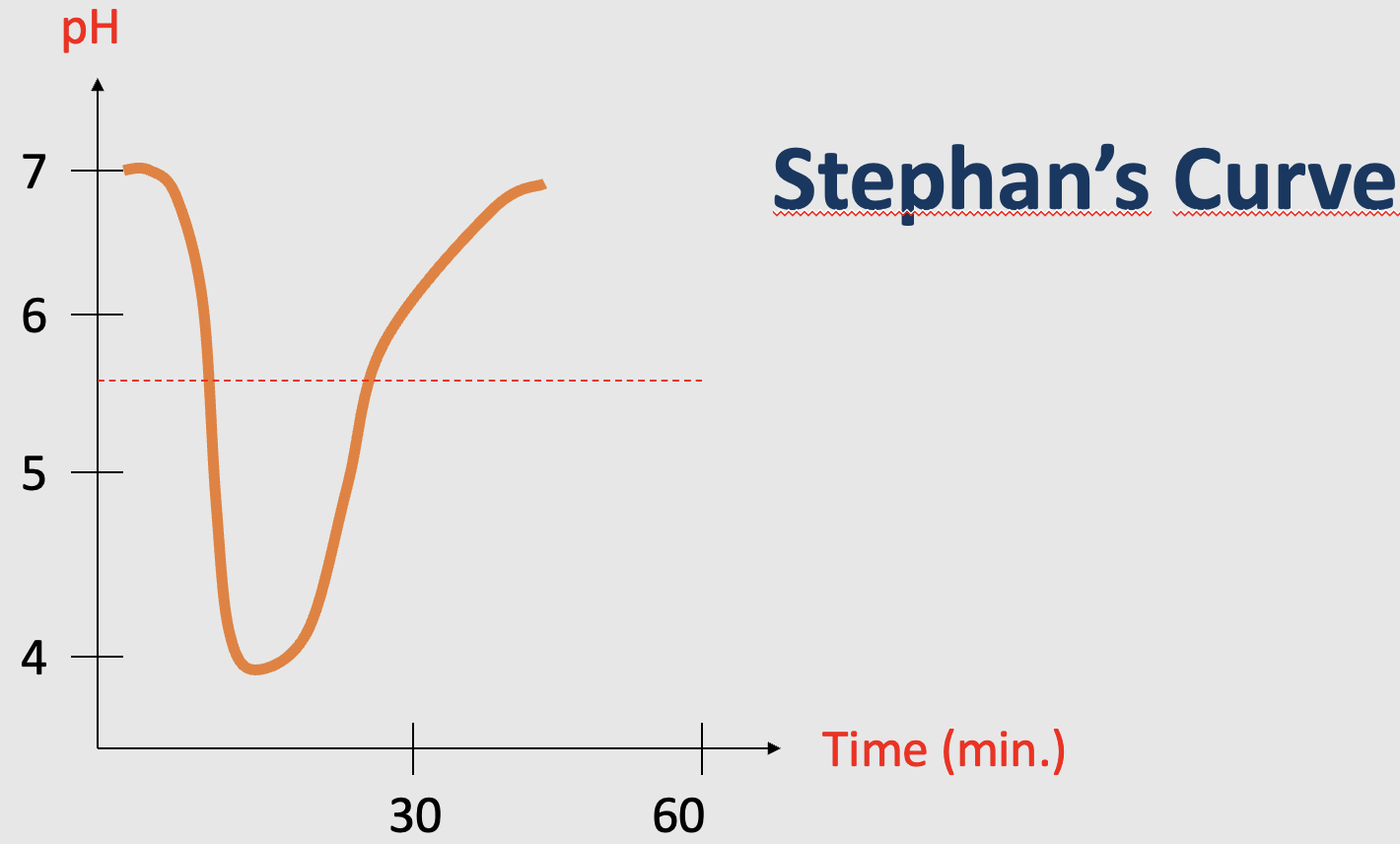
The Stephan curve
The Stephan curve shows how plaque pH changes over time after consuming fermentable carbohydrates
The plaque pH is around neutral (pH=7)
After sugar intake: Bacterial metabolism leads to acid production, which causes the pH to rapidly fall
The pH falls below the critial pH for enamel (pH~5,5): Demineralisation is favoured - minerals (Ca2+ and phosphate) releases from the enamel.
The pH can fall much further (f.eks: pH=4), but then saliva gradually buffers and removes the acids during remineralisation, causing the pH to rise again.
When the pH rises above the critical pH: Demineralisation decreases/stops, remineralisation is favoured and minerals return to the enamel.
Dentin is more vulnerable to acids than enamel, because its critical pH is 6,7. and therefore more easily demineralised: Acid attack → enamel demineralises first → caries progresses through enamel → reaches dentin → dentin demineralises more easily and caries can progress faster.
Why frequency of sugar intake matters
a) Frequent intake/snacking of fermentable carbohydrates: Plaque pH repeatedly falls below the critical pH for enamel throughout the day. There is less time for remineralisation between acid attacks
b) No snacking, and eating during main meals: There are only few large pH drops. Between these episodes, saliva has much longer periods to increase the pH by neutralising acids and promote remineralisation.
The two people may consume similar total amounts of sugar, but the person a) consumes sugar frequently throughout the day, and may experience much more time below the critical pH
