Neuropsych Exam 1
 Call Kai
 Learn
Learn Practice Test
 Spaced Repetition
 Match
Match Flashcards
 Knowt Play
1/66

There's no tags or description
Looks like no tags are added yet.
Name | Mastery | Learn | Test | Matching | Spaced | Call with Kai | Chat |
|---|
No analytics yet
Send a link to your students to track their progress
67 Terms
What is Neuropsychology? How is it Related to cognitive Neuroscience?
What is trephination/trepanning and why is it done? What theory do cultures use to justify it?
Trephination/Trepanning - drilling holes in skulls
Not really sure why they did it
Medicine → medically made brain injuries better, areas around hole showed healing
Magic → not sure if they really knew
What were the two theories about the structure where behavior originates?
Cardiac Hypothesis
Aristotle → heart causes behavior
heart is warm → actions are hot and brain evaporates heat so its cold
Brain Hypothesis
Hippocrates + Galen → brain causes behavior
Hippocrates saw brain injuries on right side caused paralysis on left
Galen saw connections between sense organs and brain
pressure to brain = death, pressure to heart was just pain
What are the two theories of function?
Humor Theory - 4 bodily fluids: blood, black bile, yellow bile, and phlegm
Ventricular Theory - placing aristotle’s categories into action
What was Galen’s knowledge of anatomy based on? Why did knowledge of anatomy
not advance much until 14 centuries later and how did Andreas Vesalius improve this?
Galen - dissected ape brains
Vesalius - dissected executed criminal brains
Weren’t allowed to dissect human for religious reasons → they wouldn’t make it to heaven without their body in tact
Vesalius dissected criminals because they weren’t going to heaven anyway
What is phrenology? Who is famous for creating localization of function in its pseudoscience and scientific forms?
Pseudoscience
Phrenology - reading the bumps/shape of brain
Franz Joeseph Gall and Johann Casper Spurzheim
Scientific
Broca and Wernickes Area - TBI impact speech based on these places
What is equipotentiality? Why did Flourens believe that? What is mass action and why did Karl Lashley believe it held the mass function view of function in the brain?
Equipotentiality - intact areas of the brain take over the functions of the damaged ones
Flourens - based on animal ablations and found recovery of function after cortical lesions
Functions are not strictly localized → cortex operates as a whole
Mass Action - efficiency of the brain depends on the total mass of the cortex
Karl Lashley - maze learning experiments in rats where deficits were proportional to lesion size not location
Complex functions like memory are widely distributed
What view did Hughlings Jackson propose with regard to the organization of the nervous system? What was Luria’s view on the organization of the nervous system?
Jackson - Hierarchy of organization
Luria - localization of function + equipotentiality
unit 1 - keeps body alive
unit 2 - receives and processes information
unit 3 - uses information to take action
What is the distinction between the central and peripheral nervous system? (Somatic vs Autonomic) (Afferent vs Efferent) (Parasympathetic vs Sympathetic)
CNS - encased in bone
Brain and Spinal Cord
PNS - communication network with rest of body
Somatic Nervous System (controllable) → Afferent (towards)/Efferent nerves (away)
Autonomic Nervous System (automatic) → A/E nerves → Parasympathetic (rest/digest)/sympathetic nervous systems (fight/flight)
How are the directions ventral, dorsal, rostral, caudal, medial, lateral, superior, inferior, anterior and posterior used?
Ventral - towards front/stomach
Rostral - towards snout/beak
Caudal - towards tail
Medial - to the middle
Lateral - to the side
Superior - above/upper
Inferior - below/lower
Anterior - in front
Posterior - behind
How are dorsal, ventral, rostral and caudal different for the head and the trunk why is that?
They started using these terms when discussing 4 legged things and animals
Same terms apply to humans even though we are bipedal
What are the sagittal, transverse, horizontal, frontal, coronal, midsagittal planes of section?
Sagittal = Midsagittal
Frontal = Coronal
Transverse = Horizontal

What are the two general classes of cells in the nervous system? What are the ranges of estimates for the quantity of each of these classes?
Neurons
36 billion - 200 billion
each have 10,000 connections
information transmission: made of dendritic tree (receive), cell body, and axon (send out)
within = electrical and between = chemical
Glial Cells
Probably = # of neurons and glial cells
physical glue and chemical support
may affect transmission?
What is a myelin sheath? What difference does it make if an axon is myelinated or not?
protective, fatty-protein layer that wraps around axon
works to speed up electrical signals
What are the types of glial cells? What is the function of microglia, astrocytes, oligodendrocytes, and Schwann cells?
Microganglia
Pacman!
Eats brain trash
Macroganglia
Oligodendrocytes - myelination in CNS
Schwann Cells - myelination in PNS
Astrocytes - most numerous + work as guards
Both oligodendrocytes and Schwann cells work to build something around axon
What is the difference in composition between white and gray matter? Where is gray matter found in the brain?
White Matter - mostly axons
Gray Matter - mostly cell bodies

What is the relationship between fibers, tracts and pathways?
Fibers: Individual nerve fibers are the long, thread-like extensions of neurons (axons) that transmit electrical signals away from the cell body
Tracts: A tract is a distinct bundle of these individual nerve fibers (axons) traveling together inside the central nervous system (brain and spinal cord)
share a common origin and destination
Pathways: entire macro-level circuit or route that information travels
often consists of one or more sequential tracts and multiple synaptic connections linking distant regions of the nervous system
What in general is multiple sclerosis? What is the general disease mechanism in MS? Which cells appear to get attacked?
Demyelination disorder
autoimmune disease → attacks oligodendricites in CNS
Possibly genetic succeptibility with environmental trigger
due to latitude (vitamin D levels) or ethinicity
What categories of symptoms occur in MS? What could be the cause of the emotional symptoms?
Sensory - vision problems etc
Motor - balance/movement issues
Cognitive - forgetting
Emotional - depression
Could be caused by the fact that MS is an upsetting disease to have to live with the rest of your life
What is the prevalence of MS in the US? What is the status of the latitude observation on the incidence of MS across the world? What is role of ethnic background to the risk of MS?
1 in 2000 in the US
Latitude has less vitamin D so they assumed some connection
However there are specific groups who live far north yet have a very low instance of MS
People now assume there may be some ethnic reason behind it
What is the usual age of onset of MS? What is the gender difference?
20-40 years old
more women than men
various forms of progression
What are the 3 characteristics of MS that get treated
Attacks
Progression
Symptoms
What are neurogenesis and migration? When does most neurogenesis take place? When does migration take place (look at the graph)?
Neurogenesis + Migration - making of new neurons and traveling to correct areas of brain
Neurogenesis + Migration → most occurs 2 months before born some increase at 12-20
What is Synaptogenesis? When does it start and how long does it continue (approximately)
Synaptogenesis - new formation of synapses
Starts at birth and goes until about 2/3 years old
Max synapse formations at 2 years old
What is myelination? When does it start and how long does it continue?
Myelination - creation of myelin on the axon
2 months before birth → 11 years old
less and less over time
What is synaptic pruning? When does it start and how long does it continue?
Synaptic Pruning - deletion of excess synapses
provides a more efficient route for information transmission
Starts at age 2/3 → about 11 years old
What three layers does the embryonic plate develop into? What major structures do these layers develop into?
Ectoderm - skin and nervous system
Mesoderm - musculoskeletal system
Endoderm - intestines
When does the neural tube close on average at the anterior and posterior pores? What are the defects that result when the neural pores do not close? Which type of nonclosure has the more serious defects?
Neuropore closing → Day 24 - 28 of pregnancy (after conception) very very early on
Anencephaly - anterior (front) neural pore does not close → no higher brain function
more serious effects - don’t really live past infancy
Spina Bifida - posterior (back) neural pore does not close → different levels
in utero operations available
bottom of spinal cord nerves → waist down mobility
What is lissencephaly (or agyria)? What is thought to be the process that is disturbed
in lissencephaly?
Lissencephaly (Agyria) - defective neuronal migration
problem with neuron migration where not as much of the outer layer of the brain gets formed
no sulic and no gyri leads to a smooth brain
What is microcephaly? Is the mechanism causing microcephaly known? What general
types of distal causes for microcephaly are known? Which virus was in the news for
its apparent relationship with microcephaly?
Microcephaly - “small head"
head circumference is a good indicator of brain size so small head → small brain
Microcephaly was in the news because of its relationship with Zika virus
Pre and Post natal causes
Name the 7 CNS structures caudal to rostral. Which structure in the adult human brain comes from the vertebrate myelencephalon? Which structures in the adult human brain come from the vertebrate metencephalon?
Cerebellum, Cortex, Corpus Callosum, Midbrain, Pons, Medulla Oblongada, Spinal Cord
Myelencephalon → Medulla oblongada, 4th ventricle, spinal cord
Metencephalon → Cerebellum, pons, 4th ventricle

What is another name for the vertebrate mesencephalon? Which structures in the adult human brain come from the vertebrate diencephalon? What structures in the adult human brain come from the vertebrate telencephalon?
Mesencephalon → Midbrain
Diencephalon → Forebrain (between brain) → thalamus, hypothalamus, pineal body, 3rd ventrical
Telencephalon → Forebrain (endbrain) → neocortex, basal ganglia, limbic system, olfactory bulb, lateral ventricles
Forebrain: telencephalon + Diencephalon
Midbrain: Mesencephalon
Hindbrain: Metencephalon + Myelencephalon

What are the names of the meningeal membranes? Know the order, and how located with respect to one another. What is the result of folding of the dura in the skull?
Dura Mater: outer layer
thick and tough membrane that protects spinal cord
dura folds divides brain into hemishperes
Arachnoid: middle layer
spider like and overlies the subarachnoid space and contains blood vessels (subarachnoid bleeds are more common)
Pia Mater: inner layer
overlies every detail of the outer brain and covers the brain exactly
like the thin hard boiled egg membrane

What is the subarachnoid space? What structures and liquid in that space? What types of brain bleed was mentioned w.r.t. its meningeal location?
Subarachnoid Space - space between arachnoid and pia mater filled with Cerebral spinal fluid to cushion the brain and circulate nutrients within the CNS
Subarachnoid brain bleed
What is cerebrospinal fluid? Where is it formed? How (in general) does it drain to
the venous system? How much of it (approximately) is formed on average per day?
How much (approximately) is present in the ventricles & subarachnoid space at any
time? What are the main functions of the CSF?
Cerebrospinal Fluid - layer of fluid around the entire brain and layer of liquid on bottom for brain to float in so the brain doesn’t smash against the skull
450 mL are produced everyday but only 150 mL can be used at a time
it is circulated and drained by the arachnoid villi
it is produced by the choroid plexus of the ventricles (ependymal cells)
deals with metabolites and removing waste products
What structures in the brain are the 4 ventricles and the cerebral aqueduct
associated with (and therefore know where the ventricles and the cerebral aqueduct
are located).
Forebrain - lateral ventricles → Telencephalon
Forebrain - Third ventricles → Diencephalon
Midbrain - Cerebral aqueduct → Mesencephalon
Hindbrain - Fourth ventricles → Metencephalon
Hindbrain - Myelincephalon

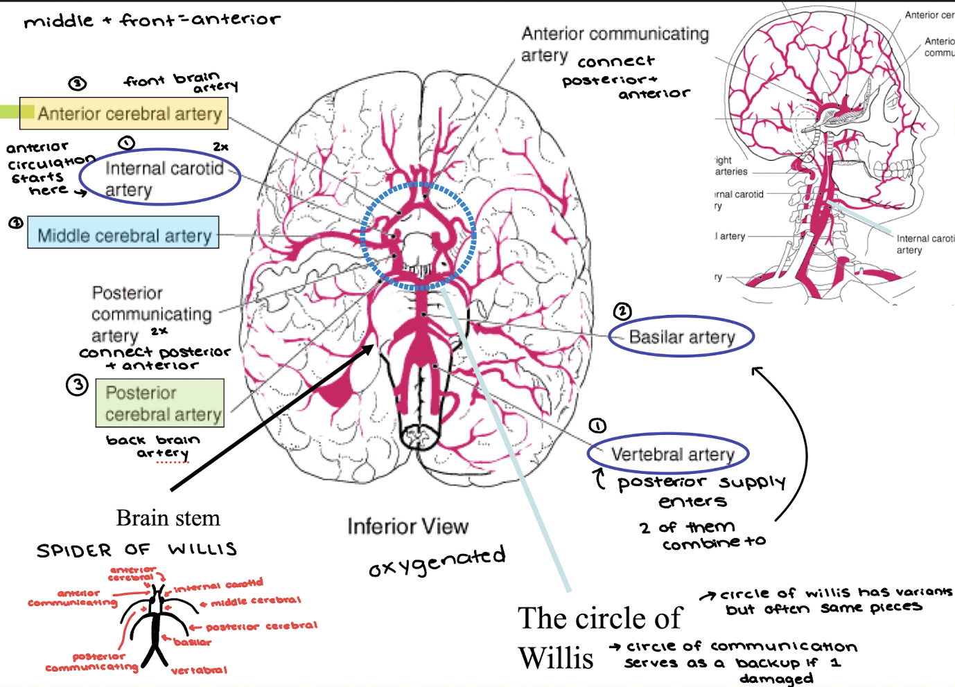
Describe the blood supply to the brain and the Circle of Willis
(internal carotid arteries, vertebral arteries, basilar artery, anterior, middle, and posterior cerebral arteries, anterior communicating artery & posterior communicating arteries)

Which arteries vascularize which lobes?
Frontal Lobe
Lateral Surface - Middle cerebral artery
Medial Surface - anterior cerebral artery
Inferior Surface - Middle + anterior cerebral arteries
Temporal Lobe
Lateral Surface - Middle cerebral artery
Medial Surface - Middle + Posterior cerebral arteries
Inferior Surface - Posterior cerebral artery
Parietal Lobe
Lateral Surface - Middle cerebral artery
Medial Surface - Anterior cerebral artery
Occipital Lobe
All Surfaces - Posterior cerebral artery
Why can a stroke in the MCA be so devastating?
Most common stroke in the middle cerebral artery
Temporal, parietal, and frontal lobes will have no blood supply
Subcortical structures get damaged over time
What is the blood-brain barrier? How do tight junctions and astrocytes contribute to the BBB? What is the point of having an incomplete BBB in places? explain how ingesting certain toxic substances involves the area postrema.
Protects the brain from any random floating thing in your blood (Major protective system)
Endothelial Cells of brain capillaries have tight junctions
tightly packed = nothing getting through (can stop medications)
Astrocyte Feet cover endothelial cells
Small uncharged molecules can pass, but others have to use active transport
Absent BBB - sampling station for what’s in the blood → check for cytotoxins, hormones, and engage feedback loops (Area Postrema - 4th ventricle → finds toxins + tells body to throw up)

What is the relationship between the spinal cord segments and spinal vertebrae? Where does the spinal cord end? How is the nomenclature of the spinal cord related to that of the spine?
Spinal Cord → encased in bone and ends at Lumbar 2, but nerves continue traveling down to exit at the bottom vertebrae
Spinal cord has theoretical sections with 2 spinal nerves out at each vertebrae
8 cervical sections, 12 thoracic sections, 5 lumbar sections, 5 sacral sections, 1 coccygeal

What are dermatomes? What is the cauda equina?
Dermatones - body segments innervated by spinal nerves
Cauda Equina - “horse tail” bundle of spinal nerve roots that extends from the lower end of the spinal cord
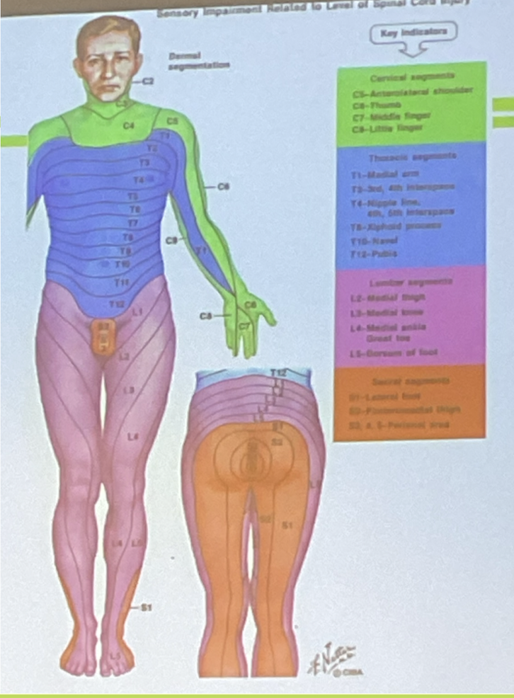
Where is a lumbar puncture done? Why is it done there? Which meninges need to be punctured? How do lumbar punctures and epidurals differ?
Lumbar Puncture - sampling cerebral spinal fluid for diagnostic purposes
done at the base of the spinal cord with a small needle so there is no risk of paralysis
Don’t hit spinal cord (L2-L4) to test same fluid washing around your brain
What are the differences between a motor neuron and a sensory neuron?
Motor Neuron - Efferent

Sensory Neuron - Afferent

What is the dorsal root ganglion? Be able to identify gray matter, white matter, dorsal root ganglion, dorsal and ventral roots of spinal nerves. Which ones are afferent? Which ones are efferent?
Dorsal Root Ganglion - where the cell bodies of neurons in Peripheral Nervous System are located (not in spinal cord) really long axons

What causes the differences in amount of white and gray matter along the length of the spinal cord?
Dispersion of motor neurons running lower through the back and legs causes more grey matter
Grey matter → cell bodies White Matter → Axons
More cell bodies + grey matter at the bottom of the spinal cord because sensory nerves have to travel all the way up
White matter at any level is info traveling up and down

What are the two factors that determine the impairments in spinal cord injuries? Explain how these affect the severity of the impairments.
Level - where on the spinal cord injury occurs
Function from injury point down becomes impaired
L1 is bad but C5 is worse
Extent - how much you can still move after
usually laceration but some fibers can still be operations
What is a paraplegic and what a quadriplegic? Which level of damage means the person with spinal cord damage will require a ventilator?
Paraplegic - loss of control over lower limbs
Quadriplegic - loss of control over all limbs
18,000 cases per year mostly C4/C5
C3 and above → lose control of your diaphragm → need ventilator
What are the top four causes of spinal cord injuries?
Motor Vehicle Accident (cars/motorcycles) - 38%
Falls (missteps/construction/stairs) - 25%
Sports (likely contact sports) - 13%
Violence - 10%
Where do the sympathetic and parasympathetic nervous system nerves exit the spinal cord? What is the function of the sympathetic nervous system? What is the function of the parasympathetic nervous system and how can you activate it?
Sympathetic
Thoracic and Lumbar spine
Engages the fight/flight response
Dialated pupils, sends blood to the muscles, inhibits salivation, increased heartrate, inhibits digestion
Parasympathetic
Cranial and Sacral spine
Engages rest, relax, and reset processes
increases salivation, reduces heart rate, stimulates digestive system → activated by deep breathing/box breathing

Where is the Medulla oblongata? Which things are regulated there? What is decussation?
Medulla Oblongata
located just above the spinal cord can be enlarged extension of spinal cord
Responsible for vital reflexes - breathing, heart rate, vomiting, salivation, coughing, and sneezing
Several tracts/axon decussate here → nerves crossing to other side of body
Compare and contrast cranial nerves and spinal nerves.
Cranial nerves and spinal nerves are functionally homologous and both have one nerve on each side of the body
Cranial - provide somatic and visceral sensory and motor function of the head and neck
Spinal Nerves - extending from spinal cord
For all the cranial nerves, know their names, numbers, functions (i.e. in general what do they do), whether they are sensory, motor or both. Know the differences between N. III, IV and VI.
I. Olfactory - Smell - sensory
II. Optic - Vision - sensory
III. Oculomotor - control of eye movements + pupil constriction - motor
IV. Trochlear - control of eye movements - motor
V. Trigeminal (x3) - skin sensations from most of the face + control of jaw muscles for chewing and swallowing - sensory and motor
VI. Abducens - control of eye movements - motor
VII. Facial - some taste, control of facial expressions, crying, salivation, and dilation of head’s blood vessels - sensory and motor
VIII. Statoacoustic - Hearing + equilibrium - sensory (vestibularcochlear, acoustic, audtitory)
IX. Glossopharyngeal - taste and some throat sensations, control of swallowing, salvation, and throat movements - sensory and motor
X. Vagus - neck sensations, control of throat, esophagus, and larynx, PSN nerves to stomach and organs (wandering nerve) - sensory
XI. Accessory - control of neck and shoulder movements - motor
XII. Hypoglossal - Control of muscles of the tongue (speech) - motor
Which cranial nerves do facial movement and sensation? Which cranial nerves serve functions to do with speech (motor to larynx, tongue)
Facial Movement and Sensation
Trigeminal - face skin sensations + control of jaw muscles
Eye Movement
Oculomotor - pupil constriction, eyelid control, lens accomodation
Trochlear - runs muscle connected to opnic nerve
Abducens - side eye muscles
Speech Functions
Hypoglossal - control of tongue muscles
Glossopharyngeal - control of swallowing, salvation, and throat movements during speech
Vagus - control of throat, esophagus, and larynx
Facial - facial expressions
Which cranial nerve heads outside the head/neck area? What does it innervate? Why has it become the focus of a lot of research in recent years?
Vagus Nerve
runs the parasympathetic nerves to stomach, intestines, and other organs
It is the cause of gallstone/kidney stone pain
Tried to cure ulcers by cutting the vegas nerve so you could no longer feel the pain sensations
Which cranial nerves have autonomic nervous system components?
Oculomotor - pupil constriction
Facial - dilation of head’s blood vessels, decrease in salivation
Glossopharyngeal - salivation control
Vagus - regulate involuntary functions of heart, lungs, and digestion
Why is breaking your skull base possibly a problem for your cranial nerves?
Nerves pass through a hole in the base of your skull so that they can connect from your brain to the rest of your body
Breaking skull base → potential to inhibit many functions of these necessary cranial nerves
Where is the pons, what structures (general) are located there?
Metencephalon - origin of neuronal activity that increases arousal/alertness
Contains - reticular formation and raphe system
Where is the cerebellum? What are the three sections (anatomical + function) of the cerebellum, what are the functions of these sections? What is the ratio of incoming vs. outgoing axons to the cerebellum?
Cerebellum - located in hindbrain (metencephalon) with many folds
3 Functional Parts
Flocculonodular Lobe - helps regulate balance and eye movements
Spinocerebellum - regulate body and limb movement
Cerebrocerebellum - planning movements and evaluating sensory information for action, has some purely cognitive functions (check system loop → movement)
40x more axons into the cerebellum than out
40-50% of neurons in the brain here, but only make up 10% of the brain

What is another name for the mesencephalon? What are the tectum and tegmentum? What sensory systems are the superior and inferior colliculi associated with? What is the connection between the substantia nigra and Parkinson’s disease? From where to where does the reticular formation extend?
Mesencephalon → Midbrain
Tectum - roof of the midbrain
Superior colliculus - swellings on each side of tectum (hills) - visual
Interior colliculus - swellings on each side of tectum (hills) - auditory
Tegmentum - intermediate level of the midbrain
Substantia Nigra - dopamine-containing pathways; “black stuff”
60% of neuron death is when parkinson’s symptoms start to appear
Reticular Formation - extends from mesencephalon through the metencephalon to myelenchepalon
descending portion - control motor neurons of spinal cord
ascending portion - increased arousal/attention
What are the two main structures in the diencephalon? Where is the thalamus located, why is the thalamus important? What is ipsilateral (same-sided) with respect to the thalamus?
Diencephalon - “subcortical” made of thalamus and hypothalamus
Thalamus - RELAY STATION → routes information to other parts of brain
paired structure - info has already been decussated when arrived at thalamus
each thalamic nucleus projects its axons to a different region of the cortex

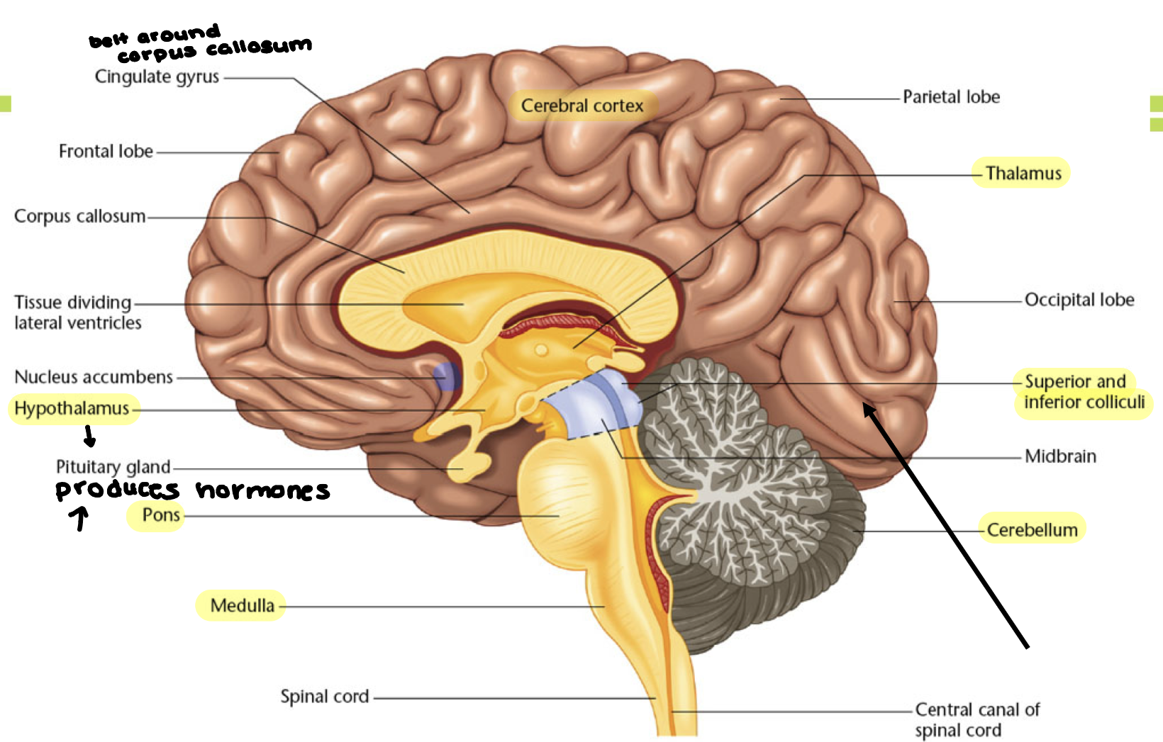
What is general is the function of the limbic system? Be able to identify the amygdala (and mental states associated), hippocampus (and function), hypothalamus (and function), pituitary gland (and function, the cingulate gyrus?
Limbic System - mostly subcortical; involved in the expression of emotions
Amygdala - expression of fear
Hippocampus - learning/memory
Hypothalamus - coveys messages to pituitary gland for hormones, in charge of behaviors that keep you alive that you must act on (hunger, thirst, reproduction)
Pituitary Gland - hormone producing gland at the base of the hypothalamus
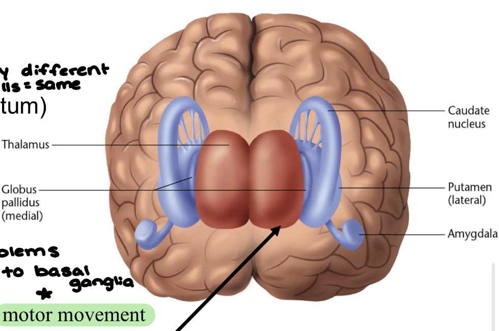
Where are the basal ganglia located in the brain? What are the three basal ganglia structures that we discussed?
Basal ganglia - (subcortical) control and coordination of movement patterns (check loop #2 of approving movement)
Caudate nucleus (striatum)
putamin (striatum)
globus pallidus
associated with motor movement, execution of automatic action sequences, aspects of memory and emotional expression, BG disease → motor issues
What are the research strategies used to figure out what different parts of the brain do?
Examine the effects of brain damage on behavior
Images of brains
Record brain activity during behaviors
Examine effects of stimulating particular parts of brain
What is inflammation? What is the function of inflammation? What are the 5 classic signs of inflammation? (Latin and English), Which ones are related to the vasodilation (Blood vessel widening)? What is the difference between acute and chronic inflammation?
Inflammation - Normal response to harmful stimuli
Rubor - redness (Vasodilation)
Calor - warmth (Vasodilation)
Tumor - swelling (Vasodilation)
Dolor - pain
Functionolasa - loss of function
Acute: call for help, fix the damage Chronic: immune system remains activated
What are tumors, what is the distinction between a malignant and a benign
tumor? What is infiltrative vs noninfiltrative tumor? What is local vs metastasized tumor?
Tumors - (“neoplasm - new tissue) Morbid enlargement or new growth of tissue, cell division and proliferation is uncontrolled
Badness → Malignant: progressive and keeps growing vs Benign: cell growth arrested (still capable of producing problems)
Infiltrative: grows into surrounding tissues vs Non-Infiltrative: gets bigger but sticks to itself
Tumor Origin → Local: stays in one location in skull vs Metastasized: arises elsewhere - other place of origin (brain tumor is secondary)