Chapter 11: Axial Musculature
 Call Kai
 Learn
 Practice Test
 Spaced Repetition
 Match
 Flashcards
 Knowt Play
1/93

There's no tags or description
Looks like no tags are added yet.
94 Terms
Endomysium
wraps individual muscle fibers
Perimysium
surrounds fascicles (bindles of cells/fibers)
Epimysium
surrounds entire muscle
Fascia
a band or sheet of fibrous connective tissue that covers, supports, and separates muscle
fusiform muscles
Thick in the middle and tapers at the end

parallel muscles
Uniform width with aligned fascicles

Triangular (convergent) muscles
Broad at one end and narrow at the other

Pennate Muscles
feather shaped

Circular muscles (sphincters)
form rings around body openings

example of fusiform muscle
biceps brachii

example of parallel muscle
rectus abdominis

example of triangular (convergent) muscle
pectoralis major

example of unipennate muscle
palmar interosseous

example of bipennate muscle
rectus femoris

example of multipennate muscle
deltoid

example of circular muscle
orbicularis oculi

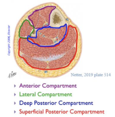
Compartments
Spaces enclosed by fasciae that contain one or more functionally related muscles, nerves and blood vessels
Fasciae
Intermuscular septa
Compartments in upper limbs
Anterior and posterior compartments
Compartments in lower limbs
Anterior, lateral, posterior deep and posterior superficial
direct (fleshy) attachment
Close association with bone and connection with collagen fibers
indirect attachment
Via tendon attaching muscle to bone and connects to periosteum and matrix
Aponeurosis
a broad, sheetlike tendon
Dermal attachments
Muscles that attach to the dermis of skin (facial muscles)
Intrinsic
Both origin and insertion contained within a particular region
Example of intrinsic muscle
O within hand and I within hand
Extrinsic
Acts upon a designated region but has its origin elsewhere
Example of extrinsic muscle
O in forearm and I within hand
Action
Effect produced by a muscle and produced or prevents movement
Prime mover (agnoist)
Produced main force of action
Synergist
Aids prime mover and may stabilize joint
Antagonist
Opposes prime mover
Antagonistic pair
act on opposite sides of joint
Fixator
Prevents bone movement
How are muscles named
Size, Shape, Location, Number of heads, Orientation of the fibers, and action
Example of size
brevis = short
Example of shape
teres = round
Example of location
Branchii = of the arm
Example of number of heads
Biceps = two heads
Example of orientation of the fibers
Rectus = straight
example of action
flexor = a muscle that flexes
Muscle innervation
Describes which nerve stimulates the muscle
Spinal nerves
Arise from spinal cord and innervate muscles below the neck
Cranial nerves
Emerge from brainstem and innervate muscles of the head and neck
Muscles of facial expression
Frontalis, orbicularis oculi, orbicularis oris, zygomaticus major, zygomaticus minor and risorius
Frontalis
elevates eyebrows

Orbicularis oculi
Closes eyes

Orbicularis oris
closes lips

Zygomaticus major and minor
raise corners of mouth for smile

Risorius
expresses horror or fake smile

Genioglossus
Protrudes tongue or moves it to one side if used unilaterally
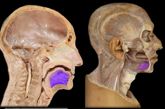
Hyoglossus
depresses tongue

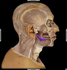
Styloglossus
Pulls tongue up and back
Muscles of the tounge
Genioglossus, hyoglossus and styloglossus
Muscles of chewing
Temporalis, masseter, medial pterygoid and lateral pterygoid
Temporalis
elevates and retracts mandible

masseter
elevates mandible

medial pterygoid
Elevate mandible and move it sideways

Lateral pterygoid
Depresses manidble and moves it sideways
Muscles of the neck
Digastric, sternohyoid and sternothyroid
Digastric
depresses mandible

Sternohyoid
depresses hyoid
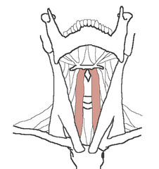
Sternothyroid
depresses larynx

suprahyoid muscles
digastric, stylohyoid, mylohyoid, geniohyoid

Infrahyoid
omohyoid, sternohyoid, sternothyroid, thyrohyoid

Sternocleidomastoid
Rotates head to side when unilaterally active, draws head forward when bilaterally active

Neck flexors
SCM and scalene group

Trapezius
Extends the neck

Neck extensors
Trapezius, splenius group and semispinalis group
Muscles of respiration
Diaphragm
Diaphragm
Prime mover of inspiration
Muscles of anterior abdominal wall
Rectus abdominis and transverse abdominal
Rectus abdominis
Flexes waist
Transverse abdominal
Compresses abdominal contents
rectus sheath
Aponeurosis of transverse abdominals, internal obliques and external obliques
Rectus muscles
Lie within sheath
external oblique
compresses abdomen (superficial)
internal oblique
flexes and rotates vertebral column (middle layer)
Transverse abdominal
compresses abdominal wall, laterally rotates waist (deep layer)
linea alba
midline tendinous seam joining the abdominal muscles
Peritoneum
membrane that lines the abdominal cavity
Parts of aponeurosis
transverse abdominal, internal oblique and external oblique
Rectus sheath
fibrous sheath formed by aponeuroses of abdominal muscles
Linea semilunaris
Two layers from rectus sheath join to form a vertical line of aponeurosis

inguinal ligament
ligament extending from pubic bone to anterior superior iliac spine, forming lower border of abdomen

Muscles of vertebral column
Erector spinae, multifidus and quadratus lumborum
Erector spinae
straightens spine

Multifidus
Stabilizes adjacent vertebrae
Quadratus lumborum
aids respiration by stabilizing the diaphragm and rib 12
Muscles of pelvic floor
Ischiocavernosus, external anal sphincter, levator ani group, piriformis
Ischiocavernosus
maintains erection of the penis or clitoris by compressing deep structures of the organ and forcing blood forward into its body

External anal sphincter
Retains feces in rectum

Levator ani group
iliococcygeus, pubococcygeus, puborectalis

Piriformis
Pear-shaped muscle that laterally rotates and abducts thigh
