CH 13 Cerebellum
 Call Kai
 Learn
 Practice Test
 Spaced Repetition
 Match
 Flashcards
 Knowt Play
1/28

There's no tags or description
Looks like no tags are added yet.
29 Terms

I LOVE YOUUUU
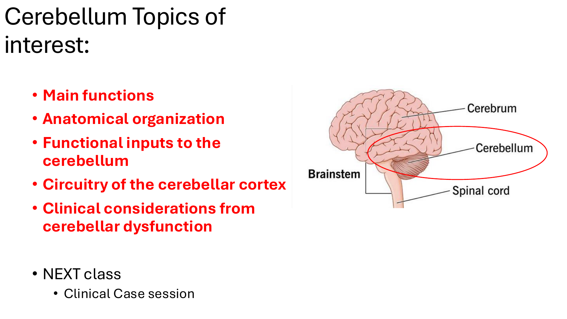
CH 13 = CEREBELLUM
We will learn what the cerebellum does → how it's organized → how information travels through it → what happens when it's damaged → cerebellar strokes.
Slide 3 → Cerebellar connections: superior, middle, and inferior peduncles
Slide 4 → Main functions: movement coordination, error correction, balance, and motor memory
Slide 5 → Folia: surface area and possible higher-level thinking functions
Slide 6 → Cerebellar regions: lateral, intermediate, vermis, and flocculonodular
Slides 7 + 8 → Cerebellar circuitry: 3 layers, mossy fibers, climbing fibers, and Purkinje cells
Slides 9 + 10 + 11 → Clinical dysfunction: ataxia, nystagmus, alcoholic degeneration, and Dandy-Walker syndrome
Slide 12 → Cerebellar blood supply: SCA, AICA, and PICA
Slides 13 + 14 + 15 → Cerebellar strokes: PICA vs. AICA vs. SCA
Slides 3–8 teach you how the cerebellum works. Slides 9–15 teach you what happens when it doesn't work properly.
When you're ready, say NEXT, and we'll begin Slide 3 — “Little Brain” using our full study format:
This slide is trying to teach you → DEFINITIONS → Tiny story → How it all connects → Focus on this: → How to remember.

This slide is trying to teach you: The cerebellum is located behind the brainstem and connects to it through 3 cerebellar peduncles, which carry information in and out.
DEFINITIONS:
Cerebellum: “Little brain”; helps coordinate movement.
Peduncles: Bundles of nerve fibers connecting the cerebellum to the brainstem.
Afferent: Information traveling INTO the cerebellum.
Efferent: Information traveling OUT OF the cerebellum.
Arbor vitae: Tree-like white matter inside the cerebellum (shown in the right-side images).
Tiny story:
Imagine the cerebellum is a movement-control center with 3 highways:
Superior highway: Mostly sends information OUT to the red nucleus and thalamus.
Middle highway: Brings information IN from the pons, carrying signals originating from the cerebral cortex.
Inferior highway: Carries information IN and OUT, including connections with vestibular nuclei.
How it all connects:
Remember the pons, medulla, and midbrain from Chapters 10–12?
The cerebellar peduncles connect the cerebellum to these brainstem regions. Now we're learning what information travels through those connections.
Focus on this:
Cerebellum = posterior to brainstem.
Superior peduncle = mainly EFFERENT → red nucleus + thalamus.
Middle peduncle = mainly AFFERENT ← pons/cortex.
Inferior peduncle = AFFERENT + EFFERENT ↔ vestibular nuclei.
Arbor vitae = tree-like cerebellar white matter.
How to remember:
S-M-I = Send, Meet, Interact
Superior = Sends OUT.
Middle = Messages come IN.
Inferior = In AND out.

This slide is trying to teach you: The cerebellum's MAIN job is to coordinate movements and correct mistakes while you're moving, so your movements stay smooth and accurate.
DEFINITIONS:
Skeletal muscles: Muscles you voluntarily control, like your arms and legs.
Motor neurons: Nerve cells that control movement.
Error correction: Adjusting a movement when it isn't going as planned.
Motor memory: Remembering learned movements, like riding a bicycle.
Tiny story:
Imagine you're learning to ride a bicycle. 🚲
Your cerebral cortex helps initiate the movement, while your cerebellum constantly adjusts your balance and muscle activity so you don't fall.
After practicing, your movements become smoother because the cerebellum helps with motor learning and memory.
How it all connects:
On Slide 3, we learned about the 3 cerebellar peduncles that carry information in and out.
Now we understand WHY those connections matter: the cerebellum uses information to coordinate and correct ongoing movements.
Focus on this:
MAIN function = coordination of skeletal muscle movements.
Works with the cerebral cortex AND basal ganglia.
Error correction = adjusts ongoing motor activity.
Also controls posture, balance, and muscle tone.
Motor memory = learned movements like cycling or dancing.
How to remember:
Cerebellum = Movement Coach 🚲
Cortex = starts the movement.
Cerebellum = makes the movement smooth and corrects mistakes.
Practice = motor memory.

This slide is trying to teach you: The cerebellum has many folds that increase its surface area, allowing it to potentially contribute to higher-level thinking, not just movement.
DEFINITIONS:
Folia: Small folds on the cerebellum that increase its surface area.
Gray matter: Brain tissue containing neuron cell bodies.
Neocortex: Outer layer of the cerebrum involved in higher-level brain functions.
Higher-level computations: Complex thinking, such as language and abstract reasoning.
Tiny story:
Imagine a large sheet of paper folded into a tiny space. 📄
That's what the cerebellum does!
Although it looks small, unfolding its surface would reveal approximately 1,600 cm², compared with 44 cm² without its folds.
The professor's slide compares this to legal paper vs. a postage stamp.
How it all connects:
Slide 4 taught us that the cerebellum coordinates physical movements.
Slide 5 introduces the possibility that it also helps coordinate mental processes, such as language and abstract reasoning.
Focus on this:
Folia = folds that increase cerebellar gray matter surface area.
Unfolded surface ≈ 1,600 cm² vs. 44 cm².
Human cerebellar surface area ≈ 80% of neocortex (33% in nonhuman primates, as reported on the slide).
Cerebellum may contribute to language and abstract reasoning, beyond movement coordination.
Higher-level cognitive role = proposed possibility, not the cerebellum's established main function.
How to remember:
Folia = Folds = More Surface Area
Cerebellum = coordinates movements AND may help coordinate thoughts.

This slide is trying to teach you: Different regions of the cerebellum have different jobs in controlling movement, posture, and balance.
DEFINITIONS:
Distal extremities: Body parts farther from the trunk, like hands and feet.
Proximal extremities: Parts closer to the trunk, like shoulders and hips.
Vermis: The middle region of the cerebellum.
Vestibular: Related to balance and sensing head movement.
Tiny story:
Imagine you're dancing. 💃
Lateral region: Plans your next movement.
Intermediate region: Helps control your hands and feet.
Medial region (vermis): Controls your trunk and nearby limbs to maintain posture.
Flocculonodular lobe: Helps you balance and coordinate your eyes with movement.
Each region has its own job, but they work together to make movement smooth.
How it all connects:
Slide 5 explained how the cerebellum has a large folded surface.
Now Slide 6 shows that different parts of that surface specialize in different functions.
Focus on this:
Lateral = motor planning.
Intermediate = distal extremities (hands and feet).
Medial (vermis) = trunk + proximal extremities.
Flocculonodular = balance + vestibular/visual coordination.
How to remember:
L-I-M-F
Lateral = Look ahead (plan).
Intermediate = Individual hands and feet.
Medial = Middle of body.
Flocculonodular = Falling prevention (balance).

This slide is trying to teach you: The cerebellar cortex has 3 layers, and each contains specific neurons that either excite or inhibit signals.
DEFINITIONS:
Histology: Study of tissues and cells.
Excitatory: Increases the activity of other neurons.
Inhibitory: Decreases the activity of other neurons.
Purkinje cells: Large neurons that send inhibitory signals to the deep cerebellar nuclei.
Deep cerebellar nuclei: Groups of neurons inside the cerebellum that receive signals from Purkinje cells.
Tiny story:
Imagine the cerebellar cortex is a 3-floor building. 🏢
Top floor — Molecular layer: Stellate and basket cells help inhibit signals.
Middle floor — Purkinje layer: Large Purkinje cells send inhibitory signals to the deep cerebellar nuclei.
Bottom floor — Granule layer: Granule cells excite, while Golgi cells inhibit.
Together, these cells help regulate cerebellar activity.
How it all connects:
Slide 6 showed us which cerebellar regions control different movements.
Slide 7 shows us how the neurons inside the cerebellar cortex are organized to process those movements.
Focus on this:
3 layers (superficial → deep) = Molecular → Purkinje → Granule.
Molecular = stellate + basket cells → inhibitory.
Purkinje = large neurons → inhibitory signals to deep cerebellar nuclei.
Granule layer = granule cells (excitatory) + Golgi cells (inhibitory).
Molecular = few cell nuclei; Granule = many cell nuclei.
How to remember:
M → P → G = Molecular, Purkinje, Granule
Inhibition: Stellate, Basket, Purkinje, Golgi.
Excitation: Granule cells.

This slide is trying to teach you: The cerebellum receives excitatory information through mossy and climbing fibers, while Purkinje cells send inhibitory signals to the deep cerebellar nuclei.
DEFINITIONS:
Mossy fibers: Major input fibers that bring information into the cerebellum.
Climbing fibers: Excitatory fibers that wrap around Purkinje cell branches.
Deep cerebellar nuclei: Structures that receive and process signals from cerebellar pathways.
Descending pathway: Information traveling outward from the cerebellar cortex.
Tiny story:
Imagine the cerebellum is a control room. 🎛
Mossy fibers: Bring information from the cerebral cortex, spinal cord, and vestibular system.
Climbing fibers: Deliver strong excitatory signals to Purkinje cells and deep cerebellar nuclei.
Purkinje cells: Act like brakes, sending inhibitory signals to the deep cerebellar nuclei.
Together, they help the cerebellum regulate movement.
How it all connects:
Slide 7 introduced the 3 layers and their neurons.
Slide 8 explains how information enters those layers and how Purkinje cells influence the cerebellum's output.
Focus on this:
Mossy fibers = primary excitatory input → granule cells, Golgi cells, and deep nuclei.
Mossy inputs originate from cortex, spinal cord, and vestibular system.
Climbing fibers = excitatory input → Purkinje cells + deep nuclei.
Purkinje cells = ONLY output from cerebellar cortex → inhibitory to deep nuclei.
Mossy + climbing = excitation; Purkinje = inhibition.
How to remember:
Mossy = Messages IN.
Climbing = Climb around Purkinje cells.
Purkinje = Put on the brakes. 🛑
This slide is trying to teach you: Damage to the cerebellum causes problems with movement coordination, muscle tone, balance, and eye movements.
DEFINITIONS:
Ataxia: Poor coordination, causing unsteady walking.
Hypotonia: Decreased muscle tone (muscles feel less firm).
Nystagmus: Involuntary, repetitive eye movements.
Flocculus: Part of the cerebellum involved in controlling eye movements and balance.
Tiny story:
Imagine someone walking down a hallway.
Ataxia: They walk unsteadily, almost like they're drunk.
Hypotonia: Their muscles have unusually low tone.
Nystagmus: Their eyes move involuntarily from side to side.
These symptoms can occur when the cerebellum is damaged.
How it all connects:
Slides 3–8 taught us how the cerebellum coordinates movement.
Now Slide 9 shows us what happens when that coordination system is damaged.
Remember Slide 6? The flocculonodular region controls balance and eye coordination, so damage involving the flocculus can cause nystagmus.
Focus on this:
Causes = toxicity (alcohol, lithium, some anticonvulsants), genetic disorders, or trauma/stroke.
Ataxia = MOST COMMON symptom → unsteady gait.
Hypotonia = decreased muscle tone, especially noted in infants.
Nystagmus = involuntary eye movements, associated with flocculus damage.
Cerebellar dysfunction = impaired motor coordination.
How to remember:
Cerebellar damage = Wobbly Walk + Loose Muscles + Moving Eyes.
Ataxia → Walk
Hypotonia → Muscles
Nystagmus → Eyes

This slide is trying to teach you: Chronic alcohol-related damage to the cerebellum causes progressive balance and walking problems, while speech, eye movements, and mental alertness may remain normal.
DEFINITIONS:
Degeneration: Gradual damage or loss of brain tissue.
Ataxia: Unsteady, poorly coordinated walking.
Romberg sign: Loss of balance when standing with eyes closed.
Wernicke's: A neurological condition associated with thiamine (vitamin B1) deficiency.
Tiny story:
Imagine someone who has experienced cerebellar damage from long-term alcohol use.
They walk unsteadily and have difficulty standing.
They may lose balance when closing their eyes.
But their speech, eye movements, and alertness remain normal.
The damage develops slowly over time, rather than suddenly.
How it all connects:
Slide 9 introduced ataxia as the most common symptom of cerebellar dysfunction.
Slide 10 gives us a specific example: alcoholic cerebellar degeneration, where ataxia is a major symptom.
The brain image on the slide shows increased spaces between the cerebellar folia, indicating tissue loss.
Focus on this:
Alcoholic cerebellar degeneration = chronic, slowly progressive cerebellar damage.
Imaging = increased spaces between folia.
Symptoms = ataxia, unsteady gait, difficulty standing.
Professor lists positive Romberg sign = balance loss with eyes closed.
Speech, eye movements, and mental status may remain normal.
Differentiate from Wernicke's encephalopathy.
How to remember:
Alcoholic cerebellar degeneration = WALKING problem, not necessarily THINKING problem.
More space between folia → less cerebellar tissue → worse balance.

This slide is trying to teach you: Dandy-Walker syndrome involves an underdeveloped cerebellum and an enlarged 4th ventricle, causing fluid buildup and problems with coordination.
DEFINITIONS:
Hydrocephalus: Excess cerebrospinal fluid (CSF) buildup in the brain.
4th ventricle: A CSF-filled space between the brainstem and cerebellum.
Fontanelle: The soft spot on a baby's skull.
Diplopia: Double vision.
Ventriculoperitoneal (VP) shunt: A tube that drains excess CSF from the brain into the abdomen.
Tiny story:
Imagine a baby born with Dandy-Walker syndrome.
Because the 4th ventricle is enlarged and the cerebellum is poorly developed:
The baby's head grows unusually fast from hydrocephalus.
They may have a bulging fontanelle, vomiting, irritability, and poor feeding.
As they grow older, they may develop balance problems, poor coordination, and double vision.
A VP shunt may be used to drain the excess fluid.
How it all connects:
Slide 10 showed us acquired cerebellar damage caused by long-term alcohol use.
Slide 11 introduces a developmental cerebellar disorder, where the cerebellum does not develop normally.
Both can cause ataxia and balance problems, but their causes are different.
Focus on this:
Dandy-Walker = poorly developed cerebellum + enlarged 4th ventricle.
Hydrocephalus = excess CSF → rapidly increasing head size in infants.
Infant symptoms = bulging fontanelle, vomiting, irritability, poor feeding.
Older children = balance problems, poor coordination, developmental delays, diplopia.
Treatment for hydrocephalus = ventriculoperitoneal (VP) shunt.
How to remember:
Dandy-Walker = BIG 4th ventricle + SMALL/underdeveloped cerebellum.
Baby → Big head.
Older child → Balance and coordination problems.

This slide is trying to teach you: The cerebellum receives blood from 3 major arteries, and damage to these arteries can affect both the cerebellum AND the brainstem.
DEFINITIONS:
Cerebellar arteries: Blood vessels that supply the cerebellum.
Cerebellar peduncles: Nerve fiber bundles connecting the cerebellum to the brainstem.
Vascular compromise: Reduced or blocked blood flow, potentially causing a stroke.
Tiny story:
Imagine the cerebellum has 3 main blood supply routes:
SCA: Superior cerebellar artery → upper cerebellum.
AICA: Anterior inferior cerebellar artery → front-lower cerebellum.
PICA: Posterior inferior cerebellar artery → back-lower cerebellum.
If one becomes blocked, the problem may extend beyond the cerebellum because these arteries also supply nearby brainstem regions and cerebellar peduncles.
How it all connects:
Remember Slide 3? The cerebellum connects to the brainstem through 3 peduncles.
Now we're learning that the arteries supplying the cerebellum can also supply those connections and parts of the brainstem.
That's why a cerebellar stroke can cause more than just balance problems.
Focus on this:
3 cerebellar arteries = SCA, AICA, PICA.
SCA = Superior Cerebellar Artery.
AICA = Anterior Inferior Cerebellar Artery.
PICA = Posterior Inferior Cerebellar Artery.
IMPORTANT: These arteries also supply cerebellar peduncles and brainstem → additional neurological symptoms.
How to remember:
S-A-P = Superior, Anterior Inferior, Posterior Inferior.
Cerebellar artery stroke = cerebellar symptoms + possible brainstem symptoms.

This slide is trying to teach you: A stroke involving the Posterior Inferior Cerebellar Artery (PICA) causes sudden dizziness, poor balance, and unsteady walking. Large strokes can become life-threatening.
DEFINITIONS:
Infarct: Brain tissue damage caused by blocked blood flow.
Ipsilateral: On the same side as the injury.
Truncal instability: Difficulty keeping your trunk/body upright.
Mass effect: Swelling that puts pressure on nearby brain structures.
Obstructive hydrocephalus: CSF buildup because its normal flow is blocked.
Foramen magnum herniation: Dangerous downward displacement of brain tissue through the opening at the base of the skull.
Tiny story:
Imagine someone suddenly experiences:
Spinning dizziness (the room feels like it's spinning).
Unsteady walking and difficulty keeping their body upright.
Back-of-head headache, nausea, and vomiting.
This could suggest a PICA stroke.
If the infarct is large, swelling can compress the 4th ventricle, blocking CSF flow and causing hydrocephalus.
How it all connects:
Slide 12 introduced the 3 cerebellar arteries.
Now we're learning how to recognize a stroke in each artery's territory, starting with PICA.
Remember Slide 11? The 4th ventricle was important in Dandy-Walker syndrome. Here, it can become compressed by swelling instead.
Focus on this:
PICA = Posterior Inferior Cerebellar Artery.
PICA causes the most common and largest cerebellar infarcts (as emphasized by your professor).
Symptoms = sudden spinning dizziness + posterior headache + nausea/vomiting.
Ipsilateral gait ataxia + truncal instability = same-side balance/coordination problems.
Large infarct → mass effect → 4th ventricle obstruction → hydrocephalus.
Foramen magnum herniation = life-threatening complication.
How to remember:
PICA = Problems with Posture + Intense spinning + Cerebellar Ataxia.
PICA stroke → Dizzy + Wobbly + Vomiting.
Large PICA stroke → Think 4th ventricle compression.

This slide is trying to teach you: An Anterior Inferior Cerebellar Artery (AICA) stroke can cause facial paralysis, hearing loss, and loss of pain/temperature sensation because it affects the cerebellum AND nearby brainstem structures.
DEFINITIONS:
AICA: Anterior Inferior Cerebellar Artery.
Facial palsy: Weakness or paralysis of facial muscles.
Deafness: Loss of hearing.
Contralateral: On the opposite side of the injury.
Flocculus: Cerebellar structure involved in balance and eye coordination.
Tiny story:
Imagine someone suddenly develops:
Facial palsy: One side of their face becomes weak.
Deafness: They lose hearing.
Contralateral pain/temperature loss: They cannot properly sense pain or temperature on the opposite side of their body.
This combination suggests an AICA stroke.
Why? Because AICA supplies areas near the pons, where important facial and hearing pathways are located.
How it all connects:
Slide 13 taught us that PICA strokes mainly produce dizziness, ataxia, and balance problems.
Slide 14 introduces AICA, where facial palsy and hearing loss become especially important clues.
Remember Chapter 11 (Pons)?
CN VII = facial movement
CN VIII = hearing and balance
These help explain the symptoms of an AICA stroke.
Focus on this:
AICA supplies anterior inferior cerebellum + flocculus + middle cerebellar peduncle.
AICA stroke → facial palsy + deafness.
Contralateral loss of pain and temperature.
Can resemble lateral medullary syndrome.
Key distinction on professor's slide: AICA does NOT affect CN IX and X.
How to remember:
AICA = Affects the Inner ear and the face.
AICA → Facial palsy + Hearing loss.
PICA → Dizziness + Ataxia + Vomiting.

This slide is trying to teach you: An SCA stroke mainly causes poor coordination of walking and limbs, along with difficulty speaking. It can also affect nearby brainstem structures.
DEFINITIONS:
SCA: Superior Cerebellar Artery.
Embolism: A clot or other material traveling through the blood and blocking an artery.
Dysarthria: Difficulty speaking clearly because of poor muscle control.
Vertigo: Spinning dizziness.
Paresis: Muscle weakness.
Horner's syndrome: A combination of:
Miosis: Small pupil.
Ptosis: Drooping eyelid.
Anhidrosis: Reduced sweating.
Tiny story:
Imagine someone suddenly develops:
Ataxia: Difficulty walking and coordinating their arms or legs.
Dysarthria: Their speech becomes slurred.
Sometimes headache and vertigo, but these are less common than in PICA strokes.
This could suggest an SCA stroke.
If the stroke extends into the upper pons or midbrain, additional neurological symptoms can appear.
How it all connects:
We now know the three major cerebellar stroke patterns:
PICA (Slide 13): Dizziness, vomiting, and balance problems.
AICA (Slide 14): Facial palsy and hearing loss.
SCA (Slide 15): Ataxia and slurred speech.
Remember Chapter 10 (Midbrain)? SCA involvement of the midbrain tegmentum can produce additional neurological symptoms, including CN IV weakness.
Focus on this:
SCA = Superior Cerebellar Artery.
Embolism = common cause emphasized by professor.
MAIN symptoms = gait/limb ataxia + dysarthria.
Headache + vertigo = less common than with PICA.
Upper pons/midbrain involvement → additional sensory, motor, and CN IV symptoms.
Horner's syndrome = miosis + ptosis + anhidrosis.
How to remember:
SCA = Speech + Coordination Affected.
PICA → Dizzy + Wobbly + Vomiting.
AICA → Face + Hearing.
SCA → Ataxia + Slurred Speech.

Part 2 I LOVE YOUUUU

This slide is trying to teach you: The brain controls movement through 3 levels: Strategy → Tactics → Execution, from deciding to move to actually moving.
DEFINITIONS:
Basal ganglia: Brain structures that help regulate movement.
Neocortical association areas: Brain regions that combine sensations and memories to help decide what to do.
Motor cortex: Brain region involved in planning and controlling voluntary movement.
Tiny story:
Imagine you're playing basketball. 🏀
Strategy: You decide to shoot the ball. WHY move?
Tactics: Your brain plans which muscles to use and when. HOW to move?
Execution: Your brainstem and spinal cord activate the motor neurons. MOVE!
How it all connects:
Remember Lecture 13? The cerebellum coordinates movement.
Now we see where it fits into the bigger picture:
Basal ganglia = strategy
Cerebellum = tactics
Brainstem + spinal cord = execution
Focus on this:
Strategy (highest) = association cortex + basal ganglia → WHY move?
Tactics (middle) = motor cortex (areas 4 and 6) + cerebellum → HOW and WHEN?
Execution (lowest) = brainstem + spinal cord → MOVE!
Motor control hierarchy = Strategy → Tactics → Execution.
How to remember:
WHY → HOW → DO
Strategy = WHY?
Tactics = HOW?
Execution = DO IT!

This slide is trying to teach you: Voluntary movement starts in the primary motor cortex (Area 4), but it also depends on Area 6 for planning and Areas 5 and 7 for sensory information.
DEFINITIONS:
Precentral gyrus: Brain region containing the primary motor cortex.
Brodmann Area 4: Primary motor cortex; contains neurons involved in direct motor control.
Brodmann Area 6: Premotor and supplementary motor areas; help prepare and plan movements.
Posterior parietal cortex (Areas 5 and 7): Processes sensory information to help guide movements.
Tiny story:
Imagine you're reaching for a glass of water. 🥛
Areas 5 and 7: Help your brain understand where the glass is.
Area 6: Helps plan how to reach for it.
Area 4: Sends motor commands to move your arm.
All three work together!
How it all connects:
Slide 2 introduced the 3 levels of motor control.
Now we're zooming into the motor cortex, which plays a major role in preparing and directing voluntary movements.
Focus on this:
Area 4 = primary motor cortex → precentral gyrus → direct motor control.
Area 6 = premotor + supplementary motor cortex → movement planning.
Areas 5 and 7 = posterior parietal cortex → sensory input for movement.
Motor control is NOT independent → these cortical areas work together.
How to remember:
4 = DO, 6 = PLAN, 5 + 7 = SENSE.
Sense → Plan → Move.

This slide is trying to teach you: The motor cortex has specific areas controlling different body parts, and movement reaches the muscles through a 2-neuron system: Upper Motor Neuron (UMN) → Lower Motor Neuron (LMN).
DEFINITIONS:
Motor homunculus: A body map showing which parts of the motor cortex control different body regions.
Corticospinal tract: Motor pathway carrying signals from the cerebral cortex toward the spinal cord.
Upper Motor Neuron (UMN): Carries motor commands from the cortex to the spinal cord.
Lower Motor Neuron (LMN): Carries motor commands from the spinal cord to skeletal muscles.
Ventral horn (VH): Region of spinal cord gray matter containing lower motor neuron cell bodies.
Tiny story:
Imagine you want to move your right hand. ✋
The hand region of your motor cortex activates.
The upper motor neuron carries the command toward the spinal cord.
The lower motor neuron carries the command from the ventral horn to your hand muscles.
Your hand moves!
If a stroke damages the hand region of the motor cortex, hand movement can be specifically affected.
How it all connects:
Slide 3 taught us that Area 4 (precentral gyrus) controls voluntary movement.
Now Slide 4 explains that Area 4 contains a body map (homunculus) and sends commands through the corticospinal tract.
Focus on this:
Motor homunculus = body-part map in the precentral gyrus.
Damage to a specific cortical area → movement problems in a specific body region.
Corticospinal tract = major descending motor pathway.
UMN = cortex → spinal cord ventral horn.
LMN = ventral horn → skeletal muscle.
How to remember:
UMN → LMN → MUSCLE
Upper = command from the brain.
Lower = delivers command to the muscle.
Homunculus = movement map.

This slide is trying to teach you: The corticospinal tract carries movement commands from the motor cortex to the spinal cord, crossing to the opposite side in the caudal medulla before reaching the muscles.
DEFINITIONS:
Internal capsule: White matter pathway carrying motor signals from the cortex toward the brainstem.
Cerebral peduncle: Midbrain structure containing descending motor fibers.
Pyramid: Structure on the front of the medulla containing corticospinal fibers.
Decussation: Crossing from one side of the nervous system to the other.
Interneurons: Neurons that connect and help regulate signals between other neurons.
Tiny story:
Imagine you want to move your right hand. ✋
Your left motor cortex sends a command:
Motor cortex → movement command begins.
Internal capsule → carries the command downward.
Cerebral peduncle (midbrain) → continues downward.
Pyramid (medulla) → motor fibers pass through.
Caudal medulla → fibers CROSS to the right side.
Spinal cord ventral horn → signal reaches motor neurons.
Skeletal muscle → your right hand moves!
How it all connects:
Slide 4 introduced the UMN → LMN → muscle system.
Slide 5 shows the exact route the upper motor neuron fibers take before activating lower motor neurons.
Remember Chapters 10 and 12?
Midbrain = cerebral peduncles.
Medulla = pyramids + pyramidal decussation.
Now we see how these structures work together!
Focus on this:
Corticospinal pathway = Cortex → Internal capsule → Cerebral peduncle → Medullary pyramid → Spinal cord → Muscle.
Decussation occurs in the CAUDAL MEDULLA.
Left motor cortex controls mainly RIGHT body movement, and vice versa.
UMN fibers synapse on motor neurons/interneurons in the spinal cord ventral horn.
LMNs carry the final command to skeletal muscles.
How to remember:
C → I → P → P → C → S → M
Cortex → Internal capsule → Peduncle → Pyramid → Cross → Spinal cord → Muscle.
The BIG exam clue: CROSSING in the caudal medulla!

This slide is trying to teach you: The corticobulbar tract carries motor commands from the cerebral cortex to motor nuclei in the pons and medulla, helping control muscles of the face and jaw.
DEFINITIONS:
Corticobulbar tract: Motor pathway connecting the cerebral cortex to motor nuclei in the brainstem.
Motor nuclei: Groups of nerve cell bodies in the brainstem that help control muscles.
Cranial nerves: Nerves that connect the brain to structures such as the face and jaw.
Tiny story:
Imagine you're smiling and chewing food. 😁
Your motor cortex sends movement commands.
The corticobulbar tract carries them toward the brainstem.
Motor nuclei in the pons and medulla receive those commands.
Cranial nerve motor pathways activate the muscles needed for smiling and chewing.
How it all connects:
Slide 5 taught us the corticospinal tract, which carries motor commands toward the spinal cord for body movement.
Slide 6 introduces the corticobulbar tract, which carries commands toward brainstem motor nuclei for movements involving the face and jaw.
Remember Chapter 11 (Pons)? We learned about CN V for chewing and CN VII for facial expression. Now we're connecting those nerves to motor commands from the cortex.
Focus on this:
Corticobulbar tract = cortex → motor nuclei in pons and medulla.
Main role on this slide = motor control of face and jaw muscles.
Corticobulbar = shorter pathway than corticospinal, but equally important.
Corticospinal → spinal cord/body; corticobulbar → brainstem/face and jaw.
How to remember:
SPINAL = Body.
BULBAR = Brainstem → Face/Jaw.
Both start from the cortex, but reach different motor targets.
This slide is trying to teach you: The rubrospinal tract helps control automatic movements, especially bending and coordinating the upper limbs.
DEFINITIONS:
Rubrospinal tract: Motor pathway from the red nucleus to the spinal cord.
Red nucleus: Structure in the midbrain involved in motor control.
Extrapyramidal system: Motor pathways involved in automatic and involuntary movement regulation.
Flexion: Bending a joint, like bending your elbow.
Extension: Straightening a joint, like straightening your elbow.
Tiny story:
Imagine you're reaching to grab a cup. ☕
Corticospinal tract: Helps you voluntarily reach for the cup.
Rubrospinal tract: Helps regulate your arm muscles while reaching.
It can promote flexion (bending your elbow and fingers) and inhibit excessive extension (straightening too much).
Together, these pathways help make your arm movement controlled and smooth.
How it all connects:
Slide 5 introduced the corticospinal tract for voluntary body movement.
Slide 6 introduced the corticobulbar tract for face and jaw movement.
Now Slide 7 introduces the rubrospinal tract, which helps regulate upper-limb movements.
Remember Chapter 10 (Midbrain)? That's where we first learned about the red nucleus!
Focus on this:
Rubrospinal tract = red nucleus (midbrain) → spinal cord.
Part of the extrapyramidal motor system → automatic motor regulation.
Targets cervical and thoracic spinal cord regions.
Facilitates upper-limb FLEXION and inhibits EXTENSION.
Helps coordinate and fine-tune reaching, grasping, and throwing.
Professor notes its exact function is still debated.
How to remember:
RUBRO = RED nucleus.
Rubrospinal = RED → SPINAL cord.
FLEX ON, EXTEND OFF. 💪

This slide is trying to teach you: Movement is NOT controlled by the motor cortex alone. The cerebellum, basal ganglia, and sensory input provide feedback to help the brain control and adjust movements.
DEFINITIONS:
Feedback loop: A system where information returns to the brain to help adjust its activity.
Descending motor system: Pathways that carry movement commands from the brain toward the spinal cord.
Sensory input: Information about body position, movement, and the environment.
Tiny story:
Imagine you're throwing a basketball. 🏀
Motor cortex: Sends the command to throw.
Basal ganglia: Help regulate the movement.
Cerebellum: Helps coordinate the movement and correct errors.
Sensory input: Tells your brain how your arm is moving.
These systems work together so your movement is smooth and accurate.
How it all connects:
Slides 5–7 introduced the corticospinal, corticobulbar, and rubrospinal pathways.
Now Slide 8 shows that these motor commands are part of a larger control system with feedback loops.
Remember Lecture 13? The cerebellum's main role was movement coordination and error correction. This is where that function fits into motor control.
Focus on this:
Motor cortex = sends descending movement commands.
Cerebellum = coordination + error correction.
Basal ganglia = regulate motor activity.
Sensory input = provides information to guide movement.
Feedback loops = integrate these systems for coordinated motor control.
How to remember:
Motor cortex = COMMAND.
Basal ganglia = REGULATE.
Cerebellum = CORRECT.
Sensory input = FEEDBACK.

This slide is trying to teach you: The basal ganglia help control movement through tonic inhibition. By reducing this inhibition, the brain can increase motor activity.
DEFINITIONS:
Tonic inhibition: Continuous inhibition that normally keeps motor activity under control.
Glutamatergic: Using glutamate, an excitatory neurotransmitter.
Globus pallidus internus (GPi): Basal ganglia structure involved in regulating thalamic activity.
Ventral lateral (VL) nucleus: Thalamic nucleus involved in motor control.
Tiny story:
Imagine your brain has a brake pedal. 🚗
Basal ganglia: Help regulate the brake.
Tonic inhibition: The brake is normally applied.
Reducing inhibition: Releases the brake, allowing more movement.
The motor system uses this regulation to control when movement should increase.
How it all connects:
Slide 8 introduced the feedback loops involved in motor control.
Slide 9 focuses on one important loop:
Cortex → Basal ganglia → Thalamus → Cortex
The professor identifies three cortical sources of input to the basal ganglia:
Prefrontal cortex
Parietal cortex
Motor areas 4 and 6
Focus on this:
Tonic inhibition = continuous braking of motor activity.
Less inhibition → increased motor activity.
Basal ganglia receive input from prefrontal cortex, parietal cortex, and Areas 4 + 6.
Thalamus (VL nucleus) → projects back to frontal cortex.
Basal ganglia–thalamus–cortex = motor feedback loop.
How to remember:
Basal ganglia = MOVEMENT BRAKES. 🚗
Brake ON → less movement.
Release brake → more movement.
Exam clarification: The slide mentions excitatory glutamatergic input alongside the GPi–thalamus connection. Keep in mind that cortical input to the basal ganglia is excitatory, while GPi output to the thalamus is inhibitory.

This slide is trying to teach you: The basal ganglia contain 3 main structures: Caudate, Putamen, and Globus Pallidus, with 2 additional structures that help regulate movement.
DEFINITIONS:
Caudate nucleus: Part of the basal ganglia involved in regulating movement.
Putamen: Helps regulate motor activity.
Globus pallidus: Helps control movement through inhibitory signals.
Subthalamic nucleus: Structure located below the thalamus that helps regulate movement.
Substantia nigra: Midbrain structure involved in motor control.
Tiny story:
Imagine the basal ganglia are a movement-control team. 🚦
Caudate + Putamen: Help process movement-related information.
Globus pallidus: Helps regulate how much movement is allowed.
Subthalamic nucleus + Substantia nigra: Help adjust the system.
All these structures work together to regulate movement.
How it all connects:
Slide 9 taught us that the basal ganglia regulate movement through tonic inhibition (braking).
Now Slide 10 introduces the structures responsible for that regulation.
Remember Chapter 10 (Midbrain)? We already learned about the substantia nigra, which is located in the midbrain!
Focus on this:
3 main basal ganglia structures = Caudate + Putamen + Globus Pallidus.
2 additional structures = Subthalamic nucleus + Substantia nigra.
Subthalamic nucleus = inferior to thalamus.
Substantia nigra = located in MIDBRAIN.
These structures work together to regulate motor activity.
How to remember:
C-P-G + S-S
Caudate Putamen Globus pallidus
Subthalamic nucleus Substantia nigra
3 main + 2 additional = 5 important structures.

This slide is trying to teach you: The basal ganglia control movement through 2 pathways: DIRECT promotes movement, while INDIRECT suppresses movement.
DEFINITIONS:
Direct pathway: Basal ganglia pathway that increases motor activity.
Indirect pathway: Basal ganglia pathway that decreases motor activity.
Striatum: Caudate nucleus + putamen.
GPi: Internal segment of the globus pallidus.
GPe: External segment of the globus pallidus.
Tiny story:
Imagine you're driving a car. 🚗
Direct pathway = GAS PEDAL: Helps your body start moving.
Indirect pathway = BRAKE PEDAL: Helps prevent unnecessary movements.
Your brain needs both to control movement properly.
How it all connects:
Slide 9 introduced tonic inhibition, the brain's continuous movement brake.
Slide 10 introduced the basal ganglia structures.
Now Slide 11 explains how those structures work together:
Direct pathway: Caudate/Putamen → GPi → promotes movement overall.
Indirect pathway: Caudate/Putamen → GPe → suppresses movement overall.
Important: Both pathways begin with inhibitory signals from the striatum. Their overall effects on movement are different.
Focus on this:
DIRECT pathway = promotes movement (overall excitatory effect).
INDIRECT pathway = suppresses movement (overall inhibitory effect).
Direct = inhibitory input from caudate/putamen → INTERNAL globus pallidus (GPi).
Indirect = inhibitory input from caudate/putamen → EXTERNAL globus pallidus (GPe).
Both pathways regulate motor activity at the cortex.
How to remember:
DIRECT = DO IT!
INDIRECT = DON'T DO IT!
GPi = Direct pathway.
GPe = Indirect pathway.

This slide is trying to teach you: Damage to Upper Motor Neurons (UMN) and Lower Motor Neurons (LMN) produces opposite clinical symptoms, helping us identify where the injury occurred.
DEFINITIONS:
Hypertonia: Increased muscle tone (stiff muscles).
Hypotonia: Decreased muscle tone (floppy muscles).
Atrophy: Loss of muscle mass (muscle shrinking).
Deep tendon reflexes: Automatic muscle responses when a tendon is tapped.
Babinski sign: Extension of the big toe when the sole of the foot is stimulated.
Tiny story:
Imagine two patients with motor neuron damage.
Patient A — Lower Motor Neuron damage:
Muscles become floppy.
Muscles begin to shrink.
Reflexes become weaker.
Patient B — Upper Motor Neuron damage:
Muscles become stiff.
Reflexes become stronger.
They show a positive Babinski sign.
These differences help doctors locate the injury.
How it all connects:
Remember Slide 4?
UMN = cortex → spinal cord.
LMN = spinal cord ventral horn → muscle.
Now we understand what happens when either part of that pathway is damaged.
Focus on this:
LMN lesion = peripheral → hypotonia + atrophy + decreased reflexes.
UMN lesion = central → hypertonia + increased reflexes + positive Babinski sign.
Babinski positive = big toe extends upward with plantar stimulation.
Peripheral nerve damage/compression may cause sensory deficits alongside LMN symptoms.
UMN vs. LMN symptoms help localize the lesion.
How to remember:
LMN = LOW
LOW muscle tone
LOW reflexes
Loss of muscle mass
UMN = UP
UP muscle tone
UP reflexes
UP big toe (Babinski)

This slide is trying to teach you: Different parts of the motor control system produce different symptoms when damaged, helping us identify whether the problem is in the descending motor pathway, cerebellum, or basal ganglia.
DEFINITIONS:
Spasticity: Abnormally increased muscle stiffness.
Ataxia: Poor coordination of movement, often causing unsteady walking.
Hyperkinetic: Excessive or involuntary movement.
Hypokinetic: Reduced or unusually slow movement.
Tremor: Involuntary, rhythmic shaking.
Tiny story:
Imagine three patients:
Patient A — Descending motor pathway damage: Their muscles become stiff and spastic.
Patient B — Cerebellar damage: They struggle to walk steadily or coordinate movements.
Patient C — Basal ganglia damage: They experience involuntary movements or tremors, especially at rest.
Each patient has a different movement problem because a different part of the motor system is affected.
How it all connects:
Slide 12 taught us how to recognize UMN vs. LMN damage.
Now Slide 13 helps us distinguish problems involving three different motor control systems.
Remember Lecture 13? We learned that cerebellar damage causes ataxia. Now we're comparing that with descending motor pathway and basal ganglia dysfunction.
Focus on this:
Descending motor pathway lesion = SPASTIC symptoms.
Cerebellar lesion = symptoms DURING movement → ataxia, gait problems, difficulty with learned motor skills.
Basal ganglia lesion = involuntary movement disturbances, often at REST.
Basal ganglia dysfunction may be HYPERKINETIC or HYPOKINETIC.
Professor's distinction: Resting tremors may decrease or disappear during voluntary movement.
How to remember:
3 locations = 3 clues
Motor pathway = STIFF.
Cerebellum = UNCOORDINATED.
Basal ganglia = TOO MUCH or TOO LITTLE movement.
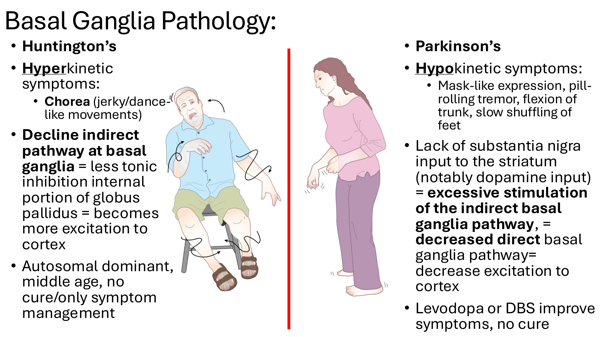
This slide is trying to teach you: Huntington's disease causes TOO MUCH movement (hyperkinetic), while Parkinson's disease causes TOO LITTLE or SLOW movement (hypokinetic) because they affect basal ganglia motor regulation differently.
DEFINITIONS:
Chorea: Involuntary, jerky, dance-like movements.
Hyperkinetic: Excessive movement.
Hypokinetic: Reduced or slow movement.
Striatum: Caudate + putamen.
Levodopa: Medication used to improve Parkinson's symptoms.
DBS (Deep Brain Stimulation): Treatment using electrical stimulation of specific brain regions.
Tiny story:
Imagine two patients:
Patient A — Huntington's disease:
Has uncontrolled, dance-like movements (chorea).
Their brain has difficulty suppressing unnecessary movements.
This happens because the indirect pathway becomes less effective.
Patient B — Parkinson's disease:
Has slow, shuffling steps.
Their face looks less expressive (mask-like face).
They may have a pill-rolling resting tremor and a flexed posture.
This happens because dopamine input from the substantia nigra decreases.
How it all connects:
Remember Slide 11?
Direct pathway = promotes movement.
Indirect pathway = suppresses movement.
Now we see what happens when those pathways are disrupted:
Huntington's → decreased indirect pathway activity → less inhibition → excessive movement.
Parkinson's → decreased dopamine → less direct and more indirect pathway influence → reduced movement.
Focus on this:
Huntington's = HYPERKINETIC → chorea (jerky, dance-like movements).
Huntington's = reduced indirect pathway → less tonic inhibition → MORE movement.
Huntington's = autosomal dominant, often begins in middle age; treatment focuses on symptoms.
Parkinson's = HYPOKINETIC → slow shuffling gait, mask-like face, pill-rolling tremor, flexed trunk.
Parkinson's = loss of substantia nigra dopamine input to striatum → LESS movement.
Parkinson's treatment = levodopa or DBS can improve symptoms but do not cure the disease.
How to remember:
Huntington's = HYPER = TOO MUCH movement. 💃
Parkinson's = HYPO = TOO LITTLE movement. 🐢
Huntington's → Indirect pathway DOWN → movement UP.
Parkinson's → Dopamine DOWN → movement DOWN.