Accumulations & Deposits
 Call Kai
 Learn
Learn Practice Test
 Spaced Repetition
 Match
Match Flashcards
 Knowt Play
1/91

There's no tags or description
Looks like no tags are added yet.
Name | Mastery | Learn | Test | Matching | Spaced | Call with Kai | Chat |
|---|
No analytics yet
Send a link to your students to track their progress
92 Terms
Intracellular Accumulations
buildup of substances that cells cannot immediately use or eliminate
Three categories of intracellular accumulations:
1.) normal cellular component accumulated in excess
2.) abnormal substance
3.) pigments
normal cellular component accumulated in excess
water, lipids, glycogen, or carbohydrates that are accumulated in excess in a cell
abnormal substance accumulations
exogenous (mineral, infectious agent) or endogenous (abnormal synthesis during metabolism)
pigment accumulations
substances located in the cytoplasm or nucleus
two fates of pigment accumulations
1.) permanent
2.) transient (temporary)
Four mechanisms of intracellular accumulations
1.) abnormal metabolism
2.) defect in protein folding + transport
3.) lack of enzyme
4.) ingestion of indigestible materials
intracellular accumulation mechanism: abnormal metabolism
Normal endogenous substance is produced at a normal or increased rate, but the rate of metabolism is inadequate to remove it from the cell
intracellular accumulation mechanism: defect in protein folding + transport
Abnormal endogenous substance accumulates due to a defect in protein folding or transport; abnormal proteins accumulate
intracellular accumulation mechanism: lack of enzyme
Normal endogenous substance accumulates due to a defect in an enzyme that is normally required for metabolism
intracellular accumulation mechanism: ingestion of indigestible materials
Abnormal exogenous substance in ingested by the cell and accumulates since the cell cannot degrade/transport it out
Five examples of intracellular accumulations
1.) Hyaline
2.) Inclusion bodies
3.) Amyloid
4.) Gout
5.) Pathologic calcification
Hyaline accumulations
protein that is deposited in tissues
Hyaline appearance using H&E stain
-homogenous
-eosinophilic (pink)
-amorphous
-glassy
*small bright pink dots in image

Two scenarios that would result in hyaline accumulations
1.) resorption of hyaline droplets due to proteinuria (such as during kidney disease)
2.) russel bodies in plasma cells
What stain can be used to better visualize hyaline droplet accumulations?
periodic acid-schiff (PAS)
*looks like dark pink dots

Pathogenesis of hyaline droplet accumulations due to kidney disease
glomerular damage --> leads to proteinuria --> protein is taken up by proximal tubular epithelial cells, forming the hyaline protein droplets
russel bodies
hyaline bodies present in the cytoplasm of some plasma cells (Mott cells)
Appearance of russel bodies
-large
-eosinophilic
-homogenous
-amorphous

What are russel bodies made of?
accumulated immunoglobulin within plasma cells
What disease is associated with russel bodies?
feline plasma cell pododermatitis
feline plasma cell pododermatitis
disease where the dermis and some of the underlying adipose tissue is infiltrated with plasma cells, some of which contain hyaline accumulations
Inclusion bodies
nuclear and cytoplasmic aggregates of material
Two types of inclusion bodies:
1.) crystalline protein inclusion bodies
2.) viral inclusion bodies
crystalline protein inclusion bodies
found in normal hepatocytes and renal tubular epithelium
crystalline protein inclusion bodies are especially found in what age/species?
old dogs
crystalline protein inclusion bodies appearance
-large
-eosinophilic
-rhomboid

viral inclusion bodies
Nuclear or cytoplasmic aggregates made of proteins created by a virus

Amyloid accumulations
Pathologic extracellular, insoluble, proteinaceous (hyaline) substance resistant to proteolysis and deposited between cells
Amyloid fybrils
abnormally folded proteins in a β- sheet confirmation; make up amyloid accumulations
What is the pathologic significance of amyloid accumulations?
amyloid accumulations mechanically interfere with the cell (by sandwiching in between cells), and thus interfere with their function
Two consequences of amyloid accumulations
1.) organ failure
2.) protein losing "organ-opathy"
What organs commonly fail due to amyloid accumulations?
kidney and liver
What organs commonly have protein losing "organ-opathies" due to amyloid accumulations?
kidney and intestines
Two types of amyloid accumulations:
1.) reactive systemic- Amyloid associated (AA)
2.) primary amyloid- Amyloid light chain (AL)
reactive systemic- Amyloid associated (AA)
amyloid accumulation composed of amyloid associated protein (AA); secondary to chronic antigen stimulation (such as due to chronic infection, neoplasia)
reactive systemic- Amyloid associated (AA) accumulations are most common in _________
animals
What canine species are reactive systemic- Amyloid associated (AA) accumulations hereditary in?
Shar peis
What feline species are reactive systemic- Amyloid associated (AA) accumulations hereditary in?
Abyssinian cats
What is the precursor protein of reactive systemic- Amyloid associated (AA) accumulations?
serum amyloid associated protein (SAA)
serum amyloid associated protein (SAA)
acute phase protein, produced during inflammation, part of the acute phase response
primary amyloid- Amyloid light chain (AL)
amyloid accumulation composed of amyloid light chain protein (AL); found in plasma cell tumors and myelomas
amyloid light chain protein (AL)
Derived from abnormal plasma cells secreting immunoglobulin light chain fragments into circulation
primary amyloid- Amyloid light chain (AL) accumulations are most common in _________
humans
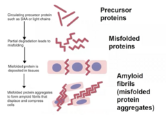
Amyloid accumulations pathogenesis (4)
1.) Precursor proteins (SAA or light chains) are in circulation
2.) partial degradation of precursor proteins occurs, causing them to be misfolded
3.) misfolded protein is deposited in the tissues, forming aggregates
4.) misfolded protein aggregates form amyloid fibrils that displace and compress cells
Five common locations of amyloid accumulation in all species:
1.) kidney
2.) liver (space of disse)
3.) spleen (germinal centers)
4.) lymph nodes (germinal centers)
5.) adrenal cortex
Where are amyloid accumulations located specific to horses?
skin and nasal septum and turbinates
Where are amyloid accumulations located specific to cats?
pancreatic islets of Langerhans
Gross morphological features of amyloid accumulations
-pale
-tan to yellow
-waxy
-swollen and enlarged

What special stain is used to grossly identify amyloid accumulations?
Lugol's iodine solution; form small black dots

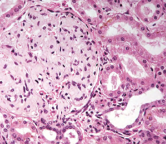
Microscopic features of amyloid accumulations
-eosinophilic
-amorphous
-looks like "cotton candy"
What special stain is used to microscopically identify amyloid accumulations?
Congo red; stains orange to red

Under fluorescence, what will amyloid accumulations look like while stained with congo red?
apple green color

Gout accumulations
deposition of sodium urate crystals or urates in the tissues
Two locations of gout accumulations
1.) viscera
2.) joints
Three species affected by gout accumulations
1.) humans
2.) birds
3.) reptiles
Why are humans, birds, and reptiles affected by gout accumulations?
they lack the enzyme uricase
uricase
aka urate oxidase; enzyme that catalyzes the oxidation of uric acid to allantoin (a substance that is more easily secreted)
Examples of things that can lead to gout accumulations
-dehydration
-high protein diets
-renal failure
**anything that disrupts excretion of uric acid!
Two reasons gout accumulations mainly occur:
1.) impaired excretion of uric acid by the kidneys (such as due to dehydration, severe renal disease, nephrotoxic drugs, etc)
2.) overproduction of uric acid (such as due to excessive protein in the diet, inherited enzyme defects)
Pathogenesis of gout accumulations
1.) species lacks the enzyme uricase
2.) leads to elevation of plasma uric acid concentration
3.) causes precipitation of urate crystals on either visceral or articular surfaces
4.) leads to chronic inflammation (usually in articular form)
In birds and reptiles, is the articular or visceral form of gout accumulations more common?
visceral form
Two organs classically affected by visceral gout accumulations
1.) heart (pericardium)
2.) kidneys
Gross appearance of gout accumulations
chalky white nodules

Microscopic appearance of gout accumulations
urate crystals surrounded by inflammatory response (macrophages, neutrophils)

Pathologic calcification accumulations
calcium deposits that occur in the soft tissues rather than in bones
Two types of pathologic calcification accumulations
1.) dystrophic calcification
2.) metastatic calcification
dystrophic calcification accumulations
calcium deposits on visibly diseased or necrotic tissue
What causes dystrophic calcification accumulations?
dead cells release phosphates which combine with calcium and form depositions
metastatic calcification accumulations
calcium deposits on visibly normal tissue
metastatic calcification accumulations are indicative of...
other diseases going on in the body; typically systemic metabolic diseases that lead to elevated serum calcium levels
Two causes of elevated serum calcium levels that lead to metastatic calcification accumulations:
1.) hyperparathyroidism (primary cause, such as PTH related protein secreting tumors)
2.) hypercalcemia (secondary to vitamin D toxicity, renal failure, etc)
Dystrophic vs metastatic calcification:
tissue type
Dystrophic calcification: necrotic tissue
metastatic calcification: normal tissue
Dystrophic vs metastatic calcification:
serum calcium levels
Dystrophic calcification: normal serum calcium
metastatic calcification: hypercalcemia
Gross appearance of pathologic calcification accumulations
gritty, hard white appearance

Microscopic appearance of pathologic calcification accumulations
calcium deposits are purple/blue with H&E stain

What special stain is used to visualize pathologic calcification accumulations?
von Kosa stain; stain black

Two pigments:
1.) cyanosis
2.) hemosiderin
cyanosis
blue color due to deoxygenated hemoglobin
What condition is cyanosis common in?
saddle thrombus in cats; blood clot prevents blood flow to rear legs and leads to blue color on paw pads

hemosiderin
iron storage pigment; golden brown color
What organ is hemosiderin mostly stored in?
spleen
What could cause increased amounts of hemosiderin?
-breakdown of erythrocytes
-hemorrhage
-congestion (slow blood flow)
-hemolysis


Case 1:
-3.5-year-old, neutered male, SharPei
-Recurrent episodes of fever of unknown origin (ie, 103°F-107°F) , with concurrent swollen, painful hocks, swelling of the muzzle, abdominal pain, diarrhea, and anorexia
Based on the histopathology, which histochemical stain would you request?
A. Von Kossa
B. Lugol's Iodine
C.Congo Red
D. Oil Red O
C. Congo Red
-amorphous
-light pink material
-looks like "cotton candy" in space of disse of liver and glomerulus in the liver,
-characteristic of amyloidosis

Case 1:
What are the structures being highlighted by this stain?
A. Renalmedulla
B. Islets
C. Spaceof Disse
D. Glomeruli
D. Glomeruli
Case 1:
What type of Amyloidosis is this?
A. Immunoglobulin‐associated (primary, AL)
B. Reactive (secondary, AA)
C. Apolipoprotein AI-derived amyloidosis
D. Islet amyloid polypeptide-derived amyloidosis
B. Reactive (secondary, AA)
-secondary to chronic antigen stimulation (such as due to chronic infection, neoplasia)
-breed disposition (Shar pei)

Based on the histopathology of amyloid accumulations in the picture, what is the likely species of the patient?
A. Horse
B. Dog
C. Cow
D. Cat
D. Cat
*islets of Langerhaans specific to cats
Case 2:
-8 day-old female alpaca cria presented with an abrupt onset of recumbency and illness.
-Clinically she was lethargic, febrile and had an elevated cardiac and respiratory rate.
-It was later revealed that she was being fed a powdered goat colostrum supplement
Gross pathology:
-Bilaterally kidneys were paler than normal
-Multifocal, gritty, hard foci on cut surface
Histopathology:
-in both kidneys, glomeruli are diffusely replaced by hard, readily fractured, intensely basophilic granular to homogeneous material consistent with mineral/calcification
Lab results:
Calcium highly elevated (13.1 compared to RI of 2-2.6)
Which type of calcification is this?
A. Metastatic
B.Dystrophic
A. Metastatic
-for metastatic, serum calcium will be high; for dystrophic, serum calcium will be normal

What is this?
A. Hyaline
B. Mott cells
C. Amyloid
D. Calcification
A. Hyaline
-homogenous
-eosinophilic (pink)
-amorphous
-glassy
*small bright pink dots in image to the right

What is the cause of this lesion?
A. Deposition of insoluble masses of glycoprotein fibrils arranged in β-pleated sheets
B. Deposition of insoluble minerals (carbonate and phosphate salts of Ca+,Mg+, and K+)
C. Deposition of an amorphous, eosinophilic protein material
D. Deposition of urate crystals on articular/synovial surfaces of joints and/or serous surfaces of viscera
D. Deposition of urate crystals on articular/synovial surfaces of joints and/or serous surfaces of viscera
Gout!!
-chalky white
-changes from gritty to pasty depending
**A is amyloid
**C is very nonspecific, could to apply to anything
**B is good differential but due to location of this deposition (serous surface of viscera) more likely to be D
Which of the following is a cause of metastatic calcification?
A Renal disease
B Vitamin D toxicity
C Paraneoplastic syndrome
D All of the above
D All of the above
What does the histochemical stain Congo Red detect?
A Lipid
B Glycogen
C Amyloid
D Mineral
C Amyloid