Body Wall and Hip
 Call Kai
 Learn
 Practice Test
 Spaced Repetition
 Match
 Flashcards
 Knowt Play
1/95

There's no tags or description
Looks like no tags are added yet.
96 Terms
How many ribs are there?
12
What do the ribs articulate with?
Posteriorly with the vertebra. Join the sternum via a costal cartilage or join a common costal cartilage which then articulates with the sternum.
What are the parts of the sternum?
Manubrium, body, xiphoid process.
What are the types of ribs?
Typical ribs - number 2 through 10. Rib 1 is an atypical shape. Ribs 11 and 12 are floating (not attached by costal cartilage to the sternum)

Parts of the vertebra [Image]
[Image]


What is the structure of each rib [Image]?
[Image]


Regions of vertebral column [Image]?
[Image]
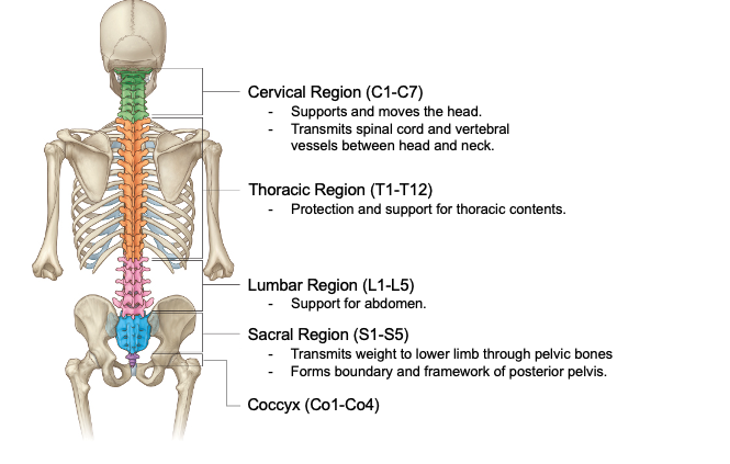
What are the 4 vertebral curvatures?
Cervical - lordosis. Thoracic - kyphosis. Lumbar - lordosis. Sacral - kyphosis.

Parts of a typical vertebrae [Image]?
[Image]
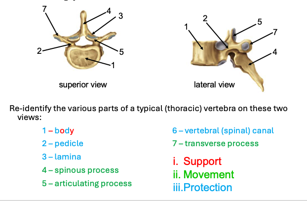
Characteristics of cervical vertebra?
(1) Transverse foramina for vertebral arteries (2) Large triangular vertebral foramen (3) Short, bifid (uncinate) spinous process (4) Zygopophyseal articulation: anterior/posterior but at ~45 degree angle
Characteristics of thoracic vertebra?
(1) Articular facets for ribs (2) Heart shaped body (3) Elongated, posteriorly deflected spinous process (4) Zygopophyseal articulation: anterior/posterior at ~90 degree angle
Characteristics of lumbar vertebra?
(1) Large, bean shaped body
(2) Shortened, squared and posteriorly projecting spinous processes (3) Zygopophyseal articulation: concave/convex
Characteristics of cervical C1; Atlas?
(1) No body. (2) Large superior articulating processes (concave) which articulate with the occipital condyles
Characteristics of cervical C2; Axis?
(1) Dens (odontoid process)

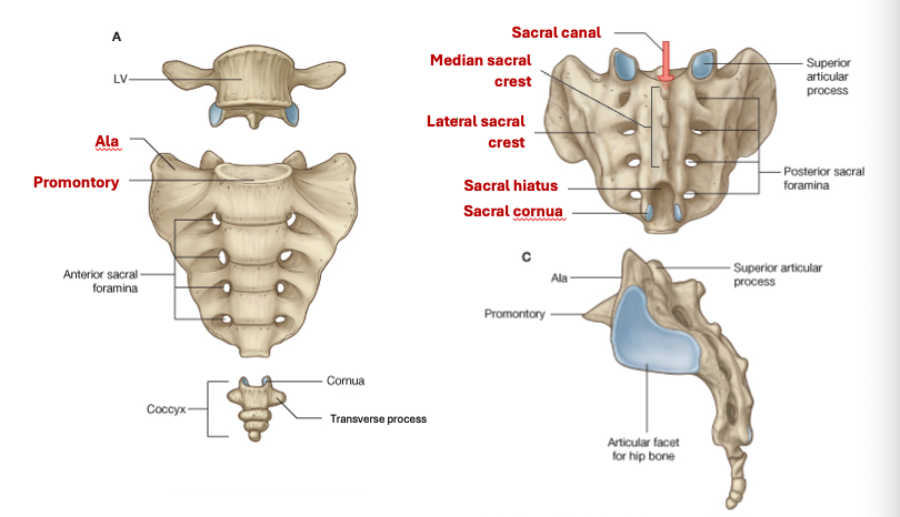
Parts of the sacrum [Image]?
[Image]

What are the intrinsic muscles of the back [Image]?
[Image]
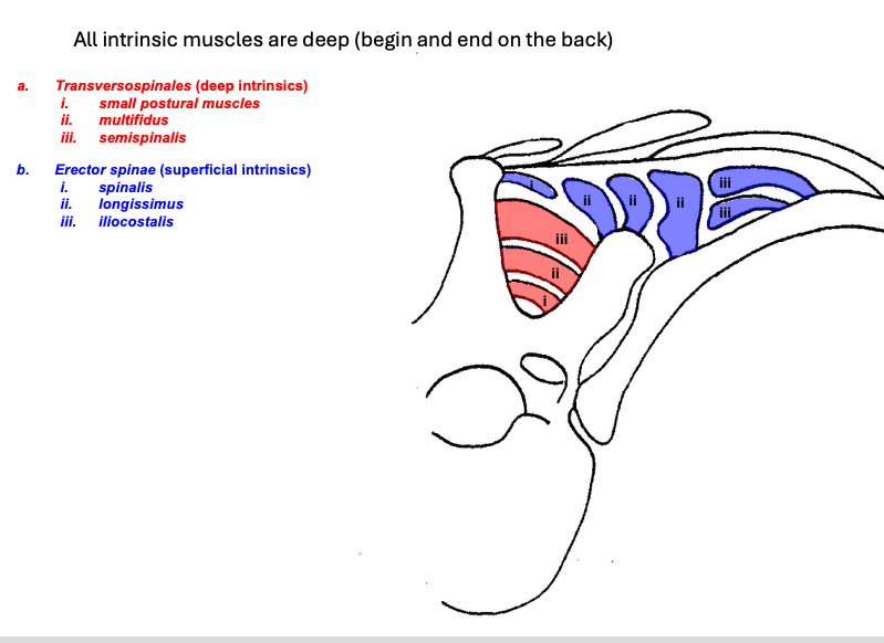
What are the deep or intrinsic back muscles innervated by?
All innervated segmentally by dorsal primary rami of spinal nerves.
What muscles make up the superficial layer of the deep intrinsics?
Erector spinae: spinalis (medial column), longissimus (intermediate column), iliocostalis (lateral column).
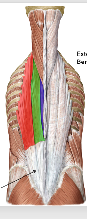
Image of superficial layer of deep intrinsics [Image]?
[Image]

What is the common origin of the erector spinae?
Common inferior origin: sacrum, iliac crest, and sacroiliac ligament. Also originate from half of the ligamentum nuchae and the spinous processes of upper thoracic.
Where does the spinalis, longissimus, and iliocostalis insert?
Spinous process. Transverse processes and ribs. Extends to angles of the ribs.
What is the action of the erector spinae?
Laterally flex the spinal column. Bilaterally extend the head and vertebral column.
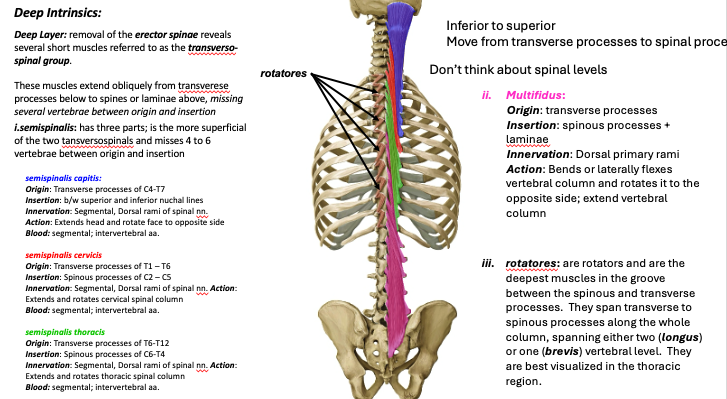
What group make up the deep layer of the deep intrinsics?
Transversospinal group.
What are the parts of the transversospinal group?
Semispinalis (more superficial and misses 4 to 6 vertebrae between origin and insertion). Multifidus. Rotatores.
What are the 3 parts of the semispinalis?
Semispinalis capitis. Semispinalis cervicis. Semispinalis thoracis.
What is the origin and insertion of semispinalis capitis?
Lower cervical/upper thoracic transverse processes. Insert base of head.
What is the origin and insertion of semispinalis cervicis?
Upper thoracic transverse processes. Insert upper cervical spinous processes.
What is the origin and insertion of semispinalis thoracis?
Lower thoracic transverse processes. Insert spinous processes of lower cervical.
What is the origin and insertion of multifidus?
Transverse processes. Spinous processes + laminae
What is the origin and insertion rotatores?
Deepest muscles. Span transverse to spinous processes along the whole column. Span either two (longus) or one (brevis) vertebral level.
What is the action of semispinalis capitis?
Extends head and rotate face to opposite side
What is the action of semispinalis cervicis?
Extends and rotates cervical spinal column
What is the action of semispinalis thoracis?
Extends and rotates thoracic spinal column
What is the action of multifidus?
Bends or laterally flexes vertebral column and rotates it to the opposite side; extend vertebral column
What is the action of rotatores?
Rotator

Image of deep layer of deep intrinsics [Image]?
[Image]
What are layers of the intercostal muscles?
External (superficial). Internal (deep). Innermost (deeper) - transversus thoracis + subcostals.
What is between the internal and innermost intercostal muscles?
Intercostal artery, vein, and nerve.
What are the orientations of intercostal muscles from rib above to rib below?
External - down and towards the midline. Internal and innermost - down and away from the midline.
What is the origin and insertion of intercostal muscles?
Inferior margin of rib above. Superior margin of rib below.
What is the innervation of the intercostal muscles?
Intercostal nerve. (ventral primary rami).
What is the action of intercostal muscles?
External - elevate ribs for inspiration. Internal + innermost - depress ribs for expiration.

Image of intercostal muscles [Image]?
[Image]


Image of surrounding structures of anterolateral abdominal wall [Image]?
[Image]

What are the actions of the anterolateral abdominal wall?
(1) Support and compress viscera (raise intra-abdominal pressure). (2) Flex vertebral column with bilateral contraction. (3) Lateral flexion of vertebral column with unilateral contraction. (4) Rotation of trunk when internal abdominal oblique and contralateral external abdominal oblique contract simultaneously (turns to side of internal abdominal oblique).
What innervates anterolateral abdominal wall?
Intercostal nerves (ventral primary rami)
What are the 3 layers of the anterolateral abdominal wall?
External abdominal oblique. Internal abdominal oblique. Transversus abdominis.
What is the origin and insertion of external abdominal oblique?
Ribs 5-12. Insert linea alba, pubic tubercle, ant 1/2 of iliac crest.
What is the origin and insertion of internal abdominal oblique?
Thoracolumbar fascia. Insert inferior borders of ribs 10-12, linea alba, pectineal line via conjoint tendon.
What is the origin and insertion of transversus abdominis?
7-12 costal cartilages, thoracolumbar fascia, iliac crest, lat ⅓ of inguinal ligament. Inserts linea alba, pectineal line via conjoint tendon.

Image of anterolateral abdominal wall musculature [Image]?
[Image]

What is the composition of the anterolateral abdominal wall sheath superior to the ASIS?
anterior rectus sheath is composed of the aponeuroses of external abdominal oblique and the half of internal abdominal oblique, while the posterior rectus sheath is composed of the other half of internal abdominal oblique and the transversus abdominis.
What is the composition of the anterolateral abdominal wall sheath inferior to the ASIS?
anterior rectus sheath is the aponeuroses of all three muscle layers; no posterior rectus sheath.
What is the separation of fascia at linea alba?
Diastasis recti.
What is the cause of diastasis recti and treatment?
Increases with abdominal strain (e.g., pregnancy).
At what week of development do flexor and extensor compartments of upper and lower limb become opposite to each other?
Upper limb rotates laterally. Lower limb rotates medially.

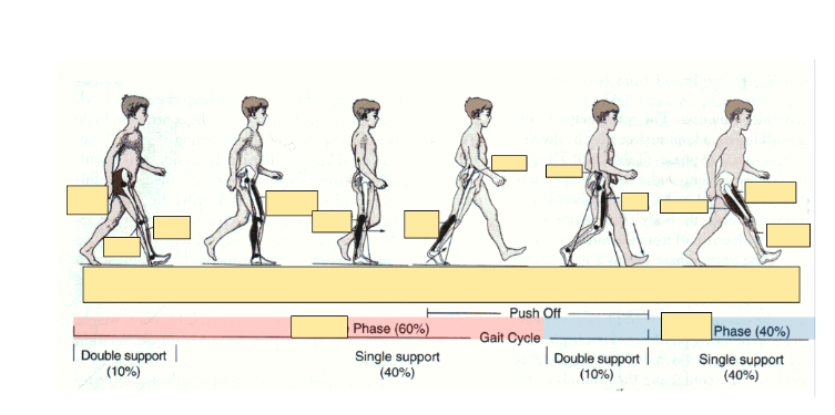
Diagram of human gait cycle [Image]?
[Image]
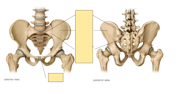
Bones of pelvic girdle [Image]?
[Image]


Image of ox coxae [Image]?
[Image]
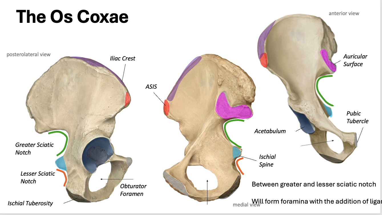

Image of proximal femur [Image]?
[Image]


Image of joints of the pelvis [Image]?
[Image]

What forms the opening for peripheral nerves that innervate muscles of the hip and the pelvic floor?
Sacropsinous + sacrotuberous ligaments.
What is the lower extremity innervated by?
Lumbosacral plexus (ventral primary rami) - L1 to L4, L4 to S3.
What nerves are from the lumbar plexus?
Obturator nerve - medial compartment of thigh. Femoral nerve - anterior compartment of thigh.
What nerves are from the sacral plexus?
Tibial nerve, common peroneal (fibular nerve) - exist as sciatic nerve above the knee. Superior gluteal nerve + inferior gluteal nerve.
What does the sacral plexus help innervate?
Innerates gluteal region, posterior compartment of thigh, all compartments of the leg, and the foot.
What to avoid during intragluteal injections?
Sciatic nerve.

Image of arteries of the thigh [Image]?
[Image]

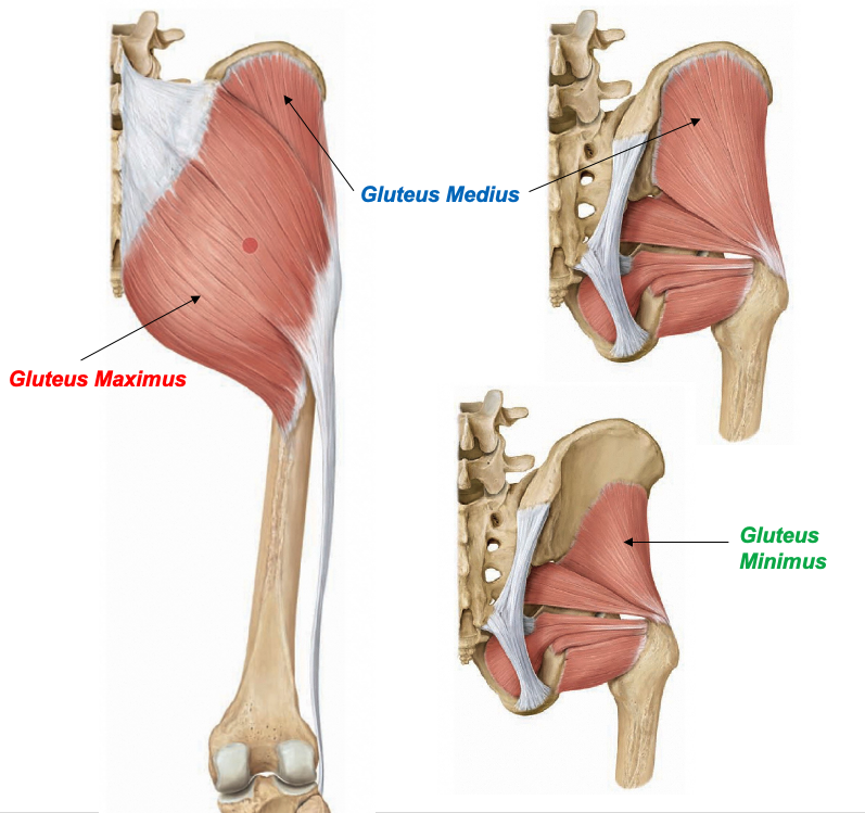
What are the gluteal muscles?
Gluteus maximus. Gluteus medius, gluteus minimus, tensor fascia latae.
What are the deep rotators?
Obturator externus, obturator internus, gemelli, piriformis, quadratus femoris.
What are the thigh muscles?
Flexors - rectus femoris, sartorius. Hamstrings - biceps femoris, semimembranosus, semitendinosus. Hip adductors - Pectineus, Adductor Longus, Adductor Brevis, Adductor Magnus, Gracilis

Image of the flexors of the thigh [Image]?
[Image]

What is the action of the iliacus?
Flexion of the thigh
What is the nerve of the iliacus?
Femoral nerve.
What is the action of the psoas major?
Flexion of the thigh.
What is the innervation of the psoas major?
Ventral rami (L1-3).
What is the action of the iliopsoas?
Bilateral contraction = trunk flexion. Fixator of the hip.
What is the function of each compartment of the thigh?
Anterior - flex thigh, extend leg. Medial - adduct thigh. Posterior - extend thigh, flex leg.
What surrounds the thigh?
Deep fascia - fascia lata. Thickened laterally and forms the iliotibial tract (IT band).
What two muscles attach to the iliotibial tract and what is their function?
Tensor fasciae latae. Gluteus maximus. Helps steady the trunk on the thigh and braces the knee of the stance foot in normal gait. Tensor fascia latae also medially rotates the lower limb.
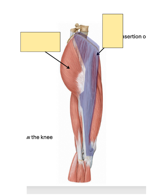
Image of the IT band [Image]?
[Image]

What is the action of the gluteus maximus?
Hyperextensor of the hip. Extends and lateral rotates thigh.
What is the nerve and blood supply of the gluteus maximus?
Inferior gluteal nerve. Sup. and inf. Gluteal arteries.
What does the gluteus maximus insert at?
IT band, gluteal line of femur.
What is the action of the gluteus medius + gluteus minimis?
Abducts and medially rotates thigh.
What is the nerve and blood supply of the gluteus medius + gluteus minimis?
Superior gluteal nerve and artery.
What is the insertion of the gluteus medius + gluteus minimis?
Greater trochanter.
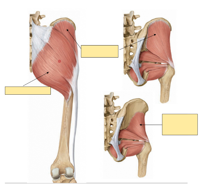
Image of gluteal region [Image]?
[Image]
What happens when the superior gluteal nerve is damaged?
Trendelenberg gait. If left side is damaged, pelvis tilts down right.
What is the action of the deep gluteal region?
Lateral rotation of the thigh.
What is the innervation of the deep gluteal region?
Branches of the sacral plexus.
What is the blood supply of the deep gluteal region? – Sup. Inf. gluteal arteries.
What is the origin and insertion of the muscles of the deep gluteal region?
Origin on pelvis. Insert onto the greater trochanter or else the intertrochanteric line or crest.

Image of the deep gluteal region [Image]?
[Image]

What is it called when the two components of the sciatic nerve pierce the piriformis muscle? What does it cause?
Piriformis syndrome. Impairments depend upon which nerve is entrapped.