Blood Vessel Anatomy, Circulation, and Major Artery Pathways
 Call Kai
 Learn
 Practice Test
 Spaced Repetition
 Match
 Flashcards
 Knowt Play
1/63

There's no tags or description
Looks like no tags are added yet.
64 Terms
General structure of a blood vessel
A tube that carries blood. The lumen is the open space containing blood, and the tunics make up the wall.
Three tunics of a blood vessel
Outside → inside: adventitia → media → intima. Capillaries only have the intima.

Tunica adventitia
Outermost connective tissue layer. Protects and anchors the vessel to nearby structures.
Tunica media
Middle smooth muscle layer. Changes vessel diameter to control blood flow and pressure.
Vasodilation
Smooth muscle relaxes → vessel gets wider → lumen increases.
Vasoconstriction
Smooth muscle contracts → vessel gets narrower → lumen decreases.
Tunica intima
Innermost layer touching the blood. Made of simple squamous epithelium and continuous with the endocardium.
Tunica intima functions
Lines vessels, allows exchange in capillaries, and helps with clotting, vessel repair, and vessel diameter.
Artery
Carries blood away from the heart. Has 3 tunics and thick, elastic walls for high pressure.
Why arteries contain elastin
Lets them stretch when high-pressure blood enters and recoil afterward.
Capillary
Tiny, one-layer vessel where substances are exchanged between blood and tissues.

Why capillaries are good for exchange
Thin + permeable + large surface area + slow blood flow = easy exchange.
Vein
Carries blood toward the heart. Thinner wall, larger lumen, less muscle and elastin. Lower-limb veins have valves.

Artery vs. vein vs. capillary
Artery: away, high pressure, thick. Vein: toward, low pressure, thin. Capillary: exchange, one layer.
What the vascular system transports
Gases, nutrients, wastes, and hormones throughout the body.
Pulmonary circuit
Heart → lungs → heart for O₂ and CO₂ exchange.

Systemic circuit
Heart → body tissues → heart.
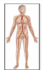
Blood vessel flow sequence
Arteries → arterioles → capillaries → venules → veins.

Arteriole
Small vessel between an artery and capillaries. Controls how much blood enters tissues.

Precapillary sphincter
Smooth muscle at the entrance to a capillary bed that opens or closes to control blood flow.
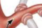
Arteriole diameter regulation
Local factors like NO, O₂, and CO₂ plus sympathetic nerves control the smooth muscle.
Sympathetic effect on blood vessels
Constricts vessels to abdominal organs while blood flow to the heart and skeletal muscle increases.
Vasa vasorum
Small vessels that supply the walls of large and medium arteries.
Capillary size
About 8 μm wide, so RBCs pass through about one at a time.
Continuous capillary
Tightest/least permeable type. Found mainly in muscle, connective tissue, and nervous system.

Fenestrated capillary
Has small pores for faster exchange. Found in kidneys, intestines, and endocrine glands.

Sinusoidal capillary
Has large gaps that allow large proteins and cells through. Found in red marrow, liver, and spleen.

Capillary types by increasing permeability
Continuous → fenestrated → sinusoidal.
Venule
Small vessel that collects blood from capillaries and carries it toward veins.
Venous system as a blood reservoir
Veins stretch easily and hold about 60% of the body's blood.
Venoconstriction
Sympathetic stimulation narrows veins, pushing stored blood toward other parts of the body.
Venous valve
Flap that prevents backflow and helps blood move toward the heart.
Varicose veins
Dilated, deformed veins associated with poor valves and increased venous pressure.

Subclavian artery
is the major upper-limb artery located under the clavicle. As it enters the axilla, it becomes the axillary artery.
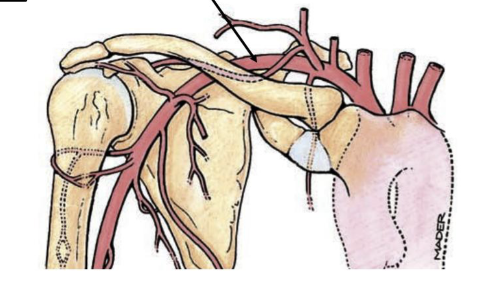
Axillary artery
is the continuation of the subclavian artery as it passes through the axilla, or armpit. It continues into the arm as the brachial artery.

Brachial artery
is the major artery of the arm. Just past the elbow, it divides into the radial and ulnar arteries.

Common iliac arteries
The right and left common iliac arteries are formed when the descending aorta bifurcates at L4. Each common iliac then divides into an internal and external iliac artery.
Femoral artery
is the major artery of the thigh and is the continuation of the external iliac artery into the lower limb. It continues toward the back of the knee.
Popliteal artery
is the continuation of the femoral artery as it passes through the popliteal fossa behind the knee. It later divides into the anterior and posterior tibial arteries.
Anterior and posterior tibial arteries
are the major arteries of the leg that branch from the popliteal artery.
Lower-limb vein order
Venous blood from the lower limb returns toward the heart in this order:Plantar arches → tibial veins → popliteal → femoral → external iliac → common iliac → IVC.
Aorta
Main systemic vessel leaving the heart. Gives rise to the body's systemic arteries.
Coronary arteries
First branches of the aorta; supply the heart itself.
Aortic arch branches
Brachiocephalic trunk → left common carotid → left subclavian.
Brachiocephalic trunk
First aortic arch branch. Splits into right common carotid + right subclavian.
Brain arterial supply
The brain receives arterial blood from two paired routes: the right and left internal carotid arteries and the right and left vertebral arteries.
Descending aorta
Continuation of the aorta through the thorax and abdomen.
Major branches of the abdominal aorta
Major abdominal aorta branches emphasized by Costa include the celiac trunk, superior mesenteric artery, inferior mesenteric artery, renal arteries, and gonadal arteries.
Celiac trunk branches
The celiac trunk is a branch of the abdominal aorta that divides into the left gastric, splenic, and common hepatic arteries.
Deep upper-limb veins
Deep upper-limb veins generally have the same names as the arteries they accompany. Examples include the radial, ulnar, and brachial veins.
Superficial upper-limb veins
The cephalic and basilic veins are major superficial veins of the upper limb. Costa notes that superficial veins are larger and fewer in number than deep veins.
Median cubital vein
Superficial vein at the elbow connecting cephalic + basilic. Common site for venipuncture.
Anastomosis
Connection between vessels that can provide an alternate route for blood.
Dural venous sinuses
Collect blood from cerebral veins → eventually drain into the internal jugular. No valves.
Head and neck venous return
The internal jugular and subclavian veins join to form the brachiocephalic veins. The right and left brachiocephalic veins then drain into the superior vena cava.
Azygos vein
Provides an alternate venous route back to the heart for blood from the trunk/lower limb.
Great saphenous vein
Superficial lower-limb vein that can be used as a graft for coronary bypass surgery.
Pulmonary circuit chamber and vessel order
RA → RV → pulmonary trunk → pulmonary arteries → lungs → pulmonary veins → LA.
Portal system
Blood passes through 2 capillary beds in a row before returning to the heart.
Three portal systems Costa lists
Costa lists the hepatic portal system from digestive organs to liver, hypophyseal portal system from hypothalamus to pituitary, and renal system from glomerulus to peritubular capillaries.
Systolic vs. diastolic pressure
Systolic pressure is arterial pressure when the ventricles contract. Diastolic pressure is arterial pressure when the ventricles relax. Blood pressure is written systolic/diastolic in mm Hg.
Where a pulse can be felt
A pulse can be felt where an artery runs near the body surface or over bone or firm tissue, making the pressure wave easier to detect.
Major limb pulse points
Major limb pulse points include the radial artery at the wrist, brachial at the elbow, popliteal behind the knee, posterior tibial behind the medial malleolus, and dorsalis pedis on the dorsum of the foot.
Arterial pressure points
Arterial pressure points are superficial arterial locations where an artery can be compressed against bone or other firm tissue to help stop arterial bleeding.