Balance Week 6
 Call Kai
 Learn
 Practice Test
 Spaced Repetition
 Match
 Flashcards
 Knowt Play
1/183

Earn XP
Description and Tags
Lung Week
184 Terms
B6A1. Trace the mechanism by which pneumonia produces alveolar consolidation and hypoxemia.
Answer: Pathogens infect the lung → local inflammation develops → inflammatory exudate fills alveoli → normal alveolar air is displaced → lung tissue consolidates → ventilation decreases while perfusion continues → low V/Q or shunt causes hypoxemia.
Extra Information:
- Consolidation means normally air-filled alveoli become occupied by inflammatory material.
- The infected region may remain perfused despite having little or no effective ventilation.
- The complete pathogen-recognition and neutrophil-recruitment pathway is not established by these sources.
Source: B6LG2 slides 5, 9–11 | First Aid Clinical Pattern Recognition (2022) p. 46
B6A1. Why does alveolar consolidation produce dullness to percussion and increased voice transmission?
Answer: Inflammatory material replaces air and increases lung tissue density, producing a dull percussion note and allowing voice vibrations to travel more effectively through consolidated tissue.
Extra Information:
- Egophony and bronchophony can indicate consolidation.
- Fever, productive cough, and focal crackles are additional pneumonia findings.
Source: First Aid Clinical Pattern Recognition (2022) pp. 46, 438
B6A2. Explain how alveolar consolidation produces a low ventilation-perfusion ratio.
Answer: Exudate fills alveoli → fresh-air delivery decreases → capillary perfusion may continue → ventilation becomes insufficient relative to blood flow → poorly oxygenated blood leaves the affected alveolar capillaries.
Extra Information:
- V refers to alveolar ventilation and Q refers to pulmonary perfusion.
- Mixing of poorly oxygenated with normally oxygenated blood decreases arterial PaO₂.
Source: B6LG2 slides 6, 8–11
B6A2. When does low V/Q become a true intrapulmonary shunt?
Answer: When ventilation to a region falls to zero while perfusion continues, producing a V/Q ratio of zero.
Extra Information:
- Blood traverses the region without exposure to fresh alveolar gas.
- A large shunt may cause severe oxygen-refractory hypoxemia.
Source: B6LG2 slides 6, 9–11, 25–28
B6A2. Why does supplemental oxygen improve low V/Q but poorly correct true shunt?
Answer: Low-V/Q units still receive some inspired air, so increasing FiO₂ raises alveolar oxygen pressure. True shunt units receive no fresh air, so increasing FiO₂ cannot directly oxygenate blood passing through them.
Extra Information:
Normal regions cannot fully compensate for a large volume of shunted blood.
Restoring alveolar aeration addresses the underlying shunt mechanism.
In a shunt, supplemental oxygen reaches healthy alveoli, but poorly oxygenated blood from unventilated alveoli mixes with oxygenated blood, causing persistent hypoxemia because hemoglobin in healthy regions is already nearly fully saturated.
Source: B6LG2 slides 8–11, 25–28
B6A2. Compare normal V/Q, low V/Q, shunt, and dead space.
Answer: Normal means ventilation and perfusion are appropriately matched. Low V/Q means relatively insufficient ventilation. Shunt is perfusion without ventilation. Dead space is ventilation without perfusion.
Extra Information:
- Shunt has V/Q = 0.
- Dead space has V/Q approaching infinity.
- Pulmonary embolism directly produces dead-space regions.
Source: B6LG2 slides 6, 25–27

B6A3. Compare the anatomic distribution of lobar pneumonia and bronchopneumonia.
Answer: Lobar pneumonia produces relatively continuous consolidation involving much of a lobe, whereas bronchopneumonia produces patchy inflammatory opacities involving scattered airspaces.
Extra Information:
- Both can produce alveolar filling and impaired gas exchange.
- The radiographic distribution does not alone establish disease severity.
Source: First Aid Clinical Pattern Recognition (2022) p. 46

B6A3. Why can lobar pneumonia and bronchopneumonia produce different V/Q patterns?
Answer: A densely consolidated lobe can create a large regional shunt or low-V/Q region, whereas scattered bronchopneumonia creates multiple regions with differing degrees of impaired ventilation.
Extra Information:
- Both patterns can increase the A–a oxygen gradient.
- The degree of hypoxemia depends on the extent of poorly ventilated but perfused lung.
Source: First Aid Clinical Pattern Recognition (2022) p. 46 | B6LG2 slides 9–11
B6A3. What sequence of pathologic stages is associated with classic lobar pneumonia?
Answer: Congestion → red hepatization → gray hepatization → resolution. CHHR
Congestion (0–24 hours): Bacteria enter → blood vessels dilate and leak → fluid and bacteria accumulate in alveoli.
Red hepatization (days 2–3): Alveoli fill with red blood cells, neutrophils, and fibrin → lung becomes red and firm like the liver.
Gray hepatization (days 4–6): Red blood cells break down → fibrin and inflammatory cells remain → lung becomes gray and stays consolidated.
Resolution (day 7 onward): Macrophages clear inflammatory debris → alveoli reopen → gas exchange improves.
Extra Information:
These stages describe changes occurring as an affected lobe becomes inflamed and then recovers.
Source: First Aid Clinical Pattern Recognition (2022) p. 46

B6A4. What normal pulmonary mechanisms help defend against inhaled microorganisms?
Answer: Mucus traps inhaled material, airway cilia move secretions toward the upper airway, and alveolar macrophages participate in immune surveillance within the distal lung.
Extra Information:
- Mucociliary clearance helps remove retained particles and pathogens.
- Distal immune defenses are especially important beyond the conducting airways.
Source: COPD Structure slides 7–10 | B6LG2 slide 5
B6A4. Explain how smoking-associated ciliary injury increases susceptibility to pneumonia.
Answer: Smoke damages airway epithelium and cilia → mucus clearance decreases → secretions and inhaled microorganisms accumulate → lower respiratory infection becomes more likely.
Extra Information:
Smoking also increases mucus production (goblet cell hyperplasia).
Squamous metaplasia may reduce normal ciliated epithelial function.
Source: COPD Structure slides 7–10

B6A4. Why does immune compromise broaden the differential diagnosis of pneumonia?
Answer: Impaired host defenses permit opportunistic organisms to produce clinically important infection in addition to conventional pneumonia pathogens.
Extra Information:
- First Aid describes organisms including Pneumocystis, Nocardia, and Aspergillus in susceptible patients.
Source: First Aid Clinical Pattern Recognition (2022) p. 45
B6A5. Explain how aspiration-associated pulmonary inflammation can lead to hypoxemia.
Answer: Material enters the lower respiratory tract → airway or alveolar injury develops → inflammation and filling impair regional ventilation → perfusion may persist → low V/Q or shunt reduces arterial oxygenation.
Extra Information:
Aspiration: Food, liquid, saliva, or vomit → trachea → lungs.
Aspiration → bacteria-containing material enters lungs → inflammation/infection → alveoli fill with exudate → impaired gas exchange.
exudate: Fluid, proteins (including fibrin), and white blood cells (especially neutrophils) accumulate in alveoli.
Inflammation → leaky capillaries + tissue factor activation → thrombin → fibrin
First Aid associates some aspiration-related infections with anaerobic organisms.
Severity depends on the amount of affected lung and resulting ventilation impairment.
Source: First Aid Clinical Pattern Recognition (2022) p. 45 | B6LG2 slides 9–11
B6A5. Which clinical history is specifically associated with aspiration-related anaerobic lung infection in First Aid?
Answer: Aspiration pneumonia occurs when oropharyngeal secretions or gastric contents enter the lower respiratory tract, introducing microorganisms that cause pulmonary infection and inflammation.
The right lower lobe is commonly affected because the right main bronchus has a steeper, more vertical orientation than the left, allowing aspirated material to enter the right lung more readily. The affected lung segments depend on patient positioning:
Upright: Posterior basal segment of the right lower lobe.
Supine: Superior segment of the right lower lobe and posterior segment of the right upper lobe.
Clinical risk factors include:
Alcohol dependence/intoxication: Depressed consciousness and impaired airway protective reflexes increase aspiration risk.
Neurologic disorders: Stroke, Parkinson disease, and other conditions causing dysphagia.
Sedation or general anesthesia: Reduced consciousness and protective airway reflexes.
Gastroesophageal reflux or esophageal motility disorders: Increased risk of regurgitation and aspiration.
Nasogastric feeding tubes: Predispose patients to aspiration.
Key takeaway: Alcohol-related impairment of airway protection increases aspiration risk, while the more vertical right main bronchus and gravity-dependent distribution explain the predominance of right lower lobe involvement.
Source: First Aid Clinical Pattern Recognition (2022) p. 45 & ChatAmboss
B6A6. Trace how alveolar exudate increases the work of breathing.
Answer: Alveolar filling and surfactant disruption → increased surface tension and reduced compliance → greater distending pressure is required for inflation → elastic work increases → breathing requires greater effort.
Extra Information:
Pneumonia → protein-rich exudate interferes with surfactant → increased surface tension → alveoli are more likely to collapse → decreased compliance.
Compliance = change in lung volume divided by change in pressure.
A stiff lung changes less in volume for a given pressure change.
Source: B6LG1 slides 12–17 | B6LG2 slide 10
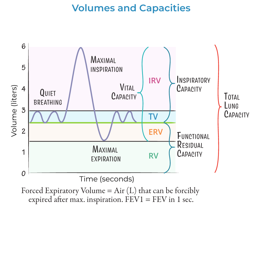
B6A6. Why can alveolar filling and instability decrease functional residual capacity?
Answer: Poorly aerated and unstable alveoli may collapse, reducing the volume of aerated lung remaining after passive expiration.
Extra Information:
- FRC = expiratory reserve volume + residual volume.
- Collapse increases the mechanical effort necessary to reopen affected lung units.
Source: B6LG1 slides 12–17 | B6LG2 slide 10
B6A6. Why might patients with low lung compliance adopt rapid, shallow breathing?
Answer: Large breaths require substantial elastic pressure when lungs are stiff, so smaller breaths can reduce elastic work per breath while respiratory frequency increases.
Extra Information:
- This compensatory pattern may reduce inspiratory elastic effort but can increase the proportion of ventilation reaching dead space.
Source: B6LG1 slides 12–15

B6A7. Which major mechanisms of hypoxemia generally respond to supplemental oxygen?
Answer: Low inspired oxygen, hypoventilation, diffusion limitation, and low-V/Q mismatch generally respond to increased FiO₂, whereas large true shunts respond poorly.
Extra Information:
- Oxygen must reach the affected alveoli to directly improve oxygenation of blood in that region.
- The provided sources do not fully specify all device-by-device FiO₂ ranges.
Source: B6LG2 slides 8–11, 25–28

B6A7. Why must the effect of supplemental oxygen be reassessed in pulmonary disease?
Answer: Oxygen response depends on the underlying gas-exchange disturbance. Improving FiO₂ helps ventilated alveoli, but severe shunt, persistent alveolar filling, or progressive ventilatory failure may require additional treatment.
Extra Information:
- Oxygen response can help distinguish low V/Q from significant true shunt.
Source: B6LG2 slides 8–11, 25–29
B6A8. What ABG abnormalities suggest severe pneumonia or pulmonary edema?
Answer: Arterial hypoxemia and an increased A–a oxygen gradient, often with early low PaCO₂ due to hyperventilation. Later ventilatory fatigue may impair CO₂ clearance.
Extra Information:
- A widened A–a gradient indicates abnormal oxygen transfer.
- Large shunts can produce oxygen-refractory hypoxemia.
Source: B6LG2 slide 11

B6A8. What does the PaO₂/FiO₂ ratio assess?
Answer: It compares measured arterial oxygen pressure with the fraction of inspired oxygen, helping characterize the severity of oxygenation impairment.
Extra Information:
- A lower PaO₂/FiO₂ ratio indicates worse oxygenation relative to delivered oxygen.
- First Aid discusses its relevance to ARDS.
Source: First Aid Clinical Pattern Recognition (2022) pp. 439–441
B6A9. How do respiratory rate and tidal volume influence carbon dioxide elimination?
Answer: Respiratory rate and tidal volume determine minute ventilation, while effective alveolar ventilation determines the amount of fresh gas reaching gas-exchanging units. Increasing alveolar ventilation generally decreases PaCO₂.
Extra Information:
- Minute ventilation = respiratory rate × tidal volume.
- Dead-space ventilation does not directly contribute to gas exchange.
Source: B6LG1 slides 4–9, 25–26
B6A9. How does positive end-expiratory pressure affect oxygenation?
Answer: PEEP helps keep recruitable alveoli open at the end of expiration, increasing aerated lung volume and potentially reducing shunt.
Extra Information:
PEEP primarily influences alveolar recruitment and oxygenation.'
PEEP keeps alveoli open at the end of expiration so they don't collapse, allowing them to participate in gas exchange.
Excessive distending pressure can also have harmful effects.
PEEP = keeps alveoli OPEN at end-expiration.
Pursed lips = keeps small airways OPEN long enough to get air OUT.
Source: First Aid Clinical Pattern Recognition (2022) p. 440
B6A9. Why are lower tidal volumes used in lung-protective mechanical ventilation?
Answer: Lower tidal volumes limit excessive expansion and mechanical stress in vulnerable alveoli, reducing the risk of additional ventilator-associated lung injury.
Extra Information:
- Ventilation must still support adequate gas exchange.
- A complete parameter-by-parameter ventilator guide is not supplied by these sources.
Source: First Aid Clinical Pattern Recognition (2022) p. 440
B6B1. Which patient-history factors help distinguish environmental and inherited COPD risk?
Answer: Cigarette exposure, pack-years, occupational dust or fumes, and biomass smoke suggest acquired contributions. Early emphysema, little smoking, and lower-lobe panacinar disease raise suspicion for alpha-1 antitrypsin deficiency.
Extra Information:
- Environmental and inherited factors may coexist.
- Obtain exposure duration and intensity rather than recording only current smoking status.
Source: B6LG3 slides 20–25 | B6LG4 slides 29–30
B6B1. How are smoking pack-years calculated?
Answer: Packs smoked per day multiplied by years of smoking.
Extra Information:
- Two packs daily for 15 years equals 30 pack-years.
- This does not quantify occupational or biomass exposure.
Source: B6LG4 slides 29–30

B6B1. When does the course recommend alpha-1 antitrypsin deficiency testing?
Answer: Testing patients with COPD at least once, particularly when emphysema occurs early, with limited smoking exposure, or in a characteristic lower-lobe panacinar pattern.
Extra Information:
- Alpha-1 antitrypsin is an antiprotease that protects alveolar elastin from elastase-mediated injury.
Source: B6LG3 slide 25 | B6LG4 slide 39
B6B2. Trace smoking-induced emphysema from cigarette exposure to air trapping.
Answer: Cigarette smoke → oxidative injury and inflammatory-cell recruitment → increased elastase activity and impaired antiprotease protection → alveolar septal destruction → loss of elastic recoil and radial traction → expiratory airway collapse → air trapping and hyperinflation.
Extra Information:
Alveolar destruction also removes gas-exchange surface area.
Bronchodilation cannot regenerate destroyed septa.
The alveolar septum is the thin wall separating two neighboring alveoli.
It contains capillaries, connective tissue, and elastic fibers.
Why it matters:
It supports the alveoli and contains blood vessels for gas exchange.
In emphysema, alveolar septa are destroyed → neighboring alveolar spaces merge → decreased gas-exchange surface area and loss of elastic recoil.
In pulmonary fibrosis, alveolar septa become thickened → oxygen has a harder time diffusing into the blood.
Source: B6LG3 slides 20–25

B6B2. What are the opposing roles of neutrophil elastase and alpha-1 antitrypsin?
Answer: Neutrophil elastase degrades elastin and supporting alveolar proteins, while alpha-1 antitrypsin inhibits elastase and protects normal lung structure.
Extra Information:
- Smoking increases protease-related inflammation.
- Inherited alpha-1 antitrypsin deficiency reduces antiprotease protection.
Source: B6LG3 slides 23–25

B6B2. Why does emphysema increase compliance while impairing expiration?
Answer: Loss of elastin makes the lung easier to inflate but also reduces the recoil pressure and radial traction that normally help keep small airways open during expiration.
Extra Information:
- Early airway closure increases residual volume.
- Greater compliance does not mean better gas exchange or airflow.
Source: B6LG3 slides 20–25

B6B2. Compare centriacinar and panacinar emphysema.
Answer: Centriacinar emphysema predominantly affects the proximal acinus and favors upper lungs with smoking. Panacinar emphysema affects the entire acinus and classically favors lower lungs with alpha-1 antitrypsin deficiency.
Extra Information:
- Both cause irreversible alveolar structural loss.
Source: B6LG3 slide 25 | COPD Structure slide 4

B6B3. What is the clinical definition of chronic bronchitis?
Answer: Productive cough for at least three months during each of two consecutive years after excluding other causes.
Extra Information:
- Mucus hypersecretion and chronic airway inflammation dominate its pathology.
Source: First Aid Clinical Pattern Recognition (2022) p. 454 | COPD Structure slides 7–9

B6B3. Compare the main structural abnormalities in chronic bronchitis, emphysema, and asthma.
Answer: Chronic bronchitis features mucus-gland enlargement and excess secretions. Emphysema features destruction of alveolar septa and elastic tissue. Asthma features airway hyperresponsiveness, inflammation, and variable bronchoconstriction.
Extra Information:
- Chronic bronchitis and emphysema often coexist within COPD.
- Asthma may have reversible airflow limitation.
Source: COPD Structure slide 2 | B6LG3 slides 9–12, 20–25

B6B3. Trace how chronic bronchitis causes productive cough and obstruction.
Answer: Repeated airway irritation → inflammation → increased mucus-producing glands and goblet cells → mucus hypersecretion and impaired clearance → narrowed airways and persistent productive cough.
Extra Information:
- Smoking-associated ciliary injury worsens mucus retention.
- Small-airway inflammation can further increase resistance.
Source: COPD Structure slides 7–10

B6B3. Why does emphysema cause expiratory airflow limitation even without major mucus obstruction?
Answer: Destruction of alveolar attachments and elastic recoil permits dynamic small-airway collapse during expiration, trapping air and reducing expiratory flow.
Extra Information:
- The dominant lesion is parenchymal destruction rather than bronchial mucus hypersecretion.
Source: B6LG3 slides 20–25
B6B3. Why does asthma often improve with bronchodilator therapy?
Answer: Part of the obstruction results from airway smooth-muscle contraction, which can be reversed through bronchodilation.
Extra Information:
- Inflammation, mucus production, and airway remodeling also contribute.
- Longstanding asthma may develop persistent airflow limitation.
Source: B6LG3 slides 9–12

B6B4. Why can COPD produce both low and high V/Q regions?
Answer: Small-airway obstruction reduces ventilation to perfused alveoli, creating low V/Q. Emphysematous capillary destruction can reduce perfusion to ventilated alveoli, creating high V/Q.
Extra Information:
- Low-V/Q regions impair arterial oxygenation.
- High-V/Q regions represent inefficient ventilation.
Source: B6LG2 slides 25–26 | B6LG3 slides 18–19

B6B4. Why is DLCO often reduced in emphysema?
Answer: Destruction of alveolar walls and capillary beds reduces the surface area available for gas diffusion.
Extra Information:
- DLCO assesses pulmonary gas-transfer capacity.
- Structural surface loss differs from isolated reversible bronchoconstriction.
Source: B6LG3 slides 18–19, 23–25

B6B4. What does an elevated A–a oxygen gradient indicate in COPD-related hypoxemia?
Answer: It suggests impaired gas exchange such as V/Q mismatch rather than low inspired oxygen or pure hypoventilation alone.
Extra Information:
- A–a gradient compares estimated alveolar oxygen pressure with arterial oxygen pressure.
- It varies with age and inspired oxygen.
Source: B6LG2 slide 8 | First Aid Clinical Pattern Recognition (2022) pp. 444–445

B6B5. How does chronic hypercapnia alter central chemoreceptor stimulation?
Answer: Sustained CO₂ elevation is accompanied by increased CSF bicarbonate, partially correcting CSF pH and reducing the central chemoreceptor response to chronic hypercapnia.
Extra Information:
- CO₂ enters CSF and influences hydrogen ion concentration.
- Central ventilatory drive is attenuated rather than abolished.
Source: B6LG1 slides 20–24

B6B5. Why can supplemental oxygen increase PaCO₂ in some patients with severe COPD?
Answer: Increased oxygen can reduce hypoxic pulmonary vasoconstriction and worsen V/Q matching. The Haldane effect and changes in ventilation can also contribute.
Extra Information:
- Oxygen-associated hypercapnia is not explained solely by loss of hypoxic drive.
- Oxygen is still necessary when clinically significant hypoxemia is present.
Source: B6LG1 slides 23–24, 30

B6B5. Why do peripheral chemoreceptors remain important during chronic CO₂ retention?
Answer: Peripheral chemoreceptors continue to detect reductions in arterial oxygen and changes in arterial pH or CO₂ even when central CSF chemoreceptor sensitivity is partially adapted.
Extra Information:
- Carotid-body stimulation increases sharply at PaO₂ below approximately 60 mm Hg.
Source: B6LG1 slides 20–24

B6B6. Explain the mechanism of barrel chest in emphysema.
Answer: Alveolar elastin destruction → reduced recoil → premature expiratory airway closure → chronic air trapping and hyperinflation → increased anteroposterior chest diameter.
Extra Information:
- Hyperinflation can flatten the diaphragm and impair its mechanical advantage.
Source: B6LG3 slides 20–21, 28 | First Aid Clinical Pattern Recognition (2022) p. 454

B6B6. Why can pursed-lip breathing improve expiration in COPD?
Answer: Exhaling through narrowed lips creates back-pressure that helps support collapsible small airways and slows expiratory airflow, reducing premature airway closure.
Extra Information:
- Pursed-lip breathing is a response to expiratory flow limitation.
- Prolonged expiration reflects difficulty moving air out of the lungs.
Source: B6LG3 slides 4, 12, 28 | First Aid Clinical Pattern Recognition (2022) p. 436
B6B6. Why are accessory respiratory muscles recruited in advanced COPD?
Answer: Increased airway resistance, hyperinflation, and reduced diaphragmatic efficiency increase the muscular effort needed for ventilation.
Extra Information:
- Accessory muscle recruitment reflects increased respiratory workload.
- Its absence does not exclude COPD.
Source: B6LG3 slides 20–21, 28

B6B7. Which post-bronchodilator spirometry finding characterizes persistent COPD obstruction?
Answer: FEV₁/FVC below 0.70 is the commonly used fixed-ratio criterion.
Extra Information:
- FEV₁ is the forced expiratory volume in one second.
- FVC is the total forced expiratory volume after maximal inspiration.
Source: B6LG3 slides 4–8, 28 | First Aid Clinical Pattern Recognition (2022) p. 454

B6B7. Why does FEV₁/FVC decrease in obstructive lung disease?
Answer: Increased airway resistance and dynamic airway collapse disproportionately decrease the amount exhaled during the first second relative to total forced vital capacity.
Extra Information:
- The expiratory limb of the flow-volume loop becomes concave or scooped.
Source: B6LG3 slides 4–8

B6B7. Compare typical TLC, RV, and FEV₁/FVC in COPD versus restrictive disease.
Answer: COPD generally reduces FEV₁/FVC and may increase RV and TLC through air trapping. Restriction decreases TLC while FEV₁/FVC is usually normal or increased.
Extra Information:
- Low FVC can occur in both obstruction and restriction.
- Confirm restriction by measuring TLC.
Source: B6LG3 slides 27–29
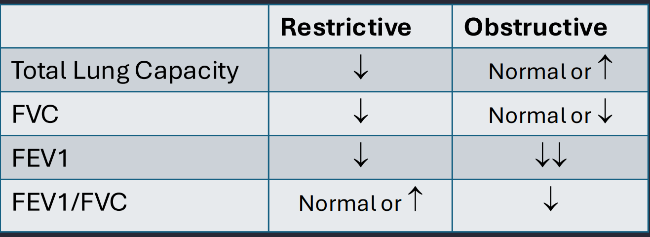
B6B7. Why can FVC decrease in severe obstruction without true restriction?
Answer: Air trapping prevents complete forced expiration, lowering the volume that can be exhaled even when TLC is normal or elevated.
Extra Information:
Low FVC alone does not confirm restrictive physiology.
YES! Obstructive lung diseases can have increased Total Lung Capacity (TLC), especially emphysema, because of increased lung compliance and air trapping.
Source: B6LG3 slides 5–8, 28–29

B6B8. Which important complications are associated with chronic COPD?
Answer: Chronic hypoxemia and pulmonary vascular abnormalities may contribute to pulmonary hypertension and cor pulmonale, while smoking is associated with lung cancer and cardiovascular disease risk.
Extra Information:
- Cor pulmonale refers to right ventricular dysfunction caused by pulmonary disease.
Source: B6LG3 slide 29 | First Aid Clinical Pattern Recognition (2022) p. 454

B6B8. Why does smoking cessation benefit patients who already have emphysema?
Answer: Removing cigarette smoke reduces ongoing inflammatory and oxidative injury, slowing future lung-function decline even though destroyed alveolar septa do not regenerate.
Extra Information:
- Smoking cessation is an essential long-term intervention.
- Supportive medication and behavioral treatment can improve cessation success.
Source: B6LG3 slide 22 | B6LG4 slides 29–40

B6B9. Explain the beta-2 agonist pathway for bronchodilation.
Answer: Beta-2 receptor activation → Gs stimulation → adenylyl cyclase activation → increased cAMP → smooth-muscle relaxation → reduced airway resistance.
Extra Information:
- Short-acting beta agonists provide rapid relief.
- Long-acting drugs provide sustained bronchodilation.
Source: B6LG4 slides 8–11
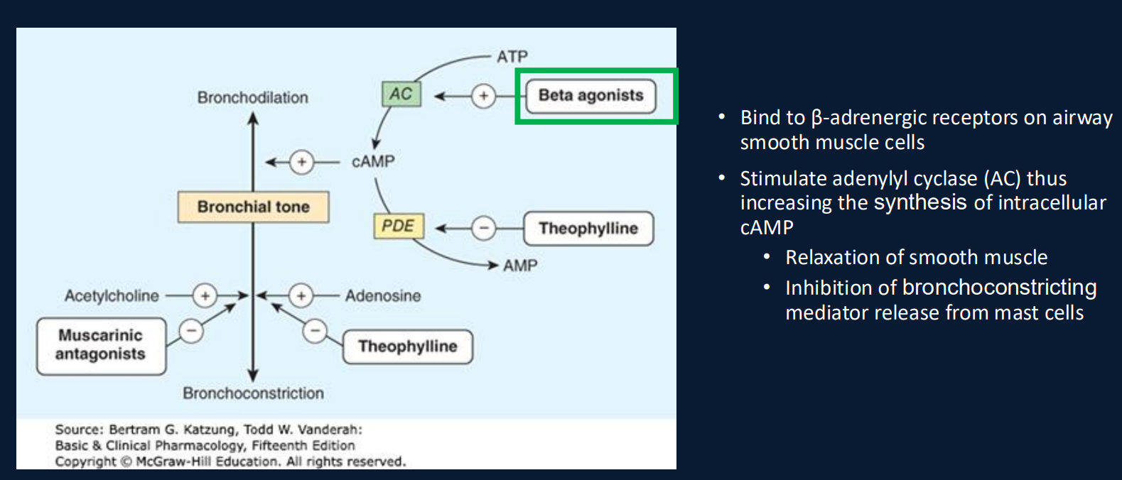
B6B9. Explain how inhaled antimuscarinic drugs produce bronchodilation.
Answer: Antimuscarinics block airway M3 receptors, preventing acetylcholine-mediated Gq–IP₃–calcium signaling and reducing bronchial smooth-muscle contraction.
Extra Information:
- Long-acting antimuscarinics are important maintenance COPD bronchodilators.
Source: B6LG4 slides 13–14, 17

B6B9. How do inhaled corticosteroids differ from LABA and LAMA therapies?
Answer: Corticosteroids reduce inflammation through glucocorticoid receptor signaling. LABAs stimulate smooth-muscle relaxation through beta-2 receptors, while LAMAs block muscarinic bronchoconstriction.
Extra Information:
- Inhaled corticosteroids do not provide immediate rescue bronchodilation.
- Their COPD use is selective.
Source: B6LG4 slides 8–18, 39

B6B10. Name the five nicotine replacement formulations and identify the sustained-delivery form.
Answer: Patch, gum, lozenge, oral inhaler, and nasal spray. The patch provides sustained transdermal nicotine delivery.
Extra Information:
- The remaining products can be used in relation to cravings.
- Different formulations have different local adverse effects.
Source: B6LG4 slides 32–33

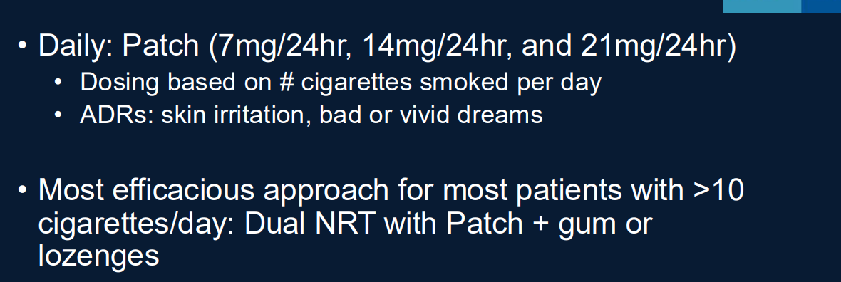
B6B10. Why can patch plus short-acting nicotine replacement therapy (NRT) work better than one formulation alone?
Answer: The patch provides steady background nicotine while gum, lozenge, or another on-demand formulation helps control breakthrough cravings.
Extra Information:
- Combination therapy addresses both continuous withdrawal and episodic craving.
- The lecture emphasizes combination therapy for heavier smokers.
Source: B6LG4 slides 33, 39–40

B6B10. What nicotine patch strengths are listed in the lecture?
Answer: 7 mg, 14 mg, and 21 mg patches over 24 hours.
Extra Information:
- Initial strength is selected according to the patient's cigarette consumption pattern.
- Patch adverse effects include skin irritation and abnormal dreams.
Source: B6LG4 slides 32–33
B6B10. How do gum and lozenges differ from nicotine patches?
Answer: Gum and lozenges provide shorter-acting oral nicotine doses for cravings, whereas patches deliver more sustained nicotine levels.
Extra Information:
- The lecture lists 2-mg and 4-mg gum and lozenge formulations.
- Time to first cigarette helps guide initial oral formulation strength.
Source: B6LG4 slides 32–33, 40
B6B10. Compare the characteristic local adverse effects of nicotine patches, oral inhalers, and nasal spray.
Answer: Patches may cause skin irritation, inhalers can cause mouth or throat irritation, and nasal spray can cause nasal irritation.
Extra Information:
- Exact onset and duration comparisons for all five formulations are not completely documented in the provided slides.
Source: B6LG4 slide 32

B6B11. Explain varenicline's smoking cessation mechanism.
Answer: Varenicline partially activates alpha-4-beta-2 nicotinic acetylcholine receptors, providing limited dopamine stimulation to reduce withdrawal while decreasing the rewarding effect of additional inhaled nicotine.
Extra Information:
- Nausea, insomnia, and abnormal dreams are emphasized adverse effects.
Source: B6LG4 slide 34

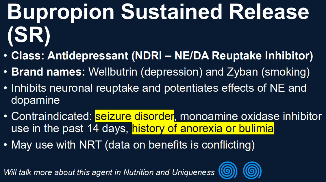
B6B11. Explain bupropion's smoking cessation mechanism.
Answer: Bupropion inhibits dopamine and norepinephrine reuptake, altering neurotransmitter signaling involved in withdrawal and craving.
Extra Information:
- Its mechanism differs from receptor agonism by nicotine or varenicline.
Source: B6LG4 slide 35

B6B11. What clinical histories contraindicate bupropion use?
Answer: Seizure disorders, anorexia or bulimia, and recent monoamine oxidase inhibitor use.
Extra Information:
- These contraindications influence selection among cessation therapies.
Source: B6LG4 slide 35
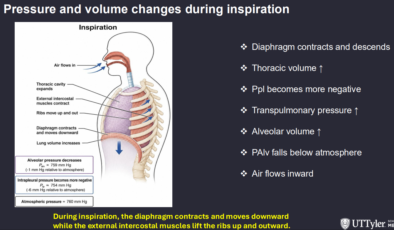
B6LG1.1. Trace the complete pressure and volume changes during quiet inspiration.
Answer: Diaphragm contracts → thoracic cavity volume increases → intrapleural pressure becomes more negative → transpulmonary pressure increases → lungs expand → alveolar pressure falls below atmospheric pressure → air enters.
Extra Information:
- Boyle's law relates increasing volume to decreasing pressure.
- Inspiration ends when alveolar pressure returns to atmospheric pressure.
Source: B6LG1 slides 4–11
B6LG1.1. Trace the pressure and volume changes during quiet expiration.
Answer: Diaphragm relaxes → thoracic volume decreases → elastic recoil decreases lung volume → alveolar pressure rises above atmospheric pressure → air flows outward.
Extra Information:
- Quiet expiration is predominantly passive.
- Forced expiration requires active expiratory muscles.
Source: B6LG1 slides 7–9

B6LG1.1. What is transpulmonary pressure and why is it important?
Answer: Transpulmonary pressure = alveolar pressure − intrapleural pressure. It is the distending pressure that opposes inward lung recoil and helps maintain inflation.
Extra Information:
At FRC, alveolar pressure equals atmospheric pressure.
FRC = Functional Residual Capacity
FRC is the amount of air remaining in your lungs after a NORMAL, passive exhalation.
It is NOT the air remaining after you forcefully exhale everything possible.
Formula
FRC = RV + RV
ERV (Expiratory Reserve Volume): Extra air you could still exhale after a normal expiration.
RV (Residual Volume): Air you cannot exhale, even with maximal effort.
Intrapleural pressure remains negative.
Source: B6LG1 slides 6, 11

B6LG1.1. Why is intrapleural pressure negative at the end of quiet expiration?
Answer: The lung naturally recoils inward while the chest wall tends to recoil outward, creating opposing forces across the pleural space.
Extra Information:
- These forces maintain positive transpulmonary pressure even when airflow is zero.
Source: B6LG1 slides 6, 11

B6LG1.2. Define compliance and elastance.
Answer: Compliance = ΔV/ΔP and describes lung distensibility. Elastance = ΔP/ΔV and describes stiffness or recoil tendency.
Extra Information:
- Emphysema increases compliance but reduces recoil.
- Fibrosis reduces compliance and increases elastic work.
Source: B6LG1 slides 12–15

B6LG1.2. What three mechanical components contribute to the work of breathing?
Answer: Elastic work, resistive work, and threshold work.
Extra Information:
- Fibrosis increases elastic work.
- Obstructive disease increases resistive work.
Source: B6LG1 slides 14–15

B6LG1.2. Which cells produce surfactant and what are its functions?
Answer: Type II pneumocytes produce surfactant, which lowers alveolar surface tension, increases compliance, and reduces the tendency of alveoli to collapse.
Extra Information:
- Surfactant is especially important for smaller alveolar units.
Source: B6LG1 slides 16–17 | First Aid Clinical Pattern Recognition (2022) p. 441
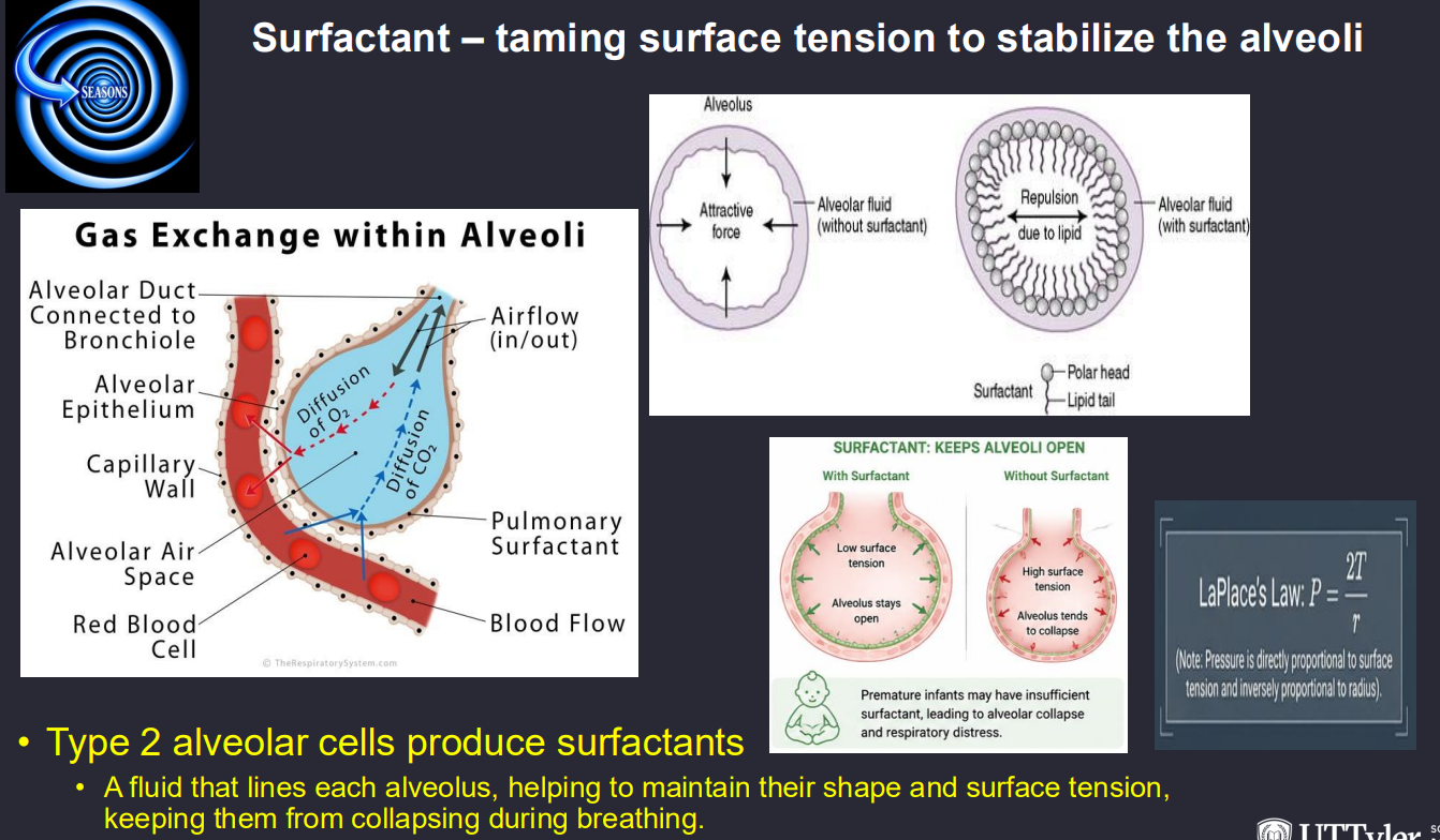
B6LG1.2. How does Laplace's relationship explain alveolar collapse without surfactant?
Answer: Collapsing pressure increases as alveolar radius decreases when surface tension is unchanged. Surfactant lowers surface tension and therefore helps stabilize smaller alveoli.
Extra Information:
- Reduced surface tension decreases the pressure needed for inflation.
Source: B6LG1 slides 16–17

B6LG1.2. Why does fibrosis increase elastic work while emphysema impairs expiration?
Answer: Fibrosis makes lung expansion difficult because compliance falls. Emphysema reduces recoil and airway-supporting traction, permitting early expiratory collapse and trapping air.
Extra Information:
- Both impair ventilatory efficiency through different mechanical mechanisms.
Source: B6LG1 slides 12–15 | B6LG3 slides 20–25

B6LG1.3. How do medullary and pontine respiratory centers cooperate?
Answer: Medullary circuits generate and organize breathing activity, while pontine networks modify respiratory timing and phase transitions. Chemoreceptor feedback adjusts breathing rate and depth.
Extra Information:
- Respiratory regulation integrates chemical and pulmonary sensory information.
Source: B6LG1 slides 19–22

B6LG1.3. What is the role of the pre-Bötzinger complex?
Answer: It is a medullary neuronal network involved in generating the automatic respiratory rhythm.
Extra Information:
- It contributes to the repetitive activity required for inspiration.
Source: B6LG1 slides 19, 26

B6LG1.3. What sensory information does the dorsal respiratory group integrate?
Answer: It receives respiratory sensory input including signals from peripheral chemoreceptors and pulmonary receptors, helping coordinate inspiratory activity.
Extra Information:
Glossopharyngeal and vagal afferents contribute to respiratory sensory signaling.
Source: B6LG1 slide 19 | Structure Respiratory Prework slides 16–18

B6LG1.4. Compare central and peripheral chemoreceptor locations and stimuli.
Answer: Central medullary chemoreceptors respond to CSF acidity driven by CO₂. Peripheral carotid and aortic bodies detect reduced arterial PO₂ and changes in arterial CO₂ or pH.
Extra Information:
- Central chemoreceptors do not directly measure arterial oxygen.
- Peripheral hypoxic drive increases strongly below PaO₂ of approximately 60 mm Hg.
Source: B6LG1 slides 20–22

B6LG1.4. Trace how elevated PaCO₂ increases central ventilatory drive.
Answer: CO₂ enters CSF → carbonic acid forms → hydrogen ion concentration increases → CSF pH decreases → central chemoreceptors stimulate respiratory networks → ventilation rises.
Extra Information:
- Increasing ventilation promotes CO₂ elimination.
Source: B6LG1 slides 20–21, 28
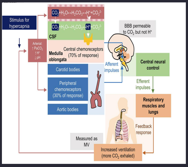
B6LG1.4. Why does chronic hypercapnia decrease central chemoreceptor sensitivity?
Answer: CSF bicarbonate adaptation partly normalizes CSF pH despite elevated PaCO₂, reducing the sustained central stimulus.
Extra Information:
- Peripheral chemoreceptors remain responsive to hypoxemia.
Source: B6LG1 slides 22–24

B6LG1.4. Trace how opioid toxicity causes respiratory depression.
Answer: Opioids activate mu-opioid receptors in respiratory-control networks → respiratory output decreases → rate and depth of breathing fall → alveolar ventilation decreases → PaCO₂ rises and hypoxemia may develop.
Extra Information:
- Severe toxicity may produce apnea and respiratory acidosis.
Source: B6LG1 slides 25–26, 32

B6LG1.5. What are the three forms of CO₂ transport in blood and their approximate percentages?
Answer: Approximately 70% travels as bicarbonate, 20–25% as carbamino compounds, and 5% physically dissolved.
Extra Information:
- Carbonic anhydrase accelerates bicarbonate formation inside red cells.
Source: B6LG1 slide 28

B6LG1.5. Trace CO₂ conversion and chloride shift in systemic capillaries.
Answer: CO₂ enters red cells → carbonic anhydrase converts it to carbonic acid → bicarbonate and H⁺ form → bicarbonate leaves through anion exchange → chloride enters to maintain electroneutrality.
Extra Information:
- The process reverses in pulmonary capillaries to support CO₂ exhalation.
Source: B6LG1 slides 28–29

B6LG1.5. Which red-cell transporter mediates the chloride shift?
Answer: Band 3, also called AE1, exchanges bicarbonate and chloride across the erythrocyte membrane.
Extra Information:
In systemic tissues, bicarbonate exits while chloride enters.
In the lungs, the transport direction reverses.
Band 3 (AE1) anion exchanger
Source: B6LG1 slide 29
B6LG1.5. Explain the Haldane effect and contrast it with the Bohr effect.
Answer: The Haldane effect means oxygenation of hemoglobin promotes CO₂ and H⁺ unloading. The Bohr effect concerns how CO₂ and H⁺ alter hemoglobin's oxygen affinity.
Extra Information:
- Deoxygenated hemoglobin carries more CO₂ and H⁺ in tissues.
- Oxygen loading promotes CO₂ removal in the lungs.
Source: B6LG1 slide 30

B6LG2.1. Predict early ABG changes in pneumonia or pulmonary edema and explain why.
Answer: Alveolar filling impairs ventilation and oxygen transfer → PaO₂ decreases and A–a gradient increases. Compensatory hyperventilation often decreases PaCO₂.
Extra Information:
- Severe shunt produces oxygen-refractory hypoxemia.
- Respiratory fatigue may eventually increase PaCO₂.
Source: B6LG2 slides 9–11

B6LG2.1. Why can early pneumonia produce respiratory alkalosis despite hypoxemia?
Answer: Hypoxemia increases respiratory drive, causing hyperventilation and increased CO₂ removal even while oxygen transfer remains impaired.
Extra Information:
- PaO₂ and PaCO₂ need not decrease or increase together.
Source: B6LG2 slide 11

B6LG2.1. Why does pulmonary edema increase breathing effort?
Answer: Alveolar flooding and surfactant disruption reduce lung compliance, making inflation more difficult while also impairing oxygenation.
Extra Information:
- Low V/Q and shunt develop when perfusion continues through poorly ventilated regions.
Source: B6LG2 slides 9–11

B6LG2.2. Trace silica exposure to pulmonary fibrosis.
Answer: Inhaled crystalline silica reaches distal lung → macrophages respond with inflammatory mediators → persistent tissue injury and fibrotic signaling develop → collagen-rich nodules form → compliance and diffusion capacity decline.
Extra Information:
- The lecture includes TNF-alpha, IL-1-beta, and reactive oxygen species in inflammatory injury.
Source: B6LG2 slides 13–14

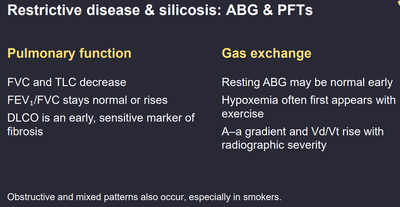
B6LG2.2. Predict PFT and gas-exchange findings in pulmonary fibrosis.
Answer: TLC and FVC decrease, FEV₁/FVC remains normal or rises, compliance decreases, and DLCO often decreases because interstitial thickening impairs diffusion.
Extra Information:
- Exercise may reveal hypoxemia before it becomes obvious at rest.
Source: B6LG2 slides 13–14 | B6LG3 slide 28

B6LG2.2. Apply Fick's law to explain diffusion defects in fibrosis versus emphysema.
Answer: Gas diffusion increases with membrane area and pressure gradient but decreases with membrane thickness. Fibrosis increases thickness while emphysema decreases surface area.
Extra Information:
- Both diseases may lower DLCO for different structural reasons.
Source: B6LG2 slide 7 | B6LG3 slides 18–19
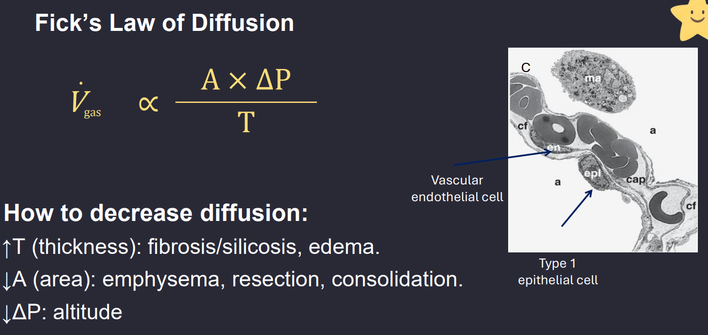
B6LG2.2. Why does exercise worsen diffusion-related hypoxemia in pulmonary fibrosis?
Answer: Increasing blood flow shortens pulmonary-capillary transit time, leaving less time for oxygen equilibration across the thickened diffusion barrier.
Extra Information:
- Exercise can reveal a gas-exchange defect that is less evident at rest.
Source: B6LG2 slide 14
B6LG2.3. Trace the acute ventilatory response to high altitude.
Answer: Barometric pressure falls → inspired oxygen pressure decreases → arterial oxygen falls → peripheral chemoreceptors stimulate hyperventilation → PaCO₂ decreases → respiratory alkalosis develops.
Extra Information:
Lower PaCO₂ partly increases alveolar PO₂ through the alveolar gas equation.
Source: B6LG2 slides 19–20

B6LG2.3. What symptoms suggest acute mountain sickness?
Answer: Headache with associated nausea, dizziness, fatigue, reduced appetite, or sleep disturbance after rapid ascent to significant altitude.
Extra Information:
Ataxia or altered mental status suggests a more serious altitude-associated cerebral complication.
Ataxia is a neurological sign and condition that causes a lack of voluntary muscle coordination, leading to clumsy and uncoordinated movements
Source: B6LG2 slide 21

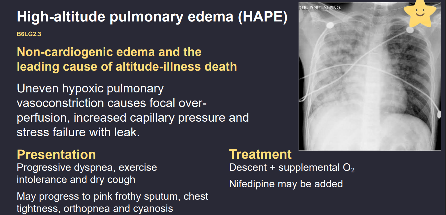
B6LG2.3. Trace the mechanism of high-altitude pulmonary edema.
Answer: Low oxygen triggers pulmonary vasoconstriction → constriction is uneven → blood is redistributed into less-constricted vessels → local capillary pressure rises → fluid leaks into lung tissue and alveoli → noncardiogenic edema worsens oxygenation.
Extra Information:
- Descent and supplemental oxygen are central management measures.
Source: B6LG2 slide 22
B6LG2.3. Why does gradual ascent reduce altitude illness risk?
Answer: Gradual exposure allows ventilatory and other physiologic acclimatization to lower inspired oxygen pressure.
Extra Information:
- Rapid altitude gain increases the risk of acute mountain illness.
Source: B6LG2 slides 20–23

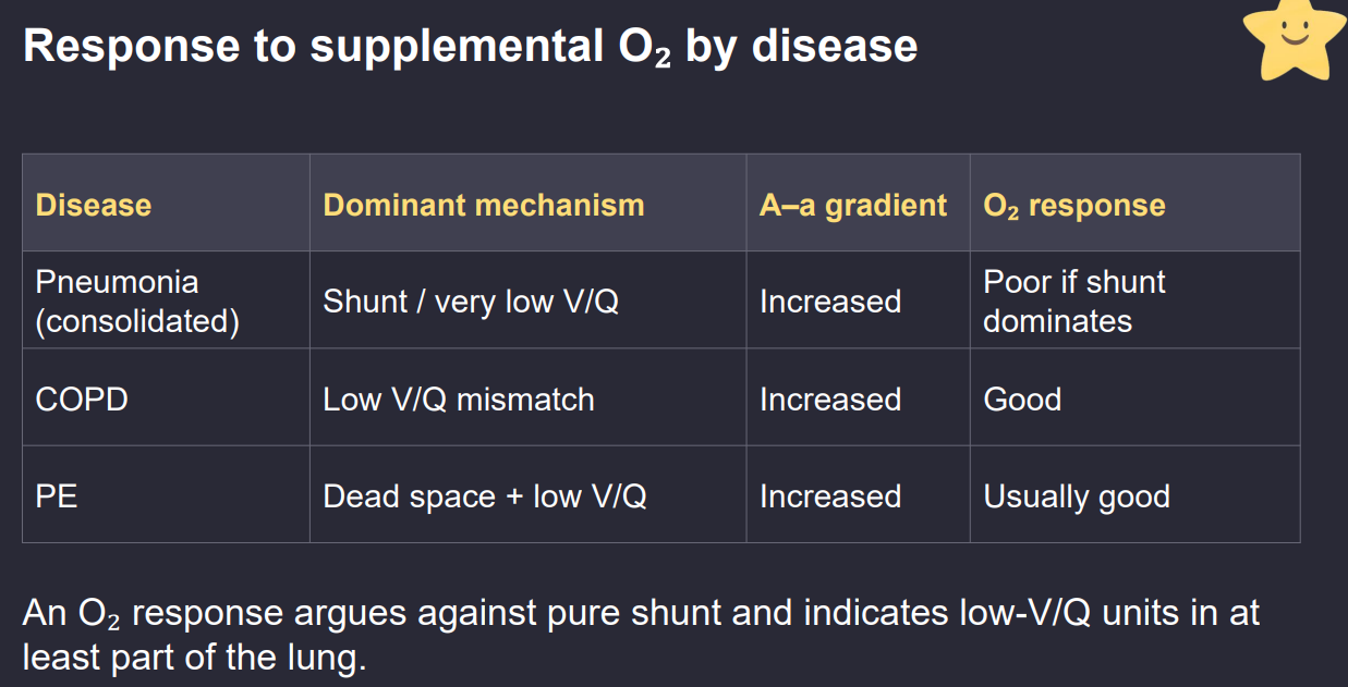
B6LG2.4. Compare V/Q abnormalities in pneumonia, COPD, and pulmonary embolism.
Answer: Pneumonia causes low V/Q or shunt, COPD produces mixed low- and high-V/Q regions, and pulmonary embolism directly creates high-V/Q dead space.
Extra Information:
- Pulmonary embolism may additionally cause low V/Q from regional blood-flow redistribution.
Source: B6LG2 slides 25–27
B6LG2.4. Why can pulmonary embolism produce hypoxemia despite causing dead space?
Answer: Although vessel obstruction creates high-V/Q regions, blood redistribution and secondary regional lung changes can produce low-V/Q regions elsewhere, lowering arterial oxygenation.
Extra Information:
- Dead-space ventilation itself is ineffective because blood is absent.
Source: B6LG2 slide 27

B6LG2.4. How does oxygen response differ between low V/Q and true shunt?
Answer: Increasing inspired oxygen generally improves low V/Q because air still reaches alveoli, whereas a true shunt responds poorly because blood passes through unventilated regions.
Extra Information:
COPD and pulmonary embolism often improve with oxygen.
Extensive consolidated pneumonia may respond less when true shunt dominates.
Source: B6LG2 slides 25–28

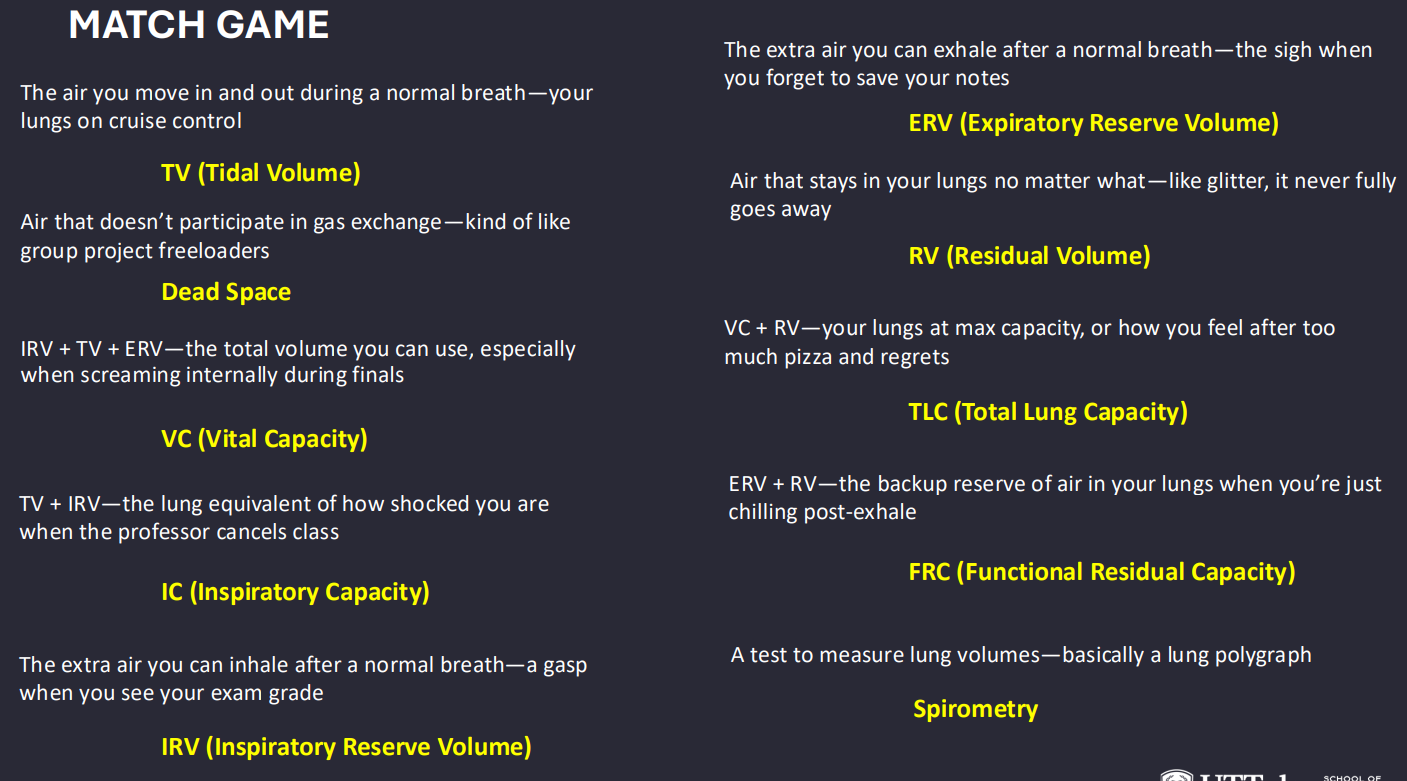
B6LG3.1. Define the four major lung volumes.
Answer: Tidal volume is air moved during quiet breathing. Inspiratory reserve volume is additional inhaled air after normal inspiration. Expiratory reserve volume is additional exhaled air after normal expiration. Residual volume remains after maximal expiration.
Extra Information:
- These volumes combine to form lung capacities.
Source: B6LG3 slide 3 | Structure PFT Prework

B6LG3.1. Give the formulas for VC, IC, FRC, and TLC.
Answer: VC = IRV + TV + ERV. IC = TV + IRV. FRC = ERV + RV. TLC = VC + RV.
Extra Information:
- FRC is the lung volume after passive expiration.
- Simple spirometry cannot directly measure residual volume.
Source: B6LG3 slide 3 | B6LG1 slide 6
B6LG3.1. Why does FEV₁/FVC decrease in obstructive pulmonary disease?
Answer: Increased resistance and expiratory airway collapse disproportionately slow the volume exhaled during the first second relative to the total forced vital capacity.
Extra Information:
- Obstructive flow-volume loops have a concave expiratory limb.
Source: B6LG3 slides 4–8

B6LG3.1. How do FEV₁/FVC and TLC distinguish obstruction from restriction?
Answer: Obstruction reduces FEV₁/FVC. Confirmed restriction reduces TLC while typically preserving or increasing FEV₁/FVC.
Extra Information:
- Low FVC alone does not prove restriction because air trapping can also reduce FVC.
Source: B6LG3 slides 4–8, 27–29
