ANT 156a Quiz 4
 Call Kai
 Learn
 Practice Test
 Spaced Repetition
 Match
 Flashcards
 Knowt Play
1/90

Earn XP
Description and Tags
pelvis!
91 Terms
Bones of The Pelvic Girdle
Os Coxae (hip bone, innominate)
Ilium
Pubis
Ischium
Sacrum (variably fused vertebrae)
Coccyx (variably fused vertebrae)

What is green?
Ilium

What is pink?
Pubis
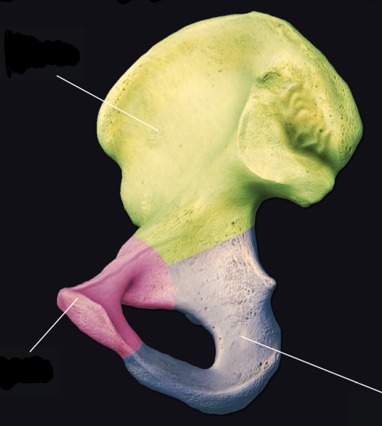
What is purple?
Ischium

What is pink (top)?
Sacrum

What is coral (bottom)?
Coccyx

Full Pelvic Girdle Which side?
Anterior

Full Pelvic Girdle Which side?
Superior
Axial or Appendicular
both, connects them functionally, develops with lower limbs
Functions of the Pelvic Girdle
Human pelvis have been shaped by
- bipedal walking & running
- giving birth to large babies
weight transfer from torso to legs
anchor abdominal and leg muscles
support and protect abdominal & pelvic organs (viscera)

What is A? (Anterior)
ilium

What is v?
iliac fossa
(whole concave surface area for iliacus muscle)
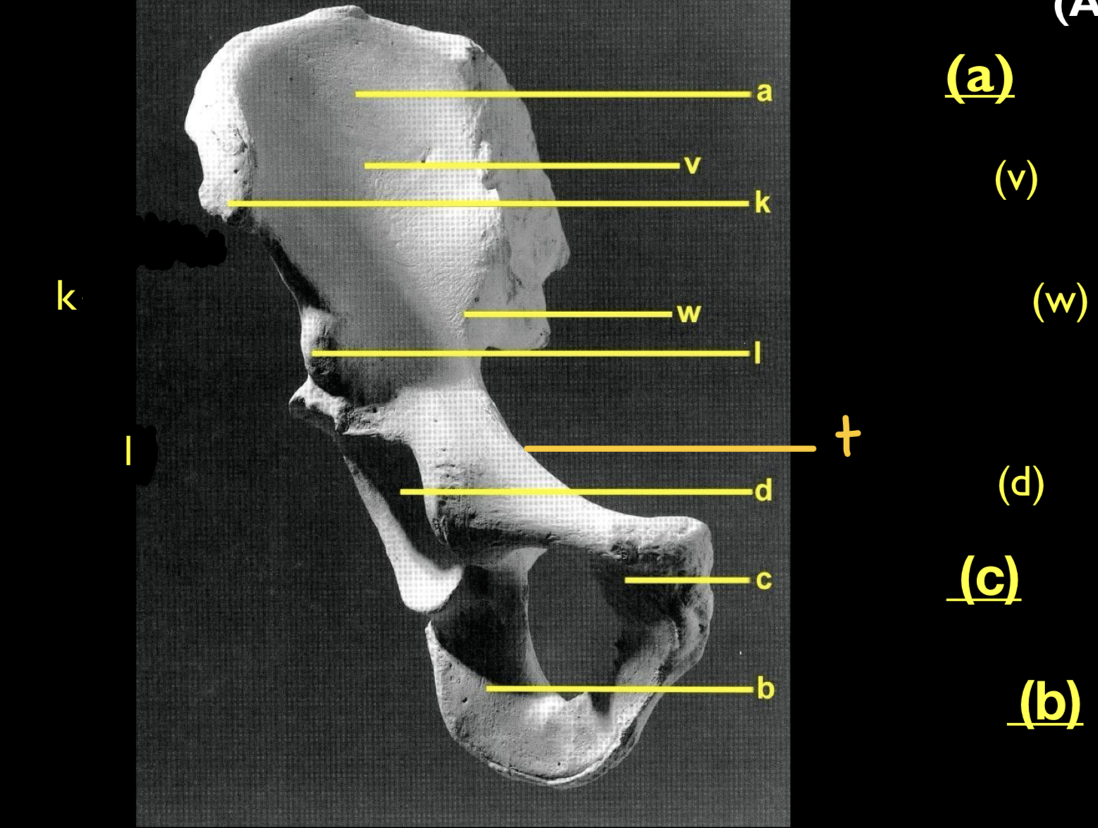
What is w?
arcuate line
(birth canal entrance)

What is t?
pectineal line
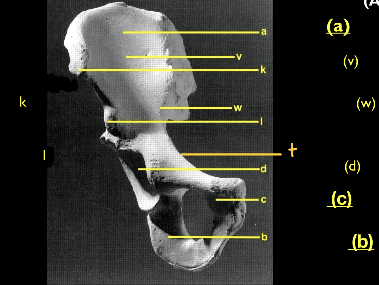
What is d?
acetabulum/ acetabula
(most lateral socket formed by all 3)
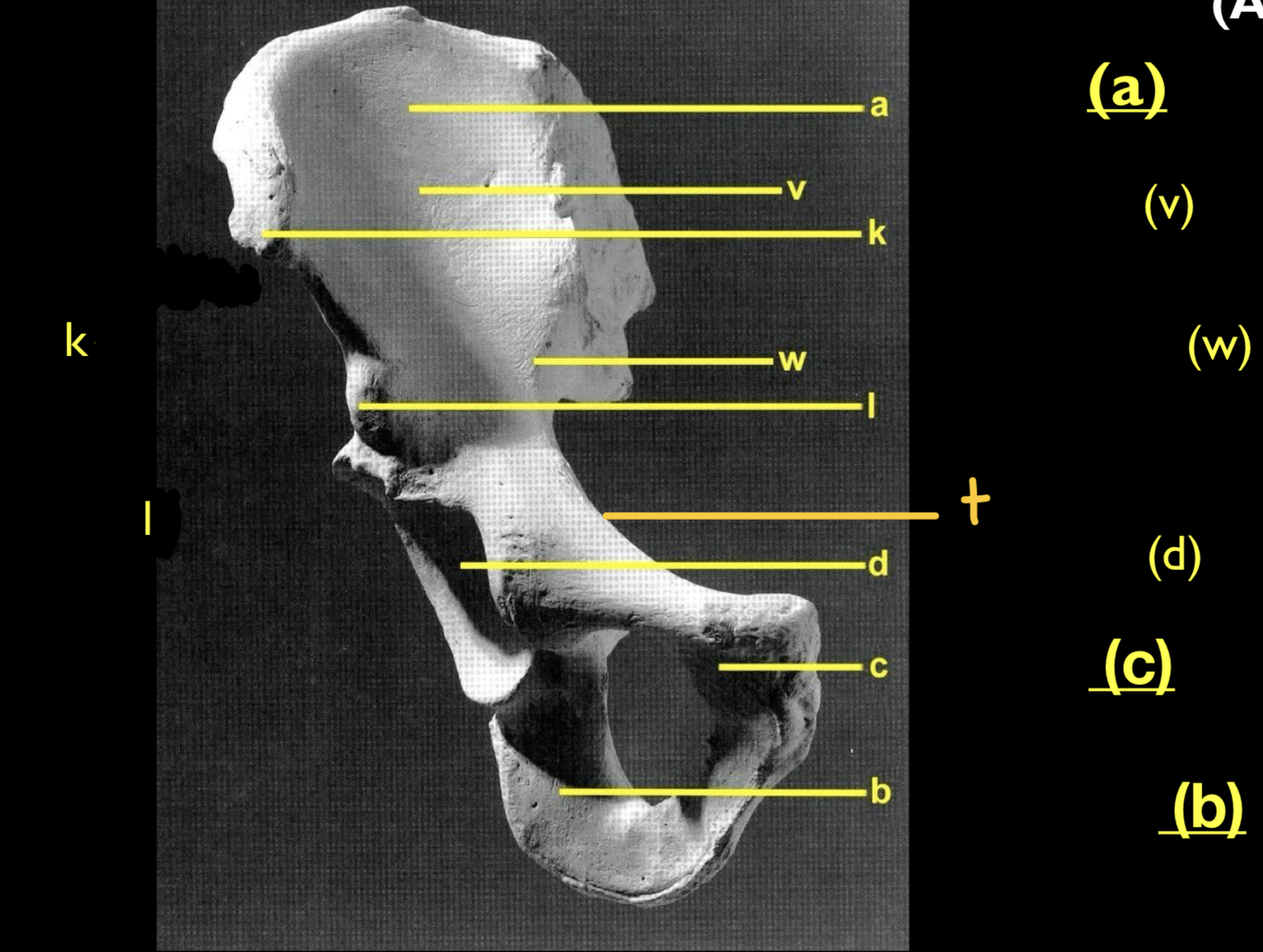
What is c?
pubis
(where they meet)

What is b?
ischium
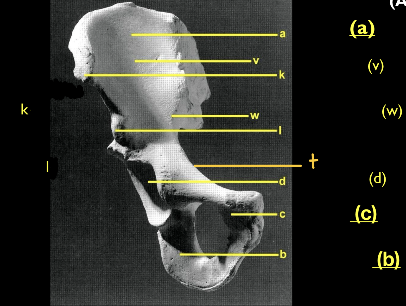
What is k?
anterior- superior iliac spine (ASIS)
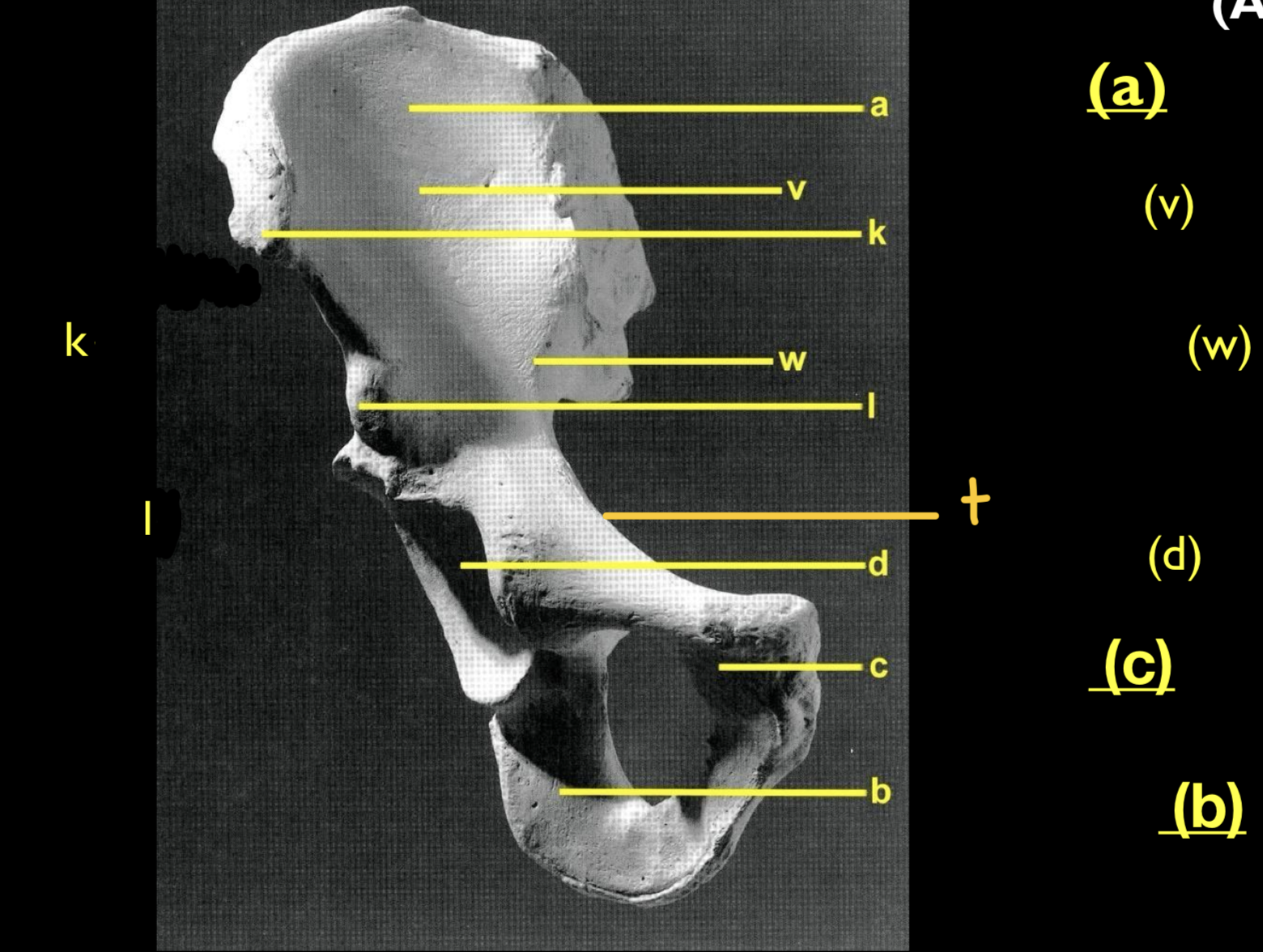
What is I?
anterior- inferior iliac spine (AIIS)
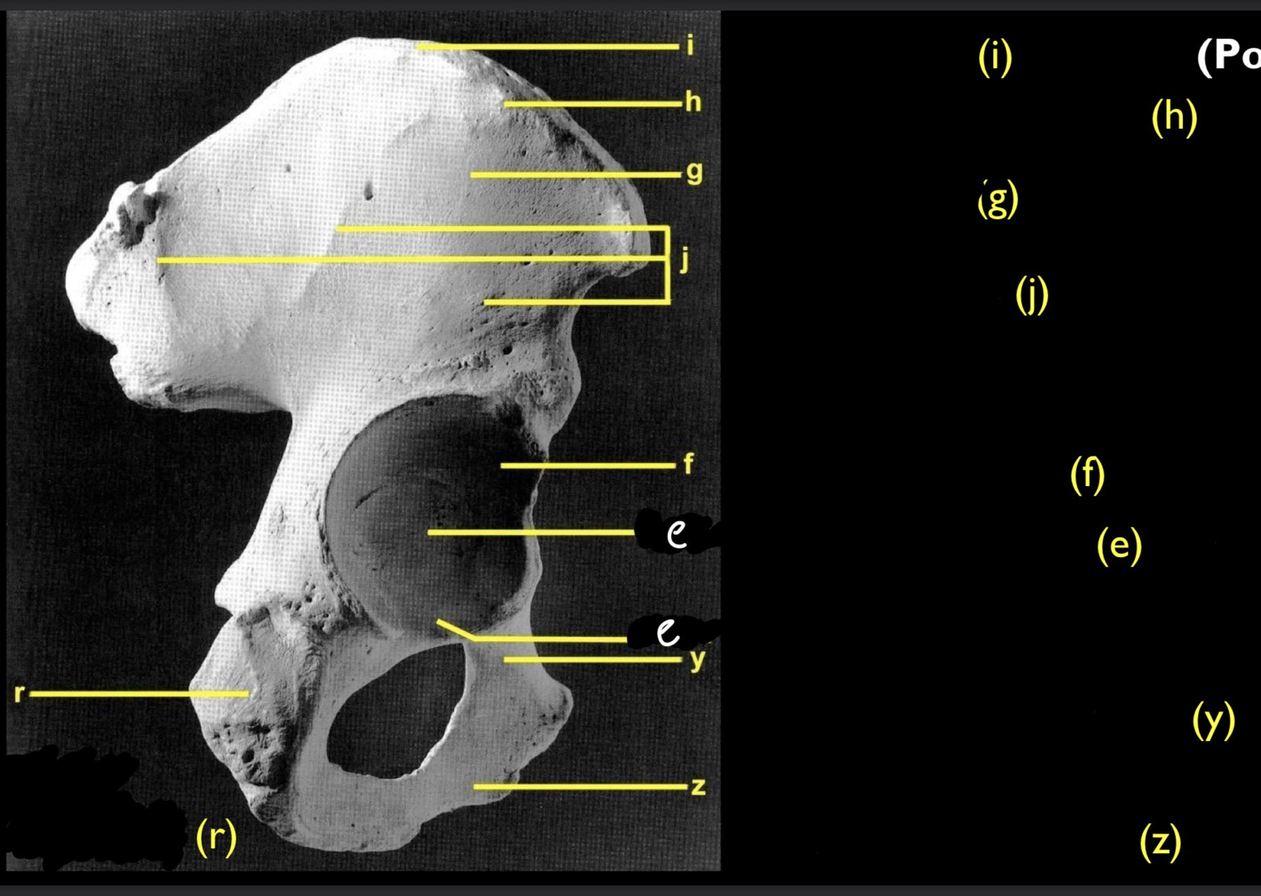
What is i? (posterolateral)
iliac crest

What is h?
iliac/cristal tubercle

What is g?
iliac pillar

What is j?
gluteal lines
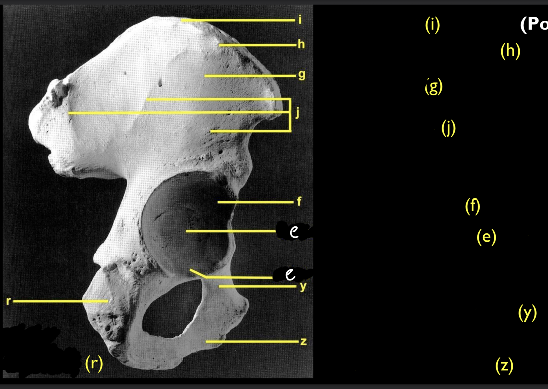
What is f?
lunate surface
(femoral head)

What is e?
acetabular fossa & acetabular notch

What is y?
superior pubic ramus

What is z?
inferior pubic ramus

What is r?
ischial tuberosity

What is t? (anteromedial)
iliac tuberosity
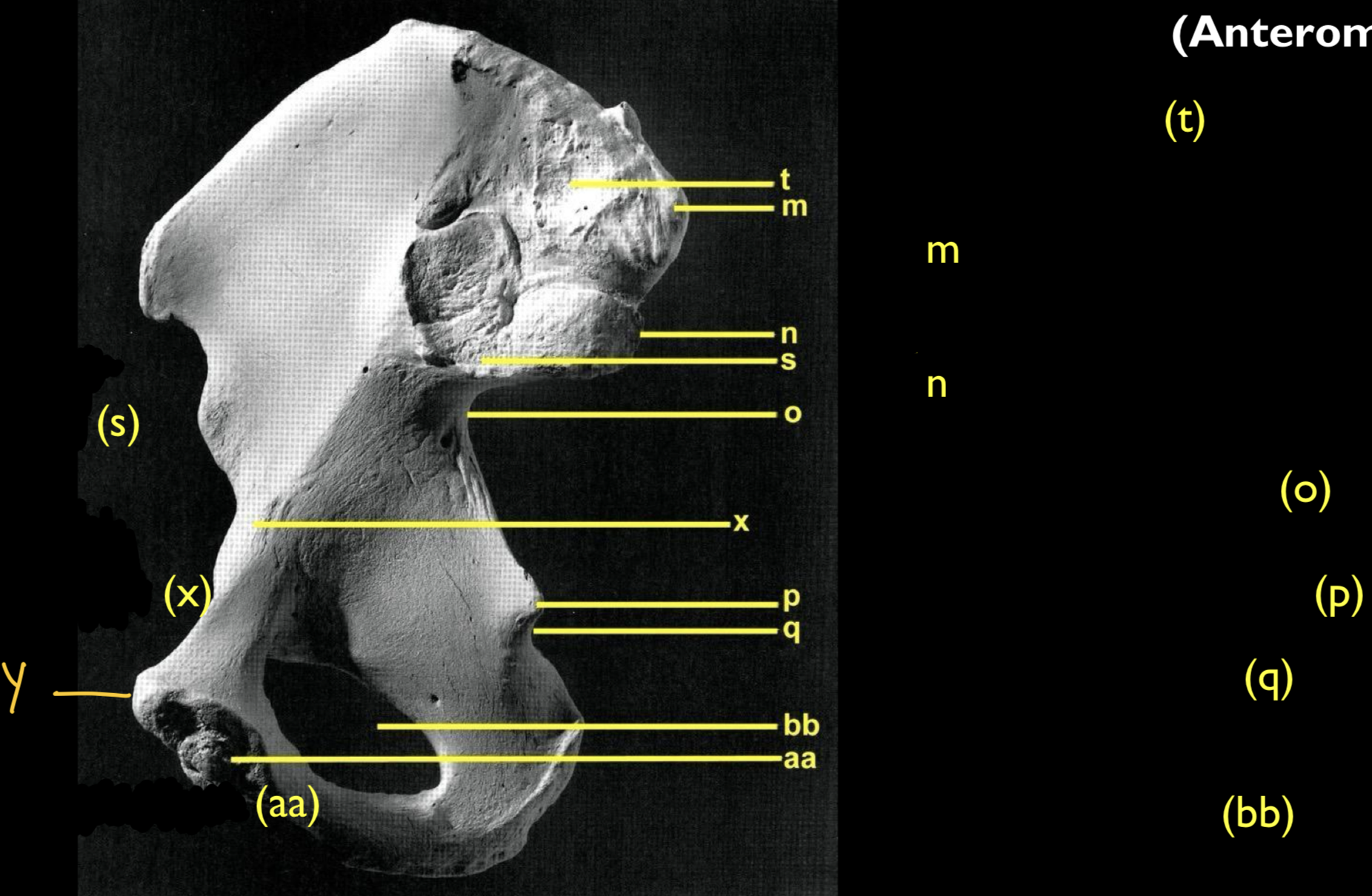
What is m?
posterior- superior iliac spine (PSIS)

What is n?
posterior- inferior iliac spine

What is o?
greater sciatic notch
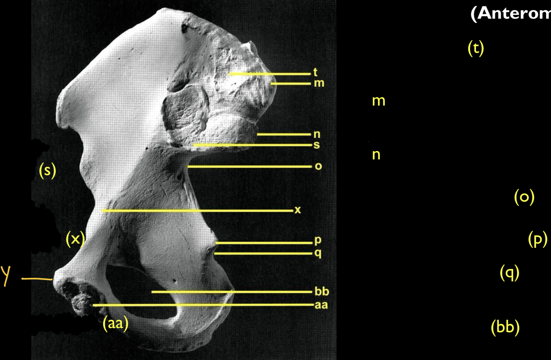
What is p?
ischial spine

What is q?
lesser sciatic notch

What is bb?
obturator foramen

What is aa?
pubic symphysis

What is x?
iliopublic eminence

What is s?
auricular surface
(latin ear shape)
(sacrum articulation)

What is y?
pubic tubercle
Soft Tissue Inside Hip Joint
transverse acetabular ligament
ligamentum teres
Sacrum, how many vertebrae are fused?
typically 5
could be more or less

What is A? (anterior sacrum)
sacral promontory
(midline, superiorly)

What is b?
ala/alae
wings
fibrous joint

What is d?
sacral foramina
(sacral nerves pass thru)

What is e?
transverse lines/ridges
(ridges down center, marks fusions)
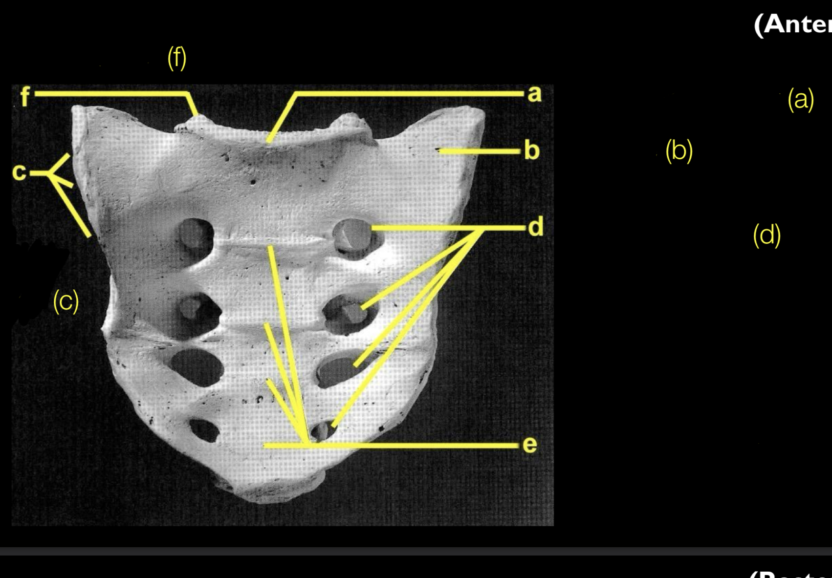
What is c?
auricular surface

What is f?
superior articular processes/facets

What is f? (posterior sacrum)
superior articular processes/facets

What is h?
median crest

What is k?
lateral crest

What is l?
sacral hiatus

What is the yellow arrow?
sacral canal

Coccyx (a,b,c)
sacral articulation
cornua
transverse process
(aka vestigial tail, tailbone, variable in 3-5 fused elements)
(fuses sometimes later in life)
Pelvic inlet and outlet

Sacral Ossification
All segments:
centrum
2 halves of vertebral arch
Segments 1-3 also have:
2 costal elements (alae and transverse processes)
Secondary Centers
2 epiphyseal rings per segment
Each auricular surface
Each infero-lateral margin
Coccyx
One primary center for each segment
NO secondary centers
What is typical age for fully fused sacrum?
26 to 30 years old
Os Coxae Ossification
Primary
Ilium
Ischium
Pubis
Secondary (5);
Ischial tuberosity
Iliac crest
Anterior-inferior iliac spine
Pubic symphysis (tubercle)
Center of acetabula fossa
Age Estimation using Os Coxae
cartilaginous joint age estimation
looking at pubic symphysis
horizontal ridges
rimmed face
depression inside rim

Sex Estimation using Os Coxae
sexual dimorphism
women have broader pelvis (for birth)
coccyx (males longer, females shorter)
greater sciatic notch shape (women= u shaped, men= j shaped)


What is purple? (ligament)
sacrotuberous
Sacrotuberous Ligament
(resists posterior rotation of the inferior sacrum)
attachments: lateral sacrum, posterior-inferior iliac spine, and internal margin of the ischial tuberosity.
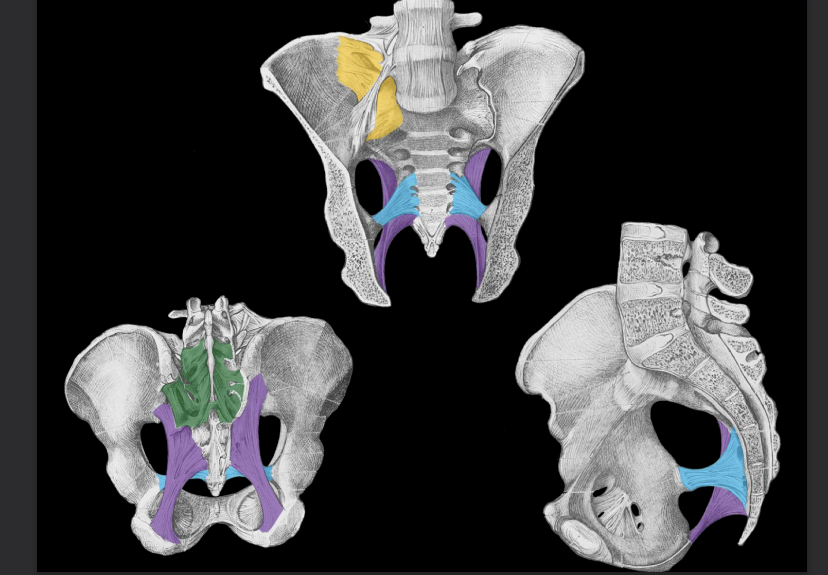
What is yellow? (ligament)
anterior sacroiliac
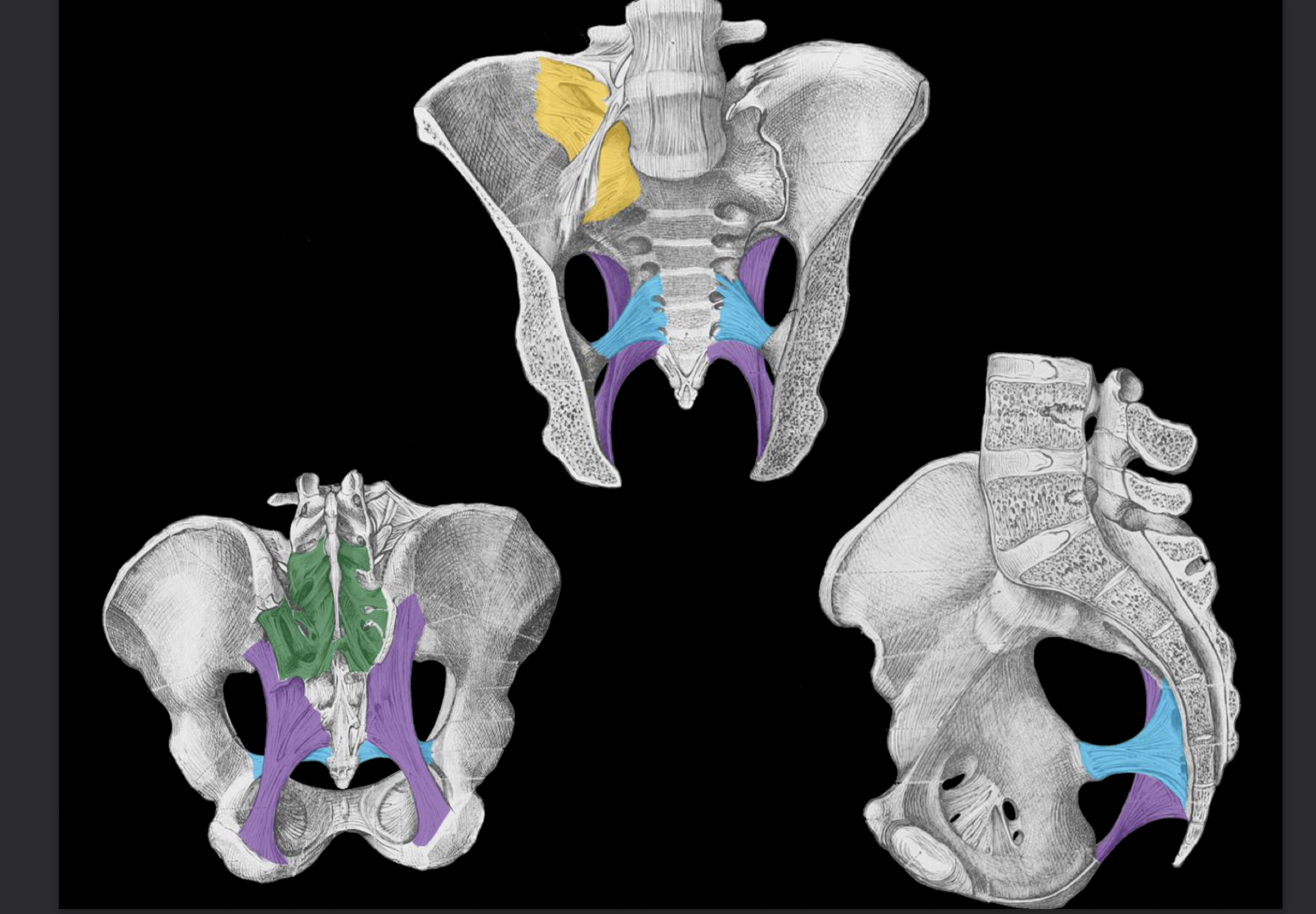
What is green? (ligament)
posterior sacrioiliac
Sacroiliac Ligaments
(strengthen the sacroiliac joint)
attachments: anterior and posterior to the sacroiliac joint, including the iliac tuberosity.

What is blue? (ligament)
sacrospinous
Sacrospinous Ligament
(resists posterior rotation of the inferior sacrum)
attachments: antero-lateral sacrum, ischial spine

What is yellow arrow?
obturator canal

What is red?
obturator membrane
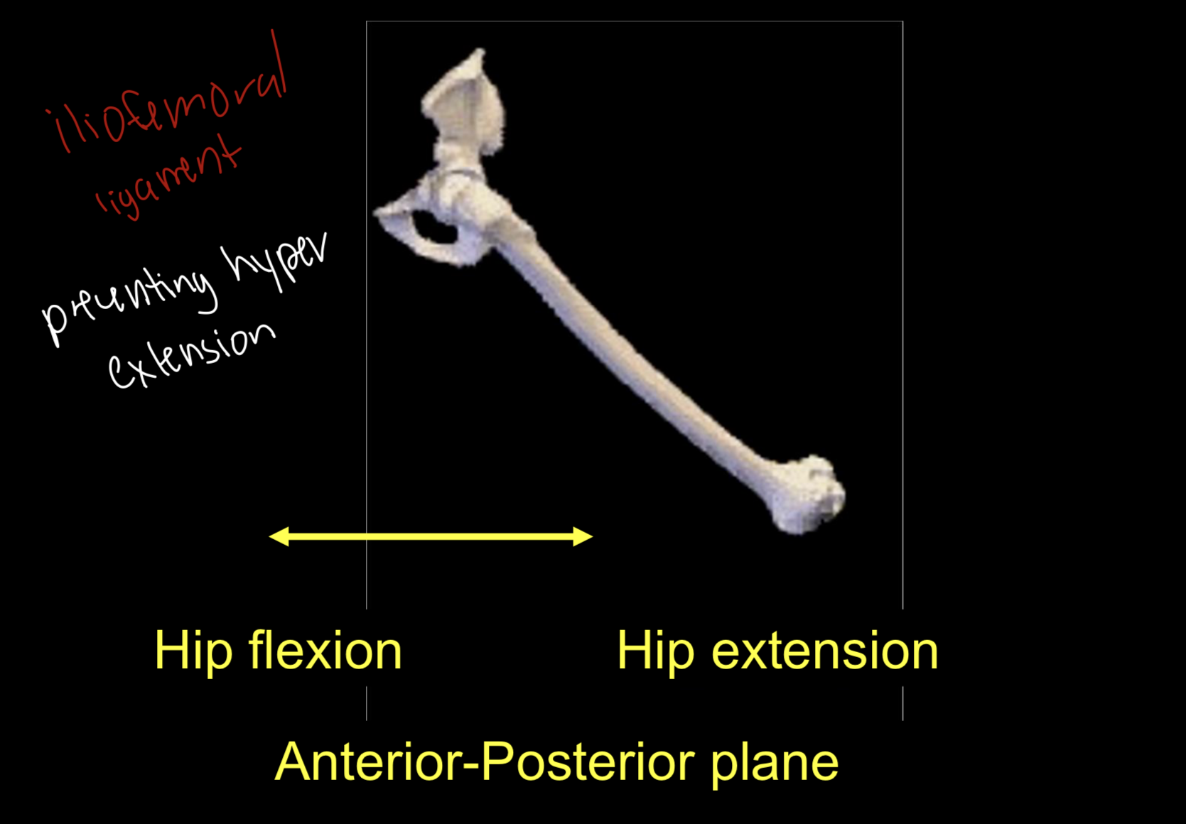
Iliofemoral Ligament
(resists hip extension)
proximal attachment: on and in the vicinity of the anterior-inferior iliac spine,
distal attachment: intertrochanteric line of the femur

Iliofemoral Ligament Movements
prevents hyper extension
Iliofemoral Ligament Movements (1)
hip flexion & hip extension = (anterior- posterior plane)
Iliofemoral Ligament Movements (2)
hip abduction & hip adduction = (medial-lateral plane)

Gluteal Muscles
Gluteus minimus, medius, maximus
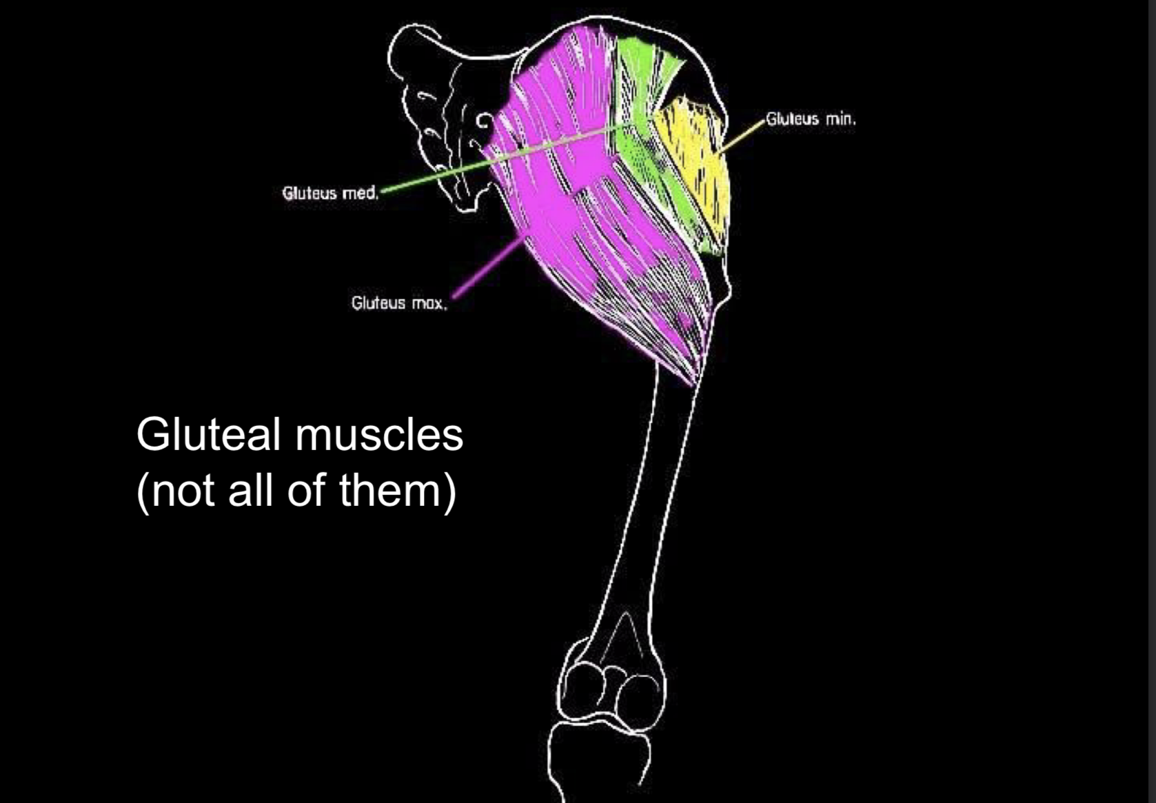

Lateral Gluteus (1)
Gluteus minimus
(hip abductor & medial rotator) –
Origin: between the anterior and inferior gluteal lines of the ilium,
Insertion: greater trochanter (anterior part) of the femur.

Lateral Gluteus (2)
Gluteus medius
(hip abductor & medial rotator) – Origin: between the iliac crest and the anterior and posterior gluteal lines of the ilium,
Insertion: greater trochanter (posterior part) of the femur.

Posterior Gluteus (3)
Gluteus maximus
(hip extensor & lateral rotator) – Origin: posterior gluteal line of the ilium, sacrum, and sacrotuberous ligament,
Insertion: gluteal line/tuberosity of the femoral shaft.
Iliopsoas Muscle Group
hip flexor
iliacus
psoas major (sometimes minor but only 50%)

Iliacus
Origin: iliac fossa, anterior sacrum, Insertion: lesser trochanter of the femur.
Psoas Major
Origin: T12-L4 (or L5), Insertion: lesser trochanter of the femur.
Sartorius
longest muscle in the human body
(hip flexor, abductor, & lateral rotator; knee flexor)
– Origin: anterior-superior iliac spine, Insertion: proximo-medial tibial shaft.

Sartorius Movement (1)
hip flexion & hip extension (anterior-posterior plane)

Sartorius Movement (2)
hip abduction & hip adduction
(medial-lateral plane)

Sartorius Movement (3)
lateral medial rotation

Hamstring Muscles
biceps femoris
semitendinosus
semimembranosus
(hip extensors, knee flexors)
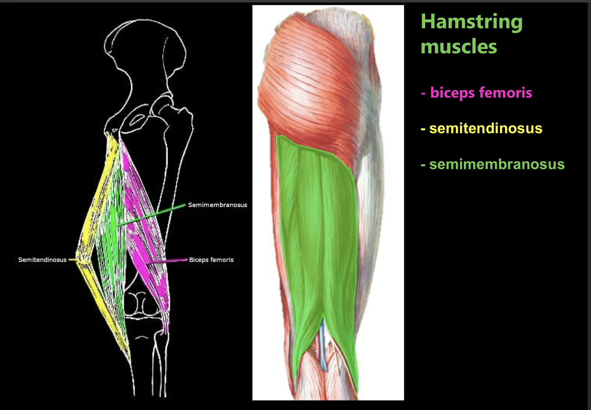
Biceps femoris
Origin: ischial tuberosity (long head; supero-medial facet), distal
femoral shaft (short head), Insertion: proximo-lateral fibula
Semitendinosus
Origin: ischial tuberosity (supero-medial facet), Insertion: proximo-
medial tibial shaft.
Semimembranosus
Origin: ischial tuberosity (supero-lateral facet), Insertion:
proximo-medial tibial condyle.
Hamstring Movement (1)
extension & flexion
knee flexors

Where is body’s center of mass?
sacroiliac joint and hip joint
Changes in bony pelvis shape
humans more wide
Rearrangement of lesser gluteals
Sagittally oriented in humans
Abduct hip during bipedal locomotion (hip abductor)
Coronally oriented in chimps
laterally rotate hip during bipedal locomotion
