Bio Exam #4
 Call Kai
 Learn
 Practice Test
 Spaced Repetition
 Match
 Flashcards
 Knowt Play
1/91

There's no tags or description
Looks like no tags are added yet.
92 Terms
Axial skeleton
Composed of the skull, vertebral column, and thoracic cage. Central axis of the body
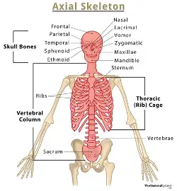
Appendicular skeleton
Bones of upper and lower limbs, Girdles of bones attach limbs to axial skeleton.

Cranial cavity
Encloses, protects, and supports the brain. Composed of the frontal bone, parietal bone, and parts of the occipital bone. Orbits (eye sockets), oral cavity, nasal cavity, paranasal sinuses.

Facial bones
form the face, protect entrances to digestive and respiratory systems, shape and individually to the face. Muscles of facial expression and chewing. Zygomatic, lacrimal, inferior nasal conchae, palatine bones, maxillae, unpaired vomer, mandible.
Cranial fossae
3 curved depressions on cranial cavity floor, house lobes of brain, anterior, middle and posterior cranial fossae.

Nasal complex
encloses nasal cavity and paranasal sinuses, bone and cartilage
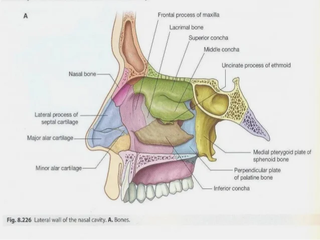
Paranasal sinuses
Air-filled chambers open into the nasal cavity. Mucous membrane humidifies and warms inhaled air. Cause skull bones to be lighter, provide respone to voice. Frontal, Ethmoid, sphenoidal, maxillary sinuses

Auditory ossicles
3 tiny ear bones, housed within petrous part of each temporal bone, malleus, incus and stapes.

Retraction
Posterior movement of a protracted body part back to the anatomic position, such as pulling the jaw inward.
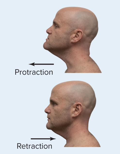
Protraction
Anterior movement of a body part from anatomic position in a horizontal plane, such as jutting the jaw forward.

Depression
The inferior movement of a body part, such as lowering the mandible when opening the mouth.
Elevation
The superior movement of a body part, such as raising the mandible when closing the mouth.

Opposition
Special movement of the thumb across the palm toward the fingertips at the carpometacarpal joint, enabling grasping of objects.

Pectoral girdle
An open skeletal ring consisting of two clavicles and two scapulae that anchors the upper limbs to the trunk while maximizing limb mobility.

Pelvic girdle
The appendicular structure formed exclusively by the right and left hip bones (ossa coxae), articulating with the lower limbs.

Bursae
A fibrous, saclike structure lined with a synovial membrane and filled with synovial fluid, situated where bones, ligaments, muscles, skin, or tendons rub together to alleviate friction.

Fat Pad
Adipose tissue located in the periphery of a synovial joint that acts as protective packing material and cushions changing joint spaces.

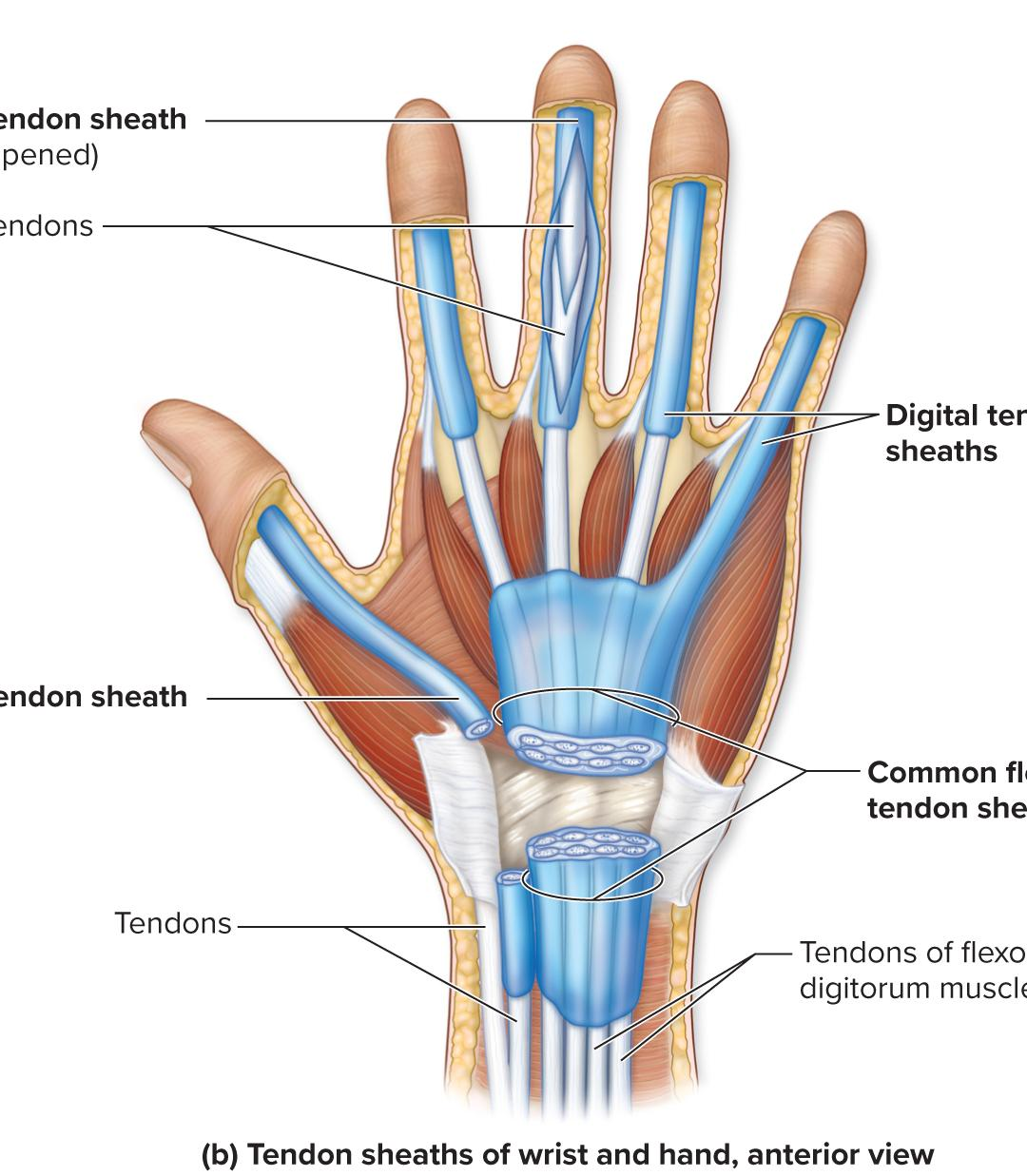
Tendon Sheath
An elongated bursae wrapped around tendons in high-friction areas such as the wrist and ankle.
Diarthrosis
A functional classification denoting a freely mobile joint; includes all synovial joints.

Amphiarthrosis
A functional classification denoting a slightly mobile joint, which can be fibrous or cartilaginous.

Synchondrosis
An immobile cartilaginous joint (synarthrosis) where bones are joined by hyaline cartilage, such as the epiphyseal plate or costochondral joints.

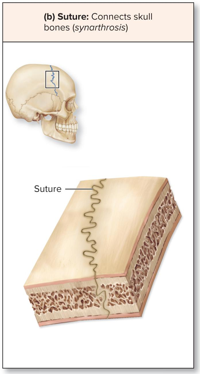
Suture
An immobile fibrous joint (synarthrosis) with interlocking, irregular edges and very short fibers found between certain skull bones.
Gomphosis
A 'peg in a socket' fibrous joint (synarthrosis) where periodontal ligaments hold the root of a tooth securely within the alveolar process of the mandible or maxilla.

Synarthrosis
A functional classification denoting an immobile joint, which can be fibrous or cartilaginous.

Ball-and-Socket Joint
The most freely mobile multiaxial joint type, formed by a spherical bone head fitting into a cuplike depression.

Saddle Joint
A biaxial synovial joint where the articulating surfaces possess matching convex and concave regions resembling a saddle. Allows flexion/extension. Abduction/adduction
Ex: thumb between the trapezium and first metacarpal.
Sternoclavicular joint- between the sternum and clavicle

Condylar Joint
A biaxial synovial joint featuring an oval, convex surface of one bone articulating with an oval, concave depression of another bone.
Ex: knuckles between the metacarpals and proximal phalanges.
Wrist- between the radius and proximal carpal bones
Atlanto-occipital joint- between the skull and first cervical vertebrae
Allows flexion, extension, abduction, adduction, and circumduction

Pivot Joint
A uniaxial synovial joint wherein a rounded bone surface fits within a ligamentous ring, allowing rotation along its longitudinal axis.
Ex: between atlas and the axis
Between the radius and ulna

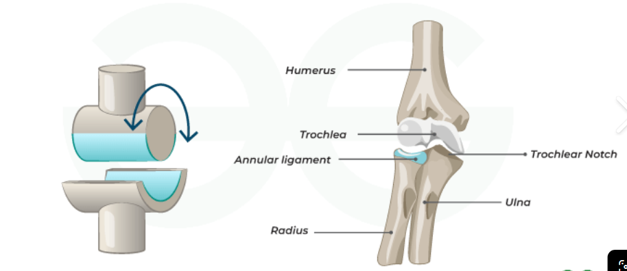
Hinge Joint
Synovial-A uniaxial synovial joint in which the convex surface of one bone fits into the concave depression of another, allowing movement in one axis.
Ex: elbow, knee. finger joints, toe joints. Only allow flexion and extension
Plane Joint
Synovial Joint--The simplest and least mobile uniaxial synovial joint, possessing flat articular surfaces that permit limited side-to-side gliding movement.
Ex: intercarpal jpints, intertarsal joints, acromiocavicular joint, between vertebrae

Multiaxial (Triaxial) Joint
A synovial joint in which bone moves in multiple planes or axes.

Biaxial Joint
A synovial joint in which bone moves in two planes or axes.

Uniaxial Joint
A synovial joint in which movement is restricted to a single plane or axis.

Dorsiflexion
Ankle joint flexion where the dorsum (superior surface) of the foot moves upward toward the anterior leg.

Plantar Flexion
Ankle joint extension where the dorsum of the foot is pointed inferiorly, such as standing on tiptoes.

Supination
Lateral rotation of the forearm such that the palm turns anteriorly or upward, returning to anatomic position.

Pronation
medial rotation of the forearm such that the palm faces posteriorly or downward.

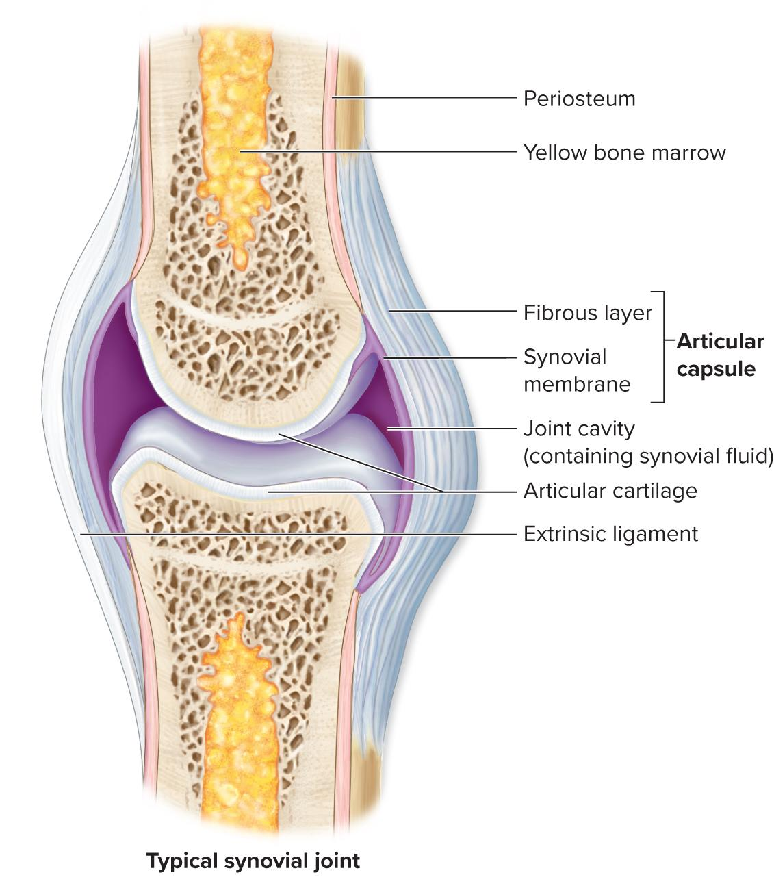
Synovial Joint
A freely mobile joint characterized by a fluid-filled joint cavity separating bone surfaces covered with articular cartilage and enclosed by a capsule.
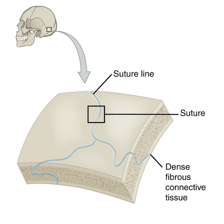
Fibrous Joint
articulating bones are held together by dense regular connective tissue without a joint cavity.
Ex: sutures, syndesmosis (between tibia and fibula), gomphosis (between teeth and their sockets in the jaw)
Cartilaginous Joint
articulating bones are united by hyaline cartilage or fibrocartilage and lack a joint cavity.
Ex:Synchondosis(hyaline cartialge) between the first rib and sternum, symphysis(fibrocartilage) between the two pubic bones

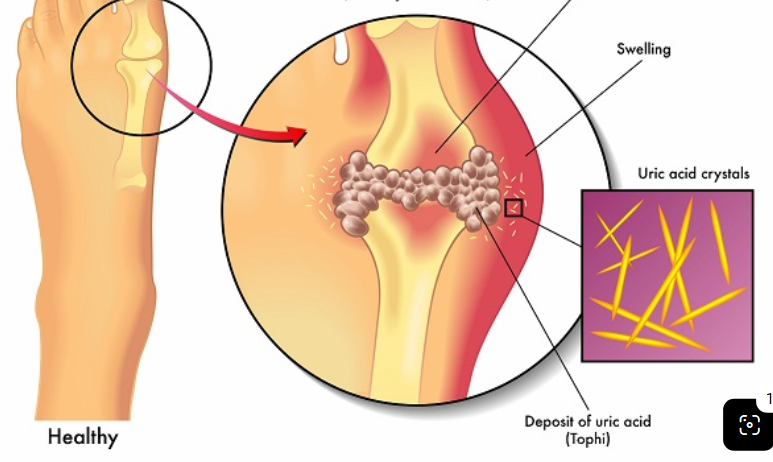
Gouty Arthritis
A form of arthritis caused by the deposition of excess uric acid crystals within joint cavities, most often occurring in middle-aged or older males.
Ankylosis
The abnormal stiffening and complete bony fusion of articulating bone ends occurring in the late stages of rheumatoid arthritis.

Rheumatoid Arthritis
An autoimmune disorder causing chronic inflammation of the synovial membrane, leading to cartilage damage, scar formation, and joint fusion.

Osteoarthritis
A degenerative joint disease typically associated with aging that is caused by mechanical wear and tear of articular cartilage.

Inversion
Movement occurring at intertarsal joints of the foot that turns the sole medially inward.

Eversion
Movement occurring at intertarsal joints of the foot that turns the sole laterally outward.

Supination
Lateral rotation of the forearm such that the palm turns anteriorly or upward, returning to anatomic position.

Axis (C2)
The second cervical vertebra, distinguished by the dens (odontoid process) that serves as a pivot point for the atlas to permit shaking the head 'no.'
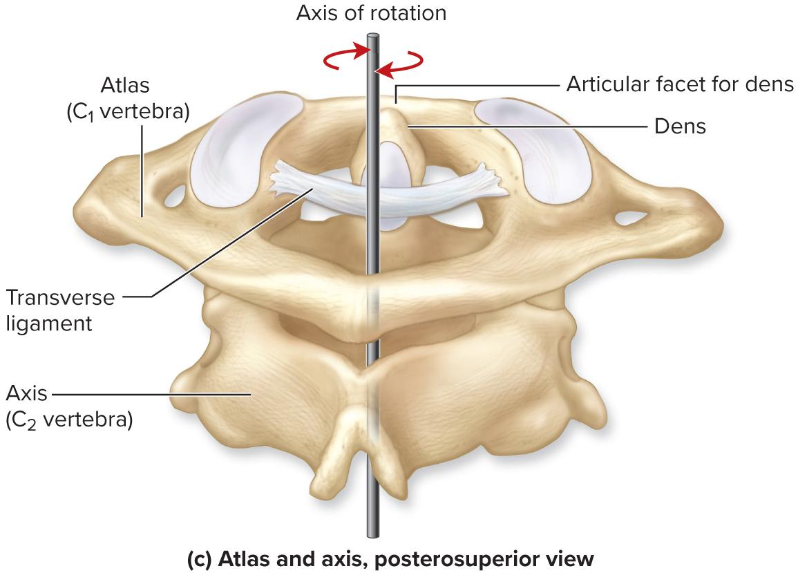
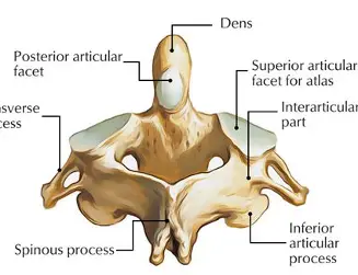
Dens
A prominent peglike projection produced by the fused vertebral body of the axis (C2) that acts as the pivot for lateral skull and atlas rotation.
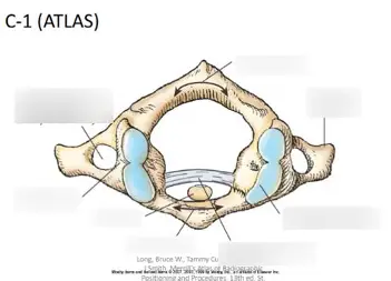
Atlas (C1)
The first cervical vertebra, which lacks a body and spinous process and features superior articular facets that articulate with occipital condyles to permit nodding 'yes.'
Nasal conchae
increase airflow turbulence through nasal cavity so air can be adequately moistened and cleaned by nasal mucosa. Reclaims moisture
Cribriform plate
Contains foramina for passageway of olfactory nerves
Ethmoidal labyrinths
contains ethmoid sinuses and nasal conchae
Sphenoid bone
houses the sphenoidal sinuses and lightens bone, moistens inhaled air, and gives resonance to voice. Protection to the pituitary gland.
Maxillary Sinus
one of the four pairs of paranasal sinuses
Location: They are air-filled chambers situated within the maxilla (the cheek region, directly inferior to the eye orbits and lateral to the nasal cavity).
Functions: Like all paranasal sinuses, they are lined with mucous membranes that help warm and humidify inhaled air, lighten the weight of the skull bones, and provide resonance to the voice.
Frontal Sinuses
The paranasal sinus located within the frontal bone superior to each orbit and deep to the superciliary arches that help to lighten the skull, humidify inhaled air, and enhance voice resonance.
Ethmoidal sinuses
A group of small interconnected air cells situated in the ethmoid bone between the orbits and the upper nasal cavity.They assist in moistening and warming inhaled air, lighten skull weight, and contribute to voice resonance.
Sphenoidal sinus
The paranasal sinus situated within the body of the sphenoid bone, positioned posterior to the eye orbits and inferior to the sella turcica. It plays a role in lightening the skull, humidifying inhaled air, and contributing to voice resonance.
Cranial sutures
Immovable joints that form rigid boundaries between cranial bones, functioning to secure the skull bones firmly together and protect the underlying brain.
Coronal suture
The immovable joint extending laterally across the superior surface of the skull that unites the anterior frontal bone with the posterior parietal bones.
Sagittal suture
The immovable joint extending along the superior midline of the skull between the coronal and lambdoid sutures, joining the right and left parietal bones
Lambdoid suture
The immovable joint extending across the posterior skull surface that articulates the two parietal bones with the occipital bone.
Squamous suture
The immovable joint located on each lateral side of the skull that articulates the temporal bone with the parietal bone.
Carpals
wrist bones, 8 bones per wrist. Permits multidirections gliding.
Tarsals
ankle bones, 7 bones per ankle, sturdy to bear body weight and form arches of the foot.
Numbering metacarpals and metatarsals
digit 1 starts on the lateral side of the hand (thumb)but the medial side of the foot(big toe)
Long bone classification
Long bones are defined as bones that are longer than they are wide, consisting of an elongated central shaft (diaphysis) and two distinct terminal ends (epiphyses). 22 long bones
Articulations of the Knee Joint
The knee is composed of two distinct articulations: - Tibiofemoral joint: between the femoral condyles and tibial condyles. - Patellofemoral joint: between the patella and patellar surface of the femur.
Patellofemoral Joint
The articulation within the knee joint formed between the posterior aspect of the patella and the smooth patellar surface on the anterior distal femur.
Bones Comprising the Elbow Joint
The humerus, radius, and ulna, all enclosed within a single articular capsule.
Articulations of the Elbow Joint
The elbow is composed of two distinct articulations: - Humeroulnar joint: between the trochlea of the humerus and the trochlear notch of the ulna. - Humeroradial joint: between the capitulum of the humerus and the head of the radius.
Bony Landmarks of the Humeroulnar Joint
The pulley-shaped trochlea of the humerus fits snugly into the deep trochlear notch of the ulna to allow hinge-like flexion and extension.
Bony Landmarks of the Humeroradial Joint
The rounded, marble-like capitulum of the humerus articulates with the cup-shaped, superior surface of the head of the radius.
Tradeoff between joint mobility and stability
The anatomical principle that as a joint's range of motion (mobility) increases, its structural stability decreases, making highly mobile joints more susceptible to dislocation and injury, whereas stable joints sacrifice movement for support.
Fibrocartilage
durable, dense form of cartilage packed with thick bundles of parallel collagen fibers. It is specialized to withstand severe compressive stress and shearing forces, serving as a heavy-duty shock absorber and stabilizing tissue in joints subjected to constant mechanical load.
Intervertebral Discs
Absorb and distribute compressive axial forces during walking, jumping, and running.Allow slight, cumulative flexibility, enabling the spinal column to bend anteriorly, posteriorly, and laterally.
Pubic Symphysis
The cartilaginous joint located along the anterior midline of the pelvis, uniting the medial symphysial surfaces of the left and right pubic bones (ossa coxae).Absorbs shearing and tensile stresses generated between the two halves of the pelvis during upright posture and locomotion.
Menisci
: C-shaped, wedge-like pads of fibrocartilage positioned on the superior articular surface (tibial plateau) of the tibia within the knee joint.Cushion the knee joint against repetitive impact during locomotion and aid in joint lubrication.
Flexion and Extension
bending the elbow, flexing the forearm at the elbow, or tilting the head forward toward the chest
Abduction and Adduction
lifting the upper limb laterally away from the torso, or spreading the fingers apart away from the middle finger
Lateral Flexion
cervical and lumbar regions
Medial (Internal) and Lateral (External) Rotation
rotating the humerus so the forearm points toward the abdomen
Circumduction
combines flexion, abduction, extension, and adduction in order.such as the head of the humerus in the glenoid cavity or the femur in the acetabulu
Tendons
Connects a muscle to a bone (or in some cases, muscle to muscle/fascia).
Ligaments
Connects a bone to another bone across a joint.
Synovial Joint
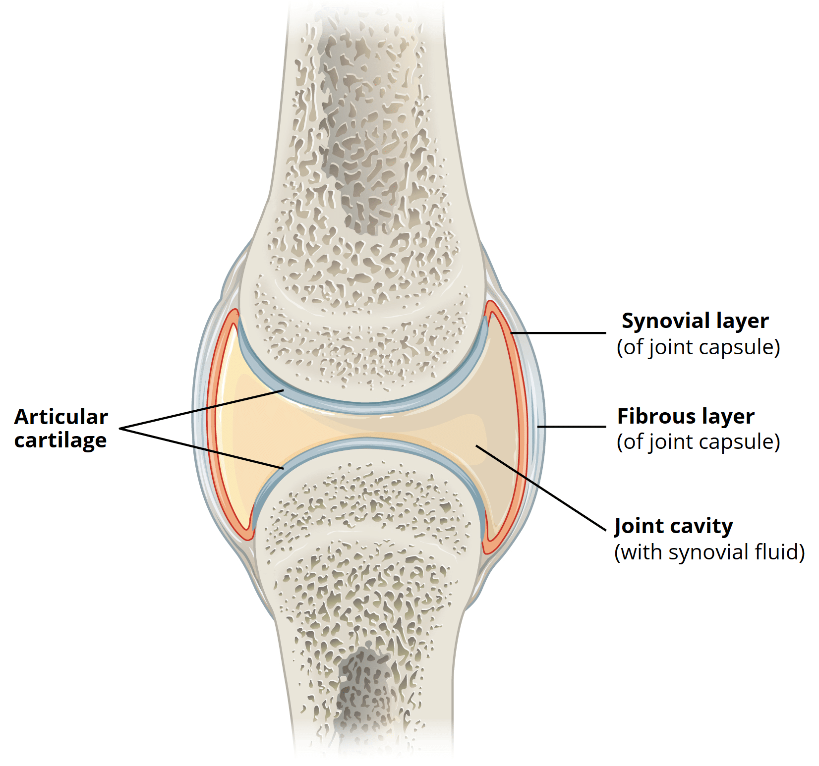
A freely mobile joint characterized by a fluid-filled joint cavity separating bone surfaces covered with articular cartilage and enclosed by a capsule.

Diarthrosis
A functional classification denoting a freely mobile joint; includes all synovial joints.

Articular Capsule
A double-layered envelope enclosing a synovial joint cavity, consisting of an outer fibrous layer of dense connective tissue and an inner synovial membrane.
Synovial Membrane
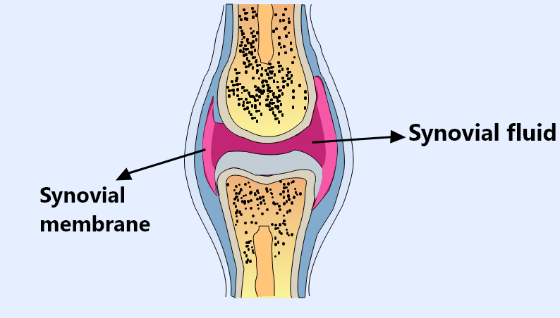
The inner layer of an articular capsule, composed predominantly of areolar connective tissue, that covers internal joint surfaces and produces synovial fluid.

Synovial Fluid
A viscous, oily substance within the joint cavity that lubricates articular cartilage, nourishes chondrocytes, and cushions against shock. Lubricates teh ends of long bones
elbow
hinge joint composed of two articulation. Humeroulnar joint with teh trochlea notch of the ulna articulating with trochlea of humerus. Humeroradial joint with capitulum of humerus articulating with head of radius.
knee
largest and most complex diarthrosis. Tibiofemoral joint- between condyles of femur and condyles of tibia. Patellofemoral joint- between patella and patellar surface of femur.