Anatomy Test 2 Prep! yayayayaya
 Call Kai
 Learn
Learn Practice Test
 Spaced Repetition
 Match
Match Flashcards
 Knowt Play
1/117

There's no tags or description
Looks like no tags are added yet.
Name | Mastery | Learn | Test | Matching | Spaced | Call with Kai | Chat |
|---|
No analytics yet
Send a link to your students to track their progress
118 Terms
What is the function of the integumentary system
To protect and insulate the body
What are the TWO PARTS of the integumentary system
The cutaneous membrane and accessory structures
What are accessory structures
Hair, nails, exocrine glands
What do melanocytes secrete?
Melanin
What is melanin, what does it do?
Melanin is a pigment that adds the brown/tan color to skin. It protects us from UV rays
What do melanocytes look like?
Octopi- the central part forms the melanin, the branches spread it out

What, other than melanocytes, can give skin its pigment?
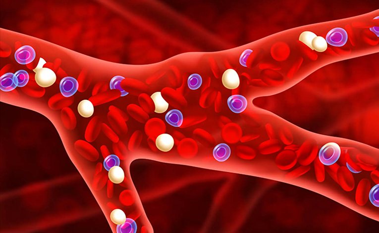
Blood can give a pink/red color (sometimes blue too)
What are the THREE big layers for the cutaneous membrane
Epidermis on top, then Dermis, and then the Hypodermis/Subcutaneous layer

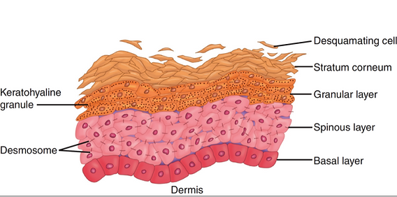
What are the layers of the Epidermis? (top-bottom)
Stratum Corneum, Stratum Lucidium, Stratum Granulosum, Stratum Spinosum, Stratum Basalae
Mnemonic is: Can Lucy Give Some Blood?

Describe the stratum corneum
Top layer of flat, dead cells that interlock with keratinocytes. It’s a dry, protective barrier.

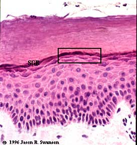
Describe the stratum granulosum
Appears as a dark line on the slide, had keratinocytes that make keratin.
Describe the stratum basale
The lowest layer of the epidermis that has constant cell division, is stratified columnar/cuboidal (Spearing says squamous???) epithelium, and is attached to the basal lamina
What does keratin do?
It is made my keratinocytes and it strengthens cells and adds water resistance
What happens when stress is put on the epidermis?
With more stress, the stratum corneum grows thicker. With less stress, the stratum corneum grows thinner. This is why old ppl often have thinner skin.
What are the two layers of the Dermis
The papillary layer and the reticular layer

What is in the papillary layer and what does it do?
It is mostly areolar connective tissue. It cushions, has a direct blood supply, and nerve endings.

What is the reticular layer and what does it do?
It has the accessory structures. The reticular layer is full of collagen fibers, accessory stuctures (glands, errector pili, etc.), and nerve endings.
Whot iz collagen?
It is the protein for elasticity
What makes up the hypodermis/subcutaneous layer, and what does it do?
This layer is below the dermis and technically isn’t part of the skin. It has large blood vessels and is mostly made of subcutaneous adipose tissue (fat). This layer insulates, cushions, and contributes to secondary s3xu@l characteristics (makes ppl know if ur male or female, it’s driven by hormones like testosterone and estrogen).

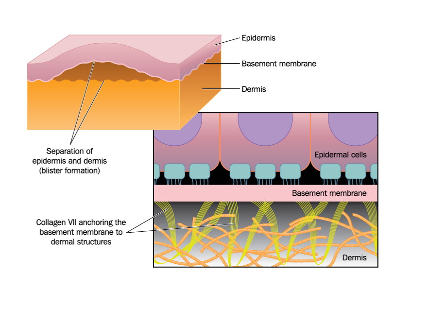
How do blisters form?
Stress hits the skin at an angle and causes the epidermis to buckle and separate from the dermis, the raised dermal cells die. Fluid rushes into the area and new layers eventually form. Stress can tear the healing dermis of a blister and fluid will rush out of the tear, this is prone to infection.
What and where are the epidermal ridges?
They’re on the inside of the hands and bottom of feet to form finger and toeprints. They develop randomly and are permanent in pattern once established. These ridges provide help with grip.

What is the function of hair?
It evolved to be an external insulator and is used for expression/communication (eyebrows).

What is hair?
A cylinder of keratin with melanin pigment that makes it any color from black to white/blonde.

How does gray and ginger hair form?
Gray hair- lack of melanin
Gingers- orange/red orange b/c of a genetic mutation (weasleys boi)

What is the hair papillae?
The living part of the hair where the keratin is produced, in the root. Has blood vessels.

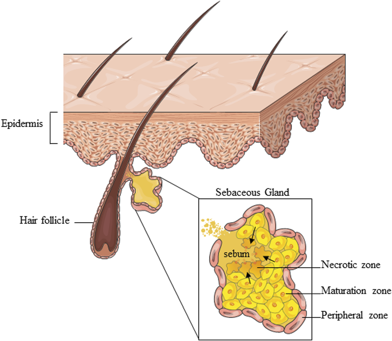
What is the sebaceous gland?
It makes sebaceous fluid to maintain the hair structure.
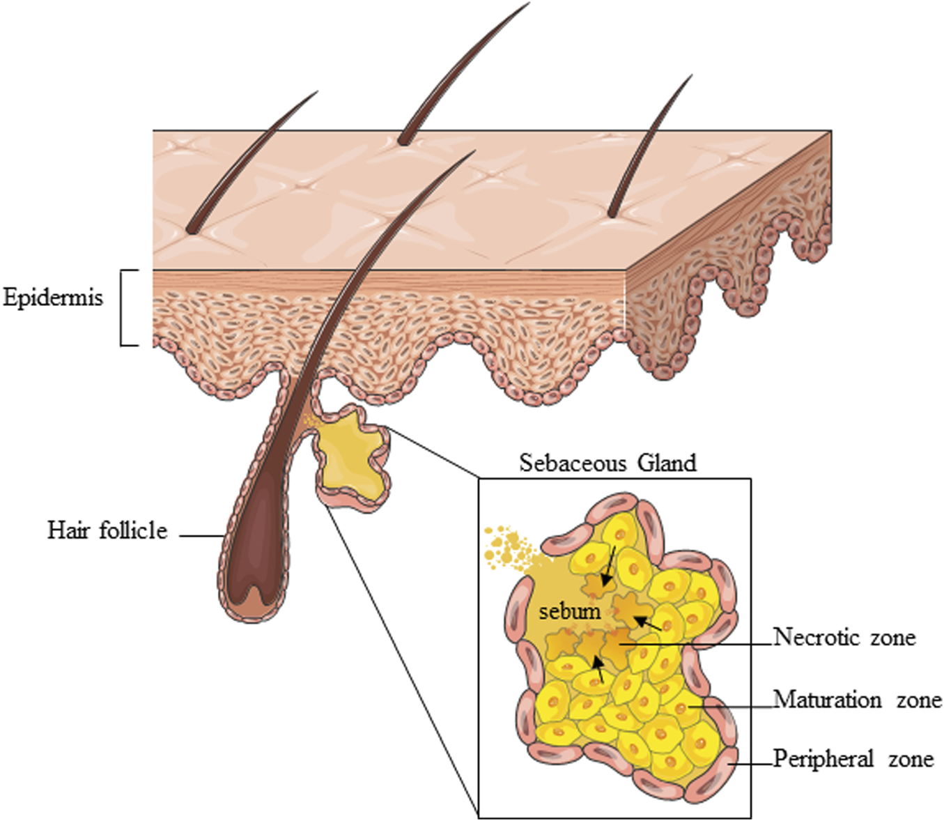
Why do split ends form?
Hair disintegrates b/c no have sebaceous fluid, so the keratin protein is broken down.

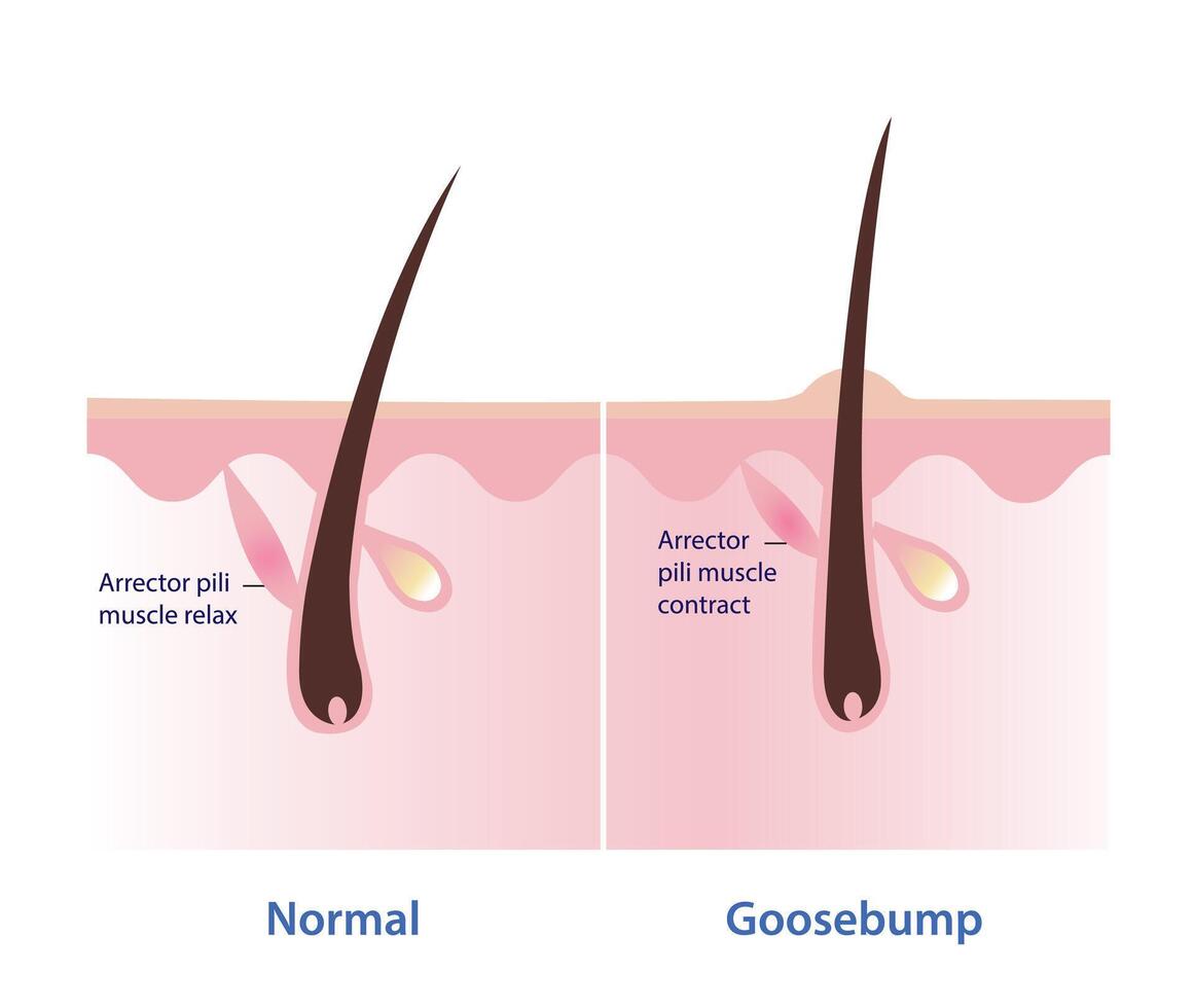
What is the arrector pili muscle?
It is connected to the hair and contracts to make goosebumps when you’re cold or afraid. Traps hair against the surface of the skin.
How does hair grow
Hair grows continuously and dies off and grows back. men can have hair loss tho

What are nails?
A keratin sheet that may exist to act as a backstop for the finger.

What is the nail root?
Where the sheet is formed.
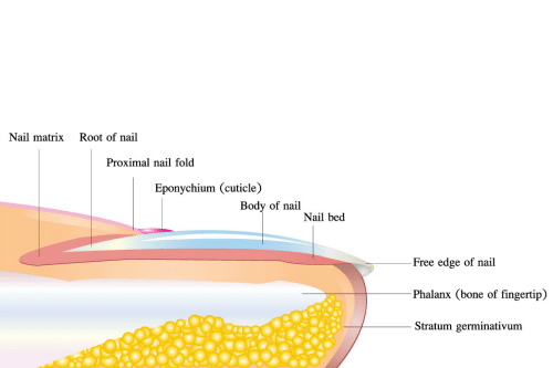
What is the nail body?
It is pressed tight to the skin, it’s the main part in the middle u can see boioioioi

What is the free edge of the nail?
The distal part of the nail that is not as tight to the skin, white in color.

What is the lunula?
The white/lighter semicircle part at the base of the nail, not as attached to the skin either.

What are the lateral nail folds?
The parts on the sides as nail meets skin.

What is the proximal nail fold?
The fold at the base of the nail.

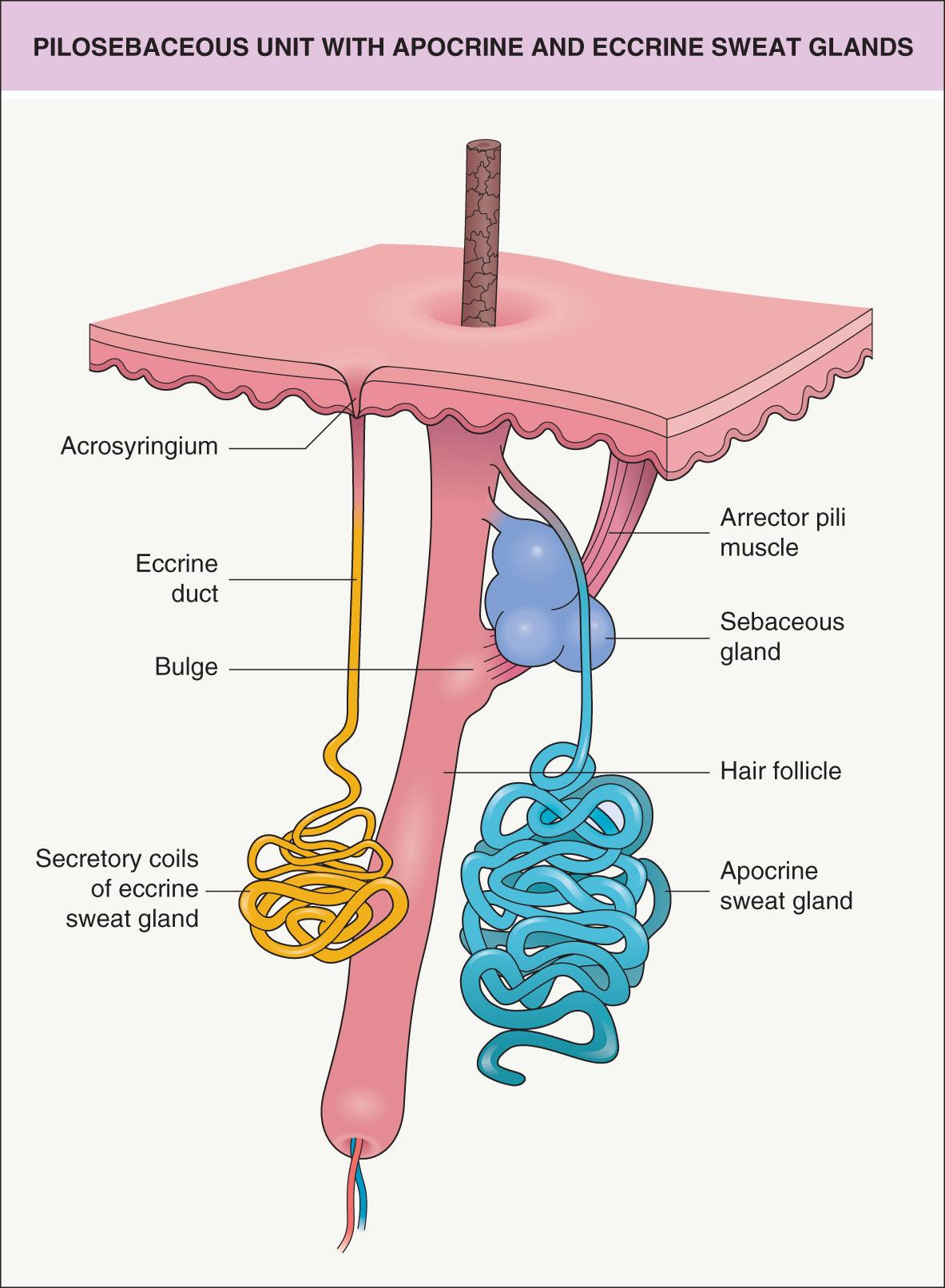
What are exocrine gland types and what do they do
Sebacceous and sweat glands. They secrete onto the surface of the skin

What do sebaceous glands secrete and where
They secrete sebaceous fluid to maintain keratin structure and provide a weak antibiotic effort. Secrete onto hair and skin surface- maintain dead keratinized cells so it don’t crack.
What are the types of SWEAT Glands?
Merrocrine and Appocrine
What are merrocrine (eccrine is the normal name tfreak) sweat glands for and where?
They’re over the entire body surface, secrete mostly water, give a weak antibacterial effort, adjust body temperature b/c of water’s high specific heat. Release onto skin with pores.

What are the 3 types of appocrine sweat glands
1.) Appocrine - pits and privates, controlled by puberty hormones, secretes odorous chemicals that may provide complex communications (pheromones)
2.) Mammary - breasts, secretes milk under proper hormonal conditions
3.) Ceruminous - auditory canals, earwax, traps pathogens like mucous does in mucosa
What are the two types of bone tissue?
Compact and Spongy
Describe compact bone.
Compressed bone for weight bearing. Made of the unit osteons that have concentric circles called lamellae.

what are Lacunae
Lacunae are where the osteocytes live

What are Canaliculi
They connect the osteocytes in the lacunae to the central canal so nutrients can pass through all the lamellae.

What is the central canal of an osteon
Where the blood vessels and nerves are

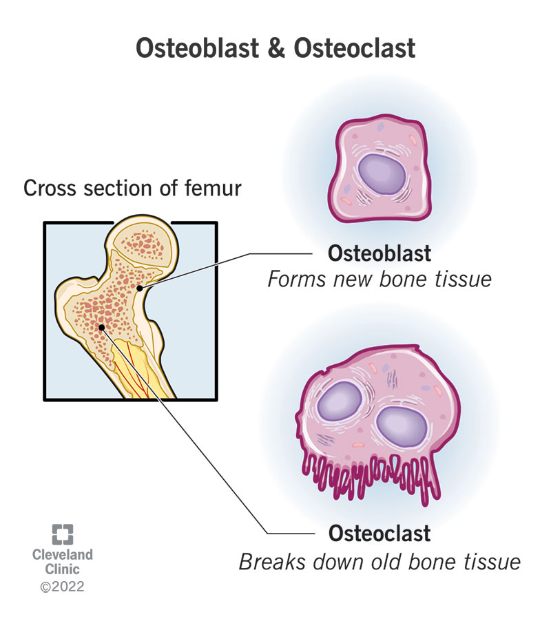
What is an osteoblast
It Builds bone tissue, it creates hydroxyapatite to make ya bone crystals.

Osteoclast
Disassembles the bone tissue
Osteocyte
The living cells of bones that keep it alive

What are the strings that make up the spongy bone
Trabeculae, and they line up with areas of highest stress, they can be easily disassembled, they have lamellae and canaliculi and osteocytes too

What surrounds the trabeculae of spongy bone
Red bone marrow

What is the medullary cavity
The space in the middle of a long bone where Yellow bone marrow is found and stores fat

Spongy bone/Red bone marrow location
The ends of long bones and in flat bones.
What does red bone marrow produce
RED BLOOD CELLS..
Where does endochondral ossification take place and what does if form
Inside cartilage to form long bones
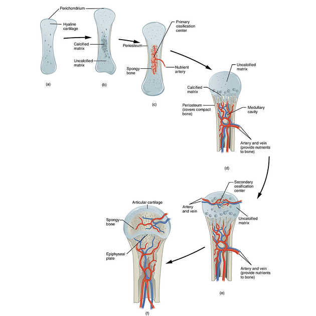
Explain endochondral ossification
Mesenchymal cells form into a long shaft of hyaline cartilage that grows until a blood vessel pierces the middle of the cartilage and primary ossification begins and the bone forms from the center out. Then, the epiphysis (ends) are pierced by secondary blood supplies and the bone grows from both ends inward. The bone meets and eventually fuse at the cartilage metaphysis.
Diaphysis
Shaft middle of long bone

Epiphysis
Ends of long bone

Metaphysis
Where the diaphysis and epiphyses fuse (growth/metaphyseal plate)

Where does intramembranous ossification occur and what does it form?
It occurs in flat bones (skull, ribs?). It forms flat bones and sutures
How does intramembranous ossification work?
Between layers of tissue, a mesenchyme clump forms until it runs into a blood vessel. From there lil spikes, spicules, grow outward from the central point. Spicules continue growing until they reach other bone’s spicules and meet to form a suture joint.

What is sesmoid ossification
When a bone forms inside of a tendon or ligament due to stress. EGZAMBL: Patella forms b/c of stress on patellar ligament. Some ppl also form extra bones on ball of foot metatarsals etc.

What happens when a bone breaks
The area is flooded with blood
The blood bulges and creates a fracture hematoma, which could tear the periosteum.
The periosteum repairs and you form disorganized bone tissue (spongy bone)
Bones begin to knit together
Can form external callus if the break is bad enough. (The bone is thicker there for a time)

Periosteum
Thin layer of tissue that surrounds the outside of the bone.

Endosteum
Thin layer of tissue on the inside of the medullary cavity so the yellow marrow doesn’t touch the bone.
Coronal suture
suture on top of head between frontal and parietal bones
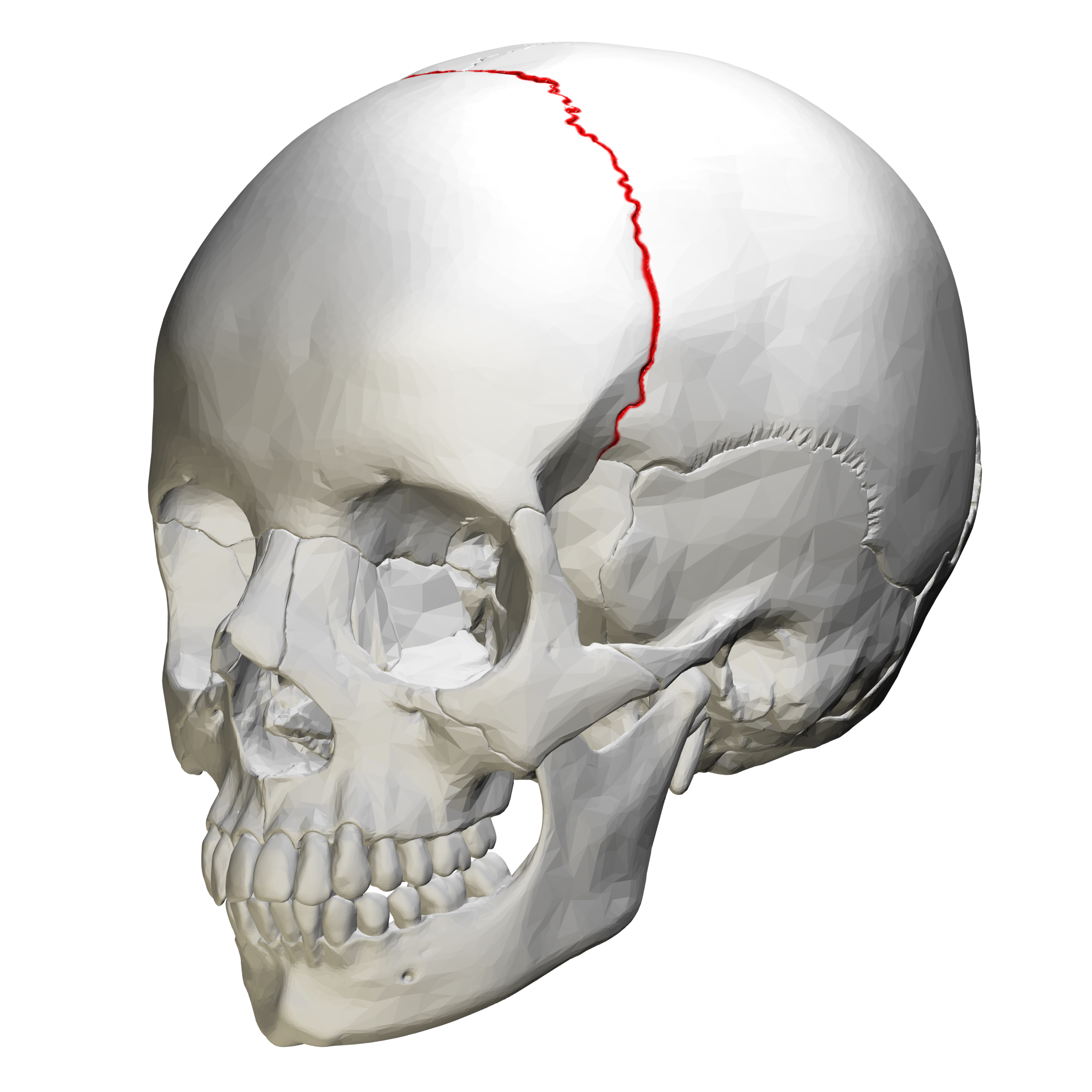
Sagittal suture
the suture on top of the head between the parietal bones


Lambdoid suture
suture in the back of the head that separates the occipital bone from the parietal bones
(worming/sutural bones develop here (???!))

Squamous suture
Sutures on sides that separate the temporal and parietal bones

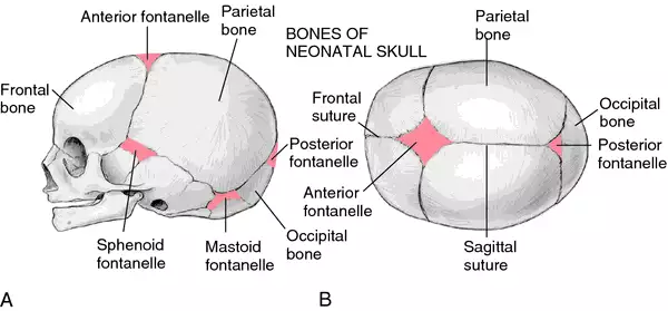
What are fontanelles
They’re the spaces in between the skull bones of a newborn baby. This is so the infant is flexible for birth and growth, they close up at around age 4. The anterior fontanelle is often called the soft spot of a baby.
why do we add heat and humidity to air?
So we don’t kill the lining of the lungs
HOW do we add humidity and heat to air?
Nasal conchae (inc surface area of nasal cavity) and nasal sinuses (holes in bones to increase the surface area)
Frontal sinuses (2)
in frontal bone, has pinhole sized openings

Maxillary sinuses (2)
in the maxilla and has a bigger opening
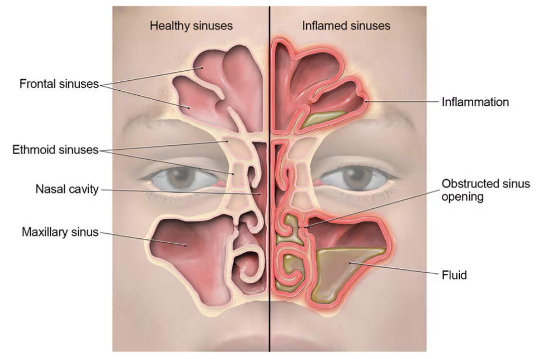
Sphenoid sinus (1)
in sphenoid bone

Ethmoid air cells (6)
In ethmoid bone

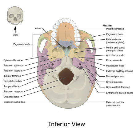
Foramen magnum
inferior skull hole for spine

2 Jugular foramen
inferior skull holes for the jugular veins, on sides of foramen magnum

2 carotid canals
Hole for carotid artery, circular, anterior to jugular foramens
Sella turcin
“Turkish Saddle”, in sphenoid bone, pituitary gland rests there

Cribiform plate
in ethmoid bone— holes for olfactory nerves

Lordosis
Concave to posterior side— lumbar and cervical experience good lordosis in moderation.

Kyphosis
concave to anterior side— sacral and thoracic experience good lordosis in moderation. Mothers have kyphosis when preggo becuzada bebe

Why does ur spine curve
As u grow it curves due to movement and gravity
Scoliosis
Lateral spine curvature, can use a brace to cure this sometimes

Slipped disc AAAHHhhhhhhhhhh!
The intervertebral disk is dislocated from btwn da vert., this is an issue if it hits a nerve root of the spinal cord bc it can cause pain or numbness.
+the discs are made of Fibrous Cartilage!

neural arch of vertebrae
lateral arch
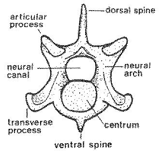
pedicle of vertebrae
connection part between centrum and posterior parts

centrum
circley body of the vertebrae

Superior and inferior articular processes
spikes on spine that articulate with other vertebrae
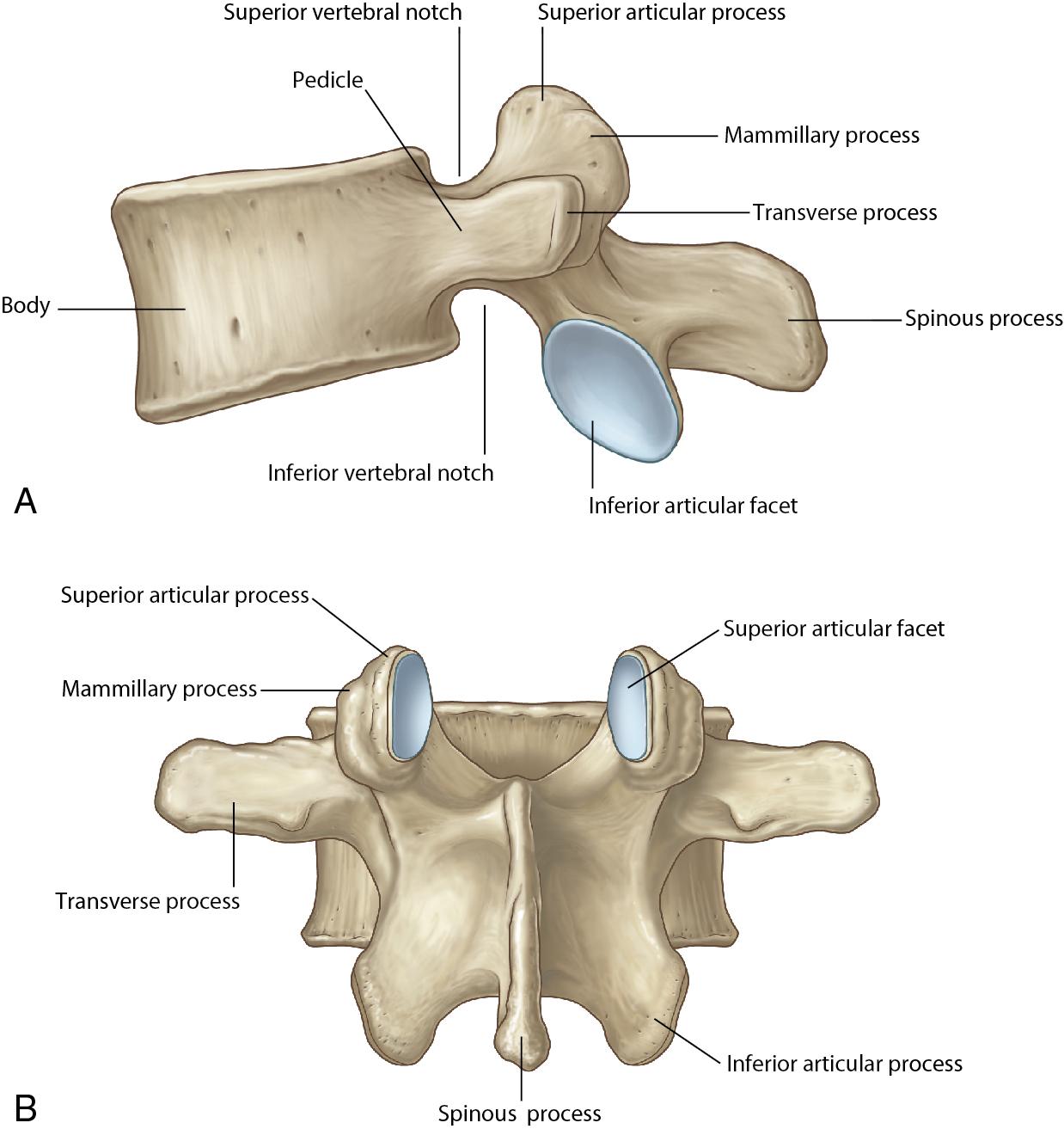
vertebral foramen
hole for spinal cord

Lamina of vertebrae
Connection parts of the processes

Cervical vert. names
C1-C7 (sup-inf)

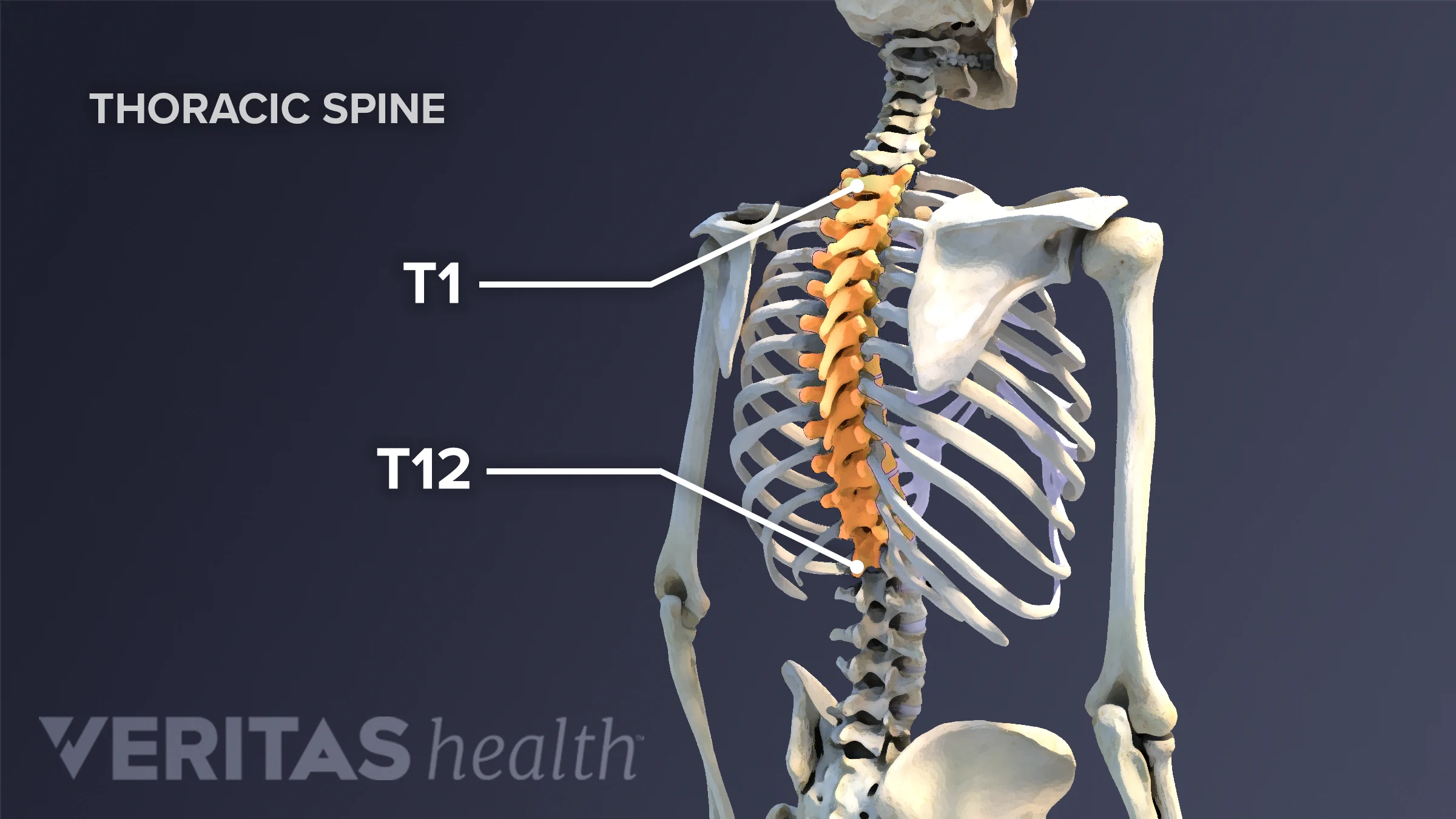
Thoracic
T1-T12
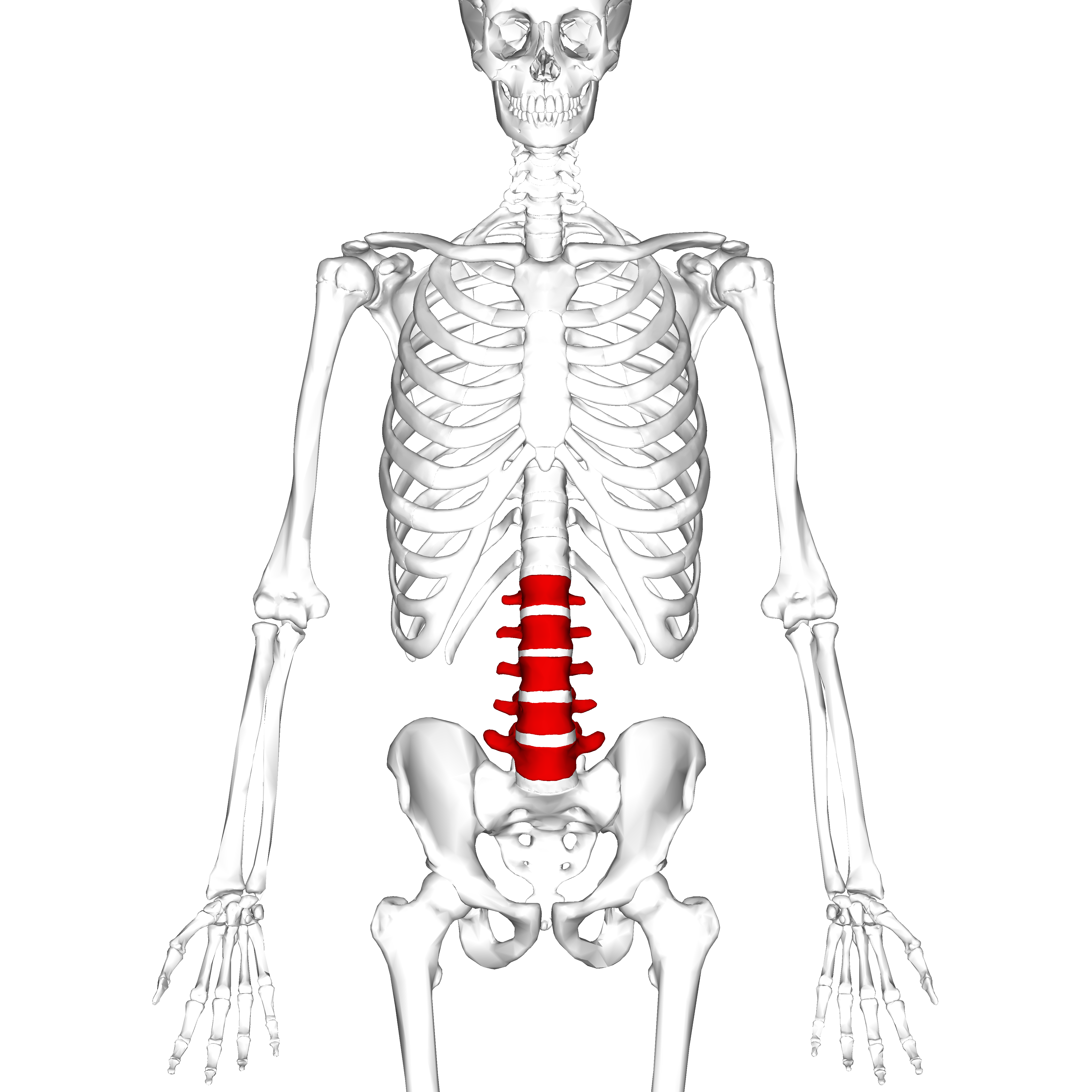
Lumbar
L1-L5
sacrum
fused bones, only L5-S1 junction issues
Coccyx
rarely, Co1-?

C1
Atlas that holds up the skull (aurafarming +1,000,000,000 aura), no centrum
C2
Axis, the pivot point that articulates with the atlas to rotate the skull L/R, has the dens/odontoid process to articulate (fuses with axis?), no centrum

Proximal rib parts:
Head: junction between the arch and body of vertebrae
Neck
Tubercle: transverse process
