Wake PA Unit 3: (PC) Comprehensive Eye Exam
 Call Kai
 Learn
Learn Practice Test
 Spaced Repetition
 Match
Match Flashcards
 Knowt Play
1/93

There's no tags or description
Looks like no tags are added yet.
Name | Mastery | Learn | Test | Matching | Spaced | Call with Kai | Chat |
|---|
No analytics yet
Send a link to your students to track their progress
94 Terms
What is the sclera?
outermost covering of the eyes; white and opaque
What attaches to the sclera?
extraocular muscles
What is the cornea?
continuation of the sclera over the anterior surface of the eye; transparent dome
What is the function of cornea?
focuses incoming visual light stimuli prior to projection onto the retina
Name the two major chambers of the eye
anterior chamber
posterior chamber
What is the anterior chamber?
anatomical space between the cornea and iris
What is the posterior chamber?
anatomical space posterior to the anterior chamber
What kind of fluid fills the anterior chamber of the eye?
aqueous humor
What is aqueous humor?
produced by cells within the ciliary body, this is a liquid that establishes pressure and maintains the integrity of the eye in the anterior chamber
What is vitreous humor?
transparent gel similar to aqueous humor but with thicker consistency that maintains the shape of the eye and exerts pressure on the retina's adherence to the choroid
What is the iris?
circular ring with musculature responsible for pupillary contraction and dilation
What is the role of the ciliary muscles?
muscles which bend the physiological lens to refract light at different angles on the posterior retina, allowing for accommodation for near vs. distance vision
What is the role of the suspensatory ligaments?
suspend the physiologic lens in place and connect to ciliary body
What is the lens?
biconvex transparent discus that is made of proteins
What is the pupuil?
circular anatomical absence of the iris that changes diameter with light exposure and accommodation
What is the retina?
innermost lining of the posterior eye; richly vascularized and innervated tissue that contains rods and cones
What is the role of cones in the eye?
receive color information (vision)
What is the role of rods in the eye?
responsible for vision at low light levels (no input on color vision)
What is the fovea?
retinal depression adjacent to the optic disc which contains the highest concentration of photoreceptors and is responsible for maximal visual acuity
What is the optic disc?
physiologic blind spot and the location of the origination of the optic nerve and vasculature supply
What is the optic nerve?
axons of retinal ganglion cells combine at this point to transmit signals from the retina to the brain
List the landmarks along the pathway of light
light source, cornea, pupil, lens, retina, optic nerve, optic chiasm, thalamus, occipital lobe
What are some basic history questions you should ask related to the eye
1. any changes or difficulty in vision (if so, is it worse up close or from a distance)?
2. is the vision loss unilateral or bilateral?
3. any pain associated with vision loss?
What is hyperopia?
difficulty with close-vision (far-sighted)
What is presbyopia?
age-related hyperopia. Patient is extending arms to attempt to read up close text
What is myopia?
difficulty with distance vision (near sighted)
What is an astigmatism?
irregularities in the lens or curvature of the cornea causing the lens to not focus light evenly onto the retina
List some painless causes of acute unilateral vision loss
- vitreous degeneration from diabetes or trauma
- macular degeneration
- retinal detachment
- retinal vein or artery occlusion
List some painless causes of bilateral vision loss
- medications (anticholinergics)
- cataracts
- macular degeneration
List some painful causes of acute unilateral vision loss
-corneal ulcer
-uveitis
-hyphema
-acute angle closure glaucoma
What are some disorders related to painful bilateral vision loss?
-acute angle closure glaucoma
-chemical or radiation exposure
What is hyphema?
bleeding in the anterior chamber of the eye
What is the etiology of hyphema?
-Result of blunt or penetrating injury to the eye
Children: sports injuries,
Teenagers & Adults: assault, air bag deployment, projectiles
-Defined as grossly visible blood in the anterior chamber
-WSpontaneous hyphema with minor trauma: diabetes, thrombocytopenia, Von willebrand disease, warfarin/aspirin use

What is the clinical presentation for hyphema?
-Vision loss with accompanied eye pain
-Nausea/vomiting, photophobia, anisocoria, elevated IOP
-Presence of blood layering in anterior chamber with penlight exam while patient upright
-CT non-contrast of orbits if concern of open globe

What are some general questions to ask a patient with a vision problem?
1. description of pain
2. are you a contact lens wearer?
3. recent eye, facial, or dental surgery?
4. any eye trauma (but this does not always mean eye pathology)
Name the tools you would use to test vision
Snellen Chart
Rosenbaum chart

How do you score a Rosenbaum Chart?
Jaeger 1+ through 16
Patient holds chart at distance they would to read (14-15 inches)
How do you score a Snellen Chart?
20/20 ratio is "normal"
If patient is standing at 20 feet away looking at the chart and they can only ready the 20/60 line, someone with normal vision standing 60 feet back from the chart could read the same line just as well.
What are the criteria for being legally blind?
- reduced central visual acuity of 20/200 or less in your best eye with use of the best eye glasses to correct your eyesight
- limitation of your field of view such that the widest diameter of the visual field in your best eye subtends an angle no greater than 20 degrees
What is a pupillary direct reaction?
pupillary constriction in the same eye the light is pointed into
What is a pupillary consensual reaction?
pupillary constriction in the opposite eye that the light is pointed into
What disease is associated with an elevated intraocular pressure?
glaucoma
What is a normal swinging light test?
swinging the light between both eyes and seeing repeated constriction of the pupil when light is directed in it
What is an abnormal swinging light test? What can cause an abnormal swinging light test?
abnormal test = "good" pupil will constrict but "bad" eye will not when light is shone into the good eye.
CVA, head trauma, multiple sclerosis
What are the "vital signs" of the eye?
1. vision
2. pupils
3. pressure
What is a tonometry pen?
applanation "flattening" tonometry which converts surface tension into an electrical signal corresponding to IOP in mm HG
What is a normal intraocular pressure?
10-21 mmHg
What is Goldmanns applanation tonometry?
based on principle that force to flatten area of cornea divided by area of flattening equals IOP
- gold standard and most acurate
Name the anterior pathway defects
glaucoma, optic neuropathy, optic neuritis, and glioma
Name the posterior pathway defects
stroke, chiasmal tumors
What is the anterior visual pathway?
origin of the optic nerve to the lateral geniculate nucleus
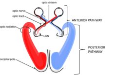
What is the posterior visual pathway?
from the lateral geniculate nucleus to the occipital lobe in the primary visual cortex
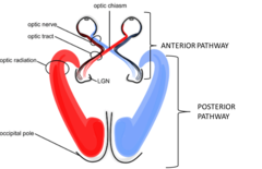
What is homonymous hemianopsia?
lesion of the optic tract interrupts fibers originating on the same side of BOTH eyes

What is bitemporal hemianopsia?
a lesion at the optic chiasm interrupts fibers crossing over to the opposite side

What is homonymous quadrantic defect?
partial lesion of the optic radiation in the temporal lobe

What are you observing during inspection of the eye?
1.Periorbital structures
2.Lashes/lids
3.Lacrimal glands
4.Conjunctiva & sclera
5.Cornea
6.Anterior chamber/Iris
7.Lens
8.Vitreous
9.Retina

What is ectropion?
condition in which the margin of lower lid is turned outward ("sagging")

What is entropion?
condition in which the margin of lid is turning inward (may cause irritation of the sclera by the eyelashes)

What is hordeolum?
also known as a stye; painful, tender, red and/or yellow/white pimple infection in a gland at the margin of the eyelid

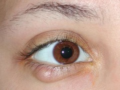
What is chalazion?
subacute, nontender, non-infectious usually painless nodule caused by a blocked meibomian gland (often occurs after stye is no longer infected but gland is still blocked)
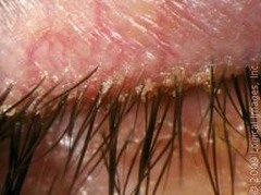
What is blepharitis?
infection/inflammation of eyelids that presents with eye irritation and redness/irritation of eyelids
What is ptosis?
drooping of the upper lid

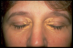
What is xanthelasma?
slightly raised, yellowish, and well-circumcized plaques that appear along the nasal portions of the eyelids
What is the etiology of viral conjunctivitis?
commonly caused by adenovirus, frequently sourced back to swimming pool exposure
What is the clinical presentation of viral conjunctivitis?
superficial pain, itching, foreign body sensation, watery discharge, erythema, typically bilateral
What is the etiology of bacterial conjunctivitis?
s. aureus, S. pneumoniae, H. flu (usually transmitted by direct contact and autoinoculation)
What is the clinical presentation of bacterial conjunctivitis?
purulent discharge, lid crusting, chemosis, mild superficial pain/itching, no visual change
What is dendritic staining?
eye drops that illuminates under blue light as green in the case of a zoster infection (a test you would do if they have shingles or another zoster infection and they have eye irritation)

What is the clinical presentation of allergic conjunctivitis?
conjunctival erythema of both eyes, cobblestone mucosa to inner/upper eyelid on eversion, symptoms of itching/tearing/redness

What is the etiology of subconjunctival hemorrhage?
- common in patientes >80 years old (daily aspirin)
- associated with HTN, minor trauma, contact lenses
- other causes: diabetes, violent coughing, vomiting, coagulopathy

What is a pinguecula?
yellowish triangular node in the bulbar conjunctiva on either side of the iris; frequently appears with age

What is a pterygium?
triangular thickening of the bulbar conjunctiva that slowly grows across the cornea and may encroach on pupil; often seen in a patient with frequent dust exposure

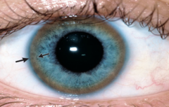
What is a corneal arcus?
thin gray/white circle around the edge of the cornea; normal in aging but can indicate hyperlipoproteinemia in younger patients

What is a Kayser=Fleischer ring?
golden/red/brown ring from copper deposition in individuals with Wilson's disease
What is the etiology of acute narrow angle closure glaucoma?
- Often precipitated by pupillary dilation which further closes the angle of aqueous humor drainage at the trabecular meshwork and canal of schlemm.
-Most common in individuals with narrow angle/large lens - such as the elderly, far-sighted, and asians
-Leading cause of preventable blindness in the US
What is the clinical presentation for acute narrow angle closure glaucoma?
headache, nausea/vomiting, blurry vision with halos around lights, peripheral vision loss, conjunctival erythema, corneal epithelial cloudiness/edema, increased IOP, mid-dilated and non-reactive/fixed pupil
What is the normal result of the corneal light reflex test?
Patient is directed to look at a penlight 2 feet away from eyes. Provider aligns eyes with light source and compares the position of the light as reflected yb the cornea of each eye.
light reflected on each cornea symmetrically and in the same relative position to the pupil
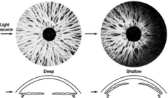
What is the Anterior Chamber Depth test? What is a normal/abnormal result?
shine pen light from temporal side of each eye at a 90 degree angle. evaluate for a crescentic shadow on the medial side of the iris?
normal: flat iris results in an open angle of the cornea causes full illumination.
abnormal: bowing abnormality of the iris forming a narrow angle with cornea creating a crescentic shadow
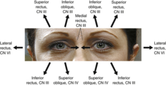
What are the six cardinal fields of gaze?
What is convergence insufficiency disorder? How do you test for it?
symptoms: headaches, eyestrain, double vision, blurry vision
- patient follows finger as you move closer to bridge of nose
- normal convergence: 5-10 cm from bridge of nose

What structures are examined in the fundoscopic exam?
lens, vitreous, and retina
Be familiar with the steps for using the ophthalmoscope
1.Dim room lights, have patient focus straight ahead behind you
2.Turn lens to the 0 diopter (adjust diopter as needed to correct your vision)
3.Hold ophthalmoscope in your RIGHT hand and use your RIGHT eye to examine the patient's RIGHT eye. (opposite to evaluate patients left eye)
4.Brace ophthalmoscope against your brow and gaze through aperture
5.Position yourself 15 inches away from patient and slightly lateral to patients' line of vision
6.Place your thumb of opposite hand on patient's eyebrow
7.Move slowly inward keeping red reflex in your gaze until you almost touch the patient's eyelashes with the ophthalmoscope
How do you differentiate between arteries and veins when looking at the retina?
arteries = light red in coloration, smaller than veins
veins = dark red in coloration, larger than arteries
How do retinal arteries appear in a patient with hypertension?
arteries show areas of narrowing with a narrow light reflex

What is "copper wiring" when looking at retinal arteries?
arteries close to the optic disc become full and tortuous developing an increased light reflex with a copper color
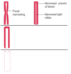
What is "silver wiring" when looking at retinal arteries?
portion of a narrowed artery develops an opaque wall where no blood is visible within it

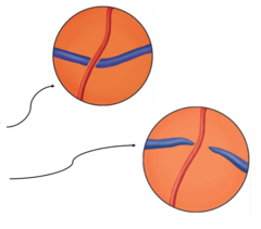
What is AV nicking?
vein appears to stop abruptly on either side of the artery
(common in hypertensive patients where arterial walls lose transparency)

What is AV tapering?
vein appears to taper down in diameter on either side of the artery
(common in hypertensive patients where arterial walls lose transparency)
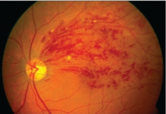
Describe the appearance of retinal hemorrhages
small, flame shaped, red streaks in the fundi
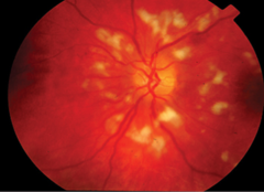
Describe the appearance of soft exudates/ cotton wool patches
white/gray ovoid lesions with soft borders
Describe the appearance of hard exudates on retinal exam
creamy yellow lesion with well-defined borders

Describe the appearance of drusen on retinal exam
yellow round spots/freckles with soft edge, typically numerous

What is strabismus?
abnormal alignment of the eyes
What is nystagmus?
involuntary eye movement