Quiz 4 chapter 5 vocab
 Call Kai
 Learn
Learn Practice Test
 Spaced Repetition
 Match
Match Flashcards
 Knowt Play
1/19

There's no tags or description
Looks like no tags are added yet.
Name | Mastery | Learn | Test | Matching | Spaced | Call with Kai | Chat |
|---|
No analytics yet
Send a link to your students to track their progress
20 Terms

Dens Evaginatus
An accessory enamel cusp found on the occlusal tooth surface
Most often seen on mandibular premolars
May cause occlusal problems
CLINICALLY: small, rounded lobe of enamel on the occlusal surface of a mandibular premolar between the buccal and lingual cusps
NO TREATMENT NEEDED

Dens Invaginatus
Occurs when the enamel organ invaginates into the crown of a tooth before mineralization
Radiographically, it appears as a tooth-like structure within a tooth
Vulnerable to caries, pulpal infection, and necrosis
ALSO KNOWN: dense in dente
Coronal dens invaginatus is seen most often in permanent maxillary lateral incisors
Radicular dens invaginatus is RARE
A nonvital dens in dente may be treated endodontically.
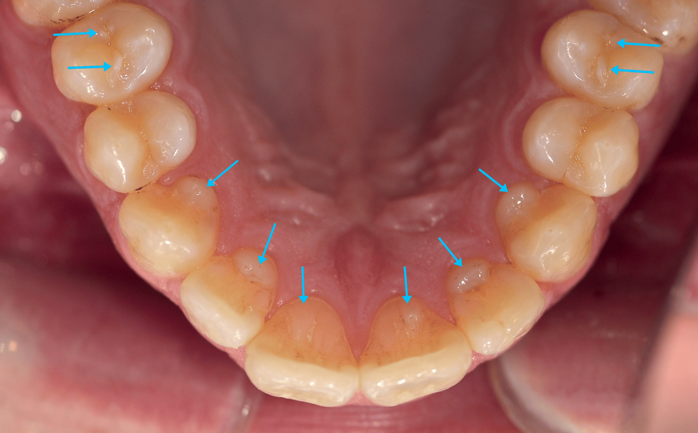
Talon Cusp
an accessory cusp located in the cingulum area of a maxillary or mandibular permanent incisor
Contains a pulp horn
May interfere with occlusion
It is said to resemble an eagle’s talon
If pulp tissue is present, endodontic therapy would be necessary

Enamel Pearl
a small, spherical enamel projection on a root surface
usually found on maxillary molars
Radiographically, it appears as a small, spherical radiopacity
Removal may be necessary if periodontal problems occur in the furcation
It is attached to cementum near the root bifurcation or trifurcation area
Uncommon finding; usually no treatment is needed

Gemination
a single tooth germ attempts to divide in two
appears as two crowns joined together by a notched incisal area
Radiographically, usually one single root and one common pulp canal exist
The patient has the full complement of teeth
TREATMENT: alteration of the tooth so that is resembles a normal tooth in size and shape

Fusion
the union of two normally separate adjacent tooth germs
Appears as a single large crown that occurs in place of two normal teeth
Radiographically, either separate or fused roots and root canals are seen
The patient is usually short one tooth
To differentiate fusion from gemination, the teeth must be counted.
Treatment involves alteration of the size and shape of the tooth and may involve replacement of one of the fused teeth

Macrodontia
one or more teeth are larger than normal
True generalized macrodontia (seen in cases of pituitary gigantism)
Relative generalized macrodontia (large teeth in a small jaw)
Macrodontia affecting a single tooth (may be seen in cases) and (may be seen in cases of facial hemihypertrophy)

Simple Bone Cyst (hemorrhagic bone cyst)
a pathological cavity in bone that is not lined with epithelium
may be associated with trauma
Radiographic: a well-defined uniocular or multiocular radiolucency, characteristically shows scalloping around roots of teeth
Treatment: curettage of the wall lining the void
Dermoid Cyst (nondontogenic cyst)
a development cyst often present at brith or noted in young children
It is usually found on the floor of the mouth when it is located in the oral cavity
May cause tongue displacement
May have a doughy consistency when palpated

Nasopalatine Duct Cyst (incisive canal cyst)
located within the nasopalatine canal or the incisive papilla
Males (40-60 years old)
usually asymptomatic
May see a small, pink bulge near the apicies and between the roots of the maxillary central incisors on the lingual surface
RADIOGRAPHIC: a well-defined, radiolucent lesion, may be oval or heart-shaped
HISTOLOGIC: lined by epithelium varying from stratified squamous to pseudostratified ciliated columnar epithelium
Treatment: surgical excision

Odontogenic Keratocyst (OKC)
Histologic: the lumen is lined by epithelium that is 8 to 19 cell layers thick and surfaced by parakeratin
Most common; often seen in the mandibular third molar region
Can move teeth and cause resorption
Treatment: because of the high recurrence rate, surgical excision and osseous curettage are recommended
Radiographic: frequently appears as well-defined, multiocular, radiolucent lesion, High recurrence rate!

Primordial Cyst (odontogenic)
Develops in the place of a tooth
Most commonly in place of a third molar
Most often seen in young adults and discovered on a radiographic examination
It originates from remnants and degeneration of the enamel organ
HISTOLOGIC: the lumen is lined by stratified squamous epithelium surrounded by parallel bundles of collagen fibers. It may prove to be an odontogenic keratocyst or a lateral periodontal cyst
TREATMENT: surgical removal, or the risk of recurrence depends on the diagnosis

Dentigerous Cyst (Follicular Cyst)
forms around the crown of an unerupted or developing tooth
The epithelial lining originates from the reduced enamel epithelium after the crown has formed and calcified
Most commonly occurs around the crown of an unerupted or impacted third molar
RADIOGRAPHIC: a well-defined, unilocular radiolucency around the crown of an unerupted or impacted tooth
HISTOLOGIC: the lumen is most chracteristically lined with cubodial epithelium surrounded by a wall of connective tissue
TREATMENT: removal of the cyst, there is some risk of cystic transformation a neoplasma

Taurodontism
the teeth have elongated pulp chambers and short roots
May occur in both deciduous and permanent dentition
Identified on radiograph
•Taurodontism has been observed in syndromes such as Down syndrome, ectodermal dysplasia, Ellis–van Creveld syndrome, Klinefelter syndrome, hypophosphatasia, and several other conditions.

Enamel Hypoplasia
the incomplete of defective formation of enamel
may be due to many factors, including:
•Amelogenesis imperfecta
•Febrile illness (measles, chickenpox, scarlet fever)
•Vitamin deficiency
•Infection of a deciduous tooth
•Ingestion of fluoride
•Congenital syphilis
•Birth injury, premature birth
•Idiopathic factors

Calcifying Odontogenic Cyst (Ghost Cells)
A nonaggressive, cystic lesion lined by odontogenic epithelium
Closely resembles an ameloblastoma
Has a characteristic feature called ghost cells

Epidermal Cyst
•A raised nodule on the skin of the face or neck
•May be noted intraorally on occasion
•Histologic: Lined by keratinizing epithelium that resembles the epithelium of the skin. The lumen is usually filled with keratin scales
•Treatment: Surgical excision

Concrescene
•Two adjacent teeth are united by cementum
•Usually discovered on a radiograph
•It generally does not require treatment.
•It is most often seen in adjacent maxillary molars and adjacent supernumerary teeth.
•If the extraction of involved teeth is difficult, fracture of the associated alveolar bone may result.

Regional Odontodysplasia (Ghost teeth)
•Exhibit a marked reduction in radiodensity and a characteristic ghostlike appearance
•Very thin enamel and dentin are present
•Usually treated by extraction
•This can affect either the deciduous or the permanent dentition.
•The cause is idiopathic.
•In adults, implants have been placed to restore function if several adjacent teeth in one quadrant are affected.

Ankylosed teeth
•Tooth cementum fused to bone
•Prevents exfoliation of the deciduous tooth and eruption of the underlying adult tooth
•The ankylosed deciduous tooth appears submerged and has a different sound when percussed (a kind of dull thud)
•The periodontal ligament space is lacking
•Difficult to extract
•Removal of deciduous teeth is necessary for eruption of the adult successor
•Removal of adult teeth may be necessary to prevent malocclusion, caries, and periodontal disease