Lecture 5: The Eye and Retina
 Call Kai
 Learn
 Practice Test
 Spaced Repetition
 Match
 Flashcards
 Knowt Play
1/41

There's no tags or description
Looks like no tags are added yet.
42 Terms
What is the term for normal vision?
Emmetropia
What is the term for nearsightedness and what is it caused by? What are some reasons a person may have nearsightedness?
Myopia
Caused by the focusing of light in front of the retina
Cornea is too thick/curved and/or the eyeball is too long
What is the term for farsightedness and what is it caused by? What are some reasons a person may have farsightedness?
Hyperopia
Focal plane is located behind the retina
Cornea is not curved enough and/or eyeball is too short
What is Snell's law?
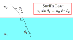
What is the refractive index of:
1. air
2. water
3. cornea
4. lens
n(air) = 1.003
n(water) = 1.33
n(cornea) = 1.38
n(lens) = 1.42
Why do we say the cornea has greater focusing power than the lens even though the lens has a higher refractive index?
focusing power is the ratio of the refractive indices of the second medium and the first (n2/n1)
n(cornea) / n (air) = 1.38
n(lens) / n(water) = 1.42/1.33 = 1.07
thus, most of the focusing is done by the cornea
Why do we have a lens if the cornea does the majority of the focusing? What does the lens do that the cornea doesn't and what other structures in the eye play a role in this?
Lens allows us to focus on objects at varying distances through accommodation (the changing of the lens' shape)
Possible due to the ciliary controlled by the autonomic nervous system
What changes in the eye when looking at near objects vs looking at far ones? Why?
Close objects: lens rounds more to increase refraction (moves plane of focus forward to fall on retina)
Far objects: lens flattens to decrease refraction (moves plane of focus farther back so it falls on the retina)
What vision condition can people get as a result of aging? What causes it?
Presbyopia
with age, the lens hardens and loses ability to "round up", making it more difficult to see objects up close
Describe how an image falls on the retina. What are the disorders associated with this?
Image is upside down and left right reversed on the retina
There are no disorders where people see the reversed image
What are all the layers of the retina starting from closest to the cornea? What is the full pathway of a phototransduced signal?
nerver fiber layer-> retinal gaglion cells -> bipolar cells -> photoreceptors -> pigment epithelium
photoreceptors-> bipolar cells -> retinal ganglion cells -> optic nerve -> optic disc -> thalamus
In the X and O blind spot experiment done in class, why does the disappearing point have to be located on the right for your right eye and vice versa for your left eye? (i.e. where is the blind spot located on the retina?)
blind spot is located on the nasal retina, so light from the lateral field of view will hit it
Why do we need to move the page to the right distance to have the O fall on the blindspot?
distance from eye determines where the O falls on the retina: closer = more medial, farther= more lateral (i.e., light from O subtends a smaller angle as page moves away)
Why aren't we usually able to see our blindspot?
Information from two eyes is usually enough for the brain to infer what is in the blind spot
Anatomy of a photoreceptor (just two main regions)
outer segment (closer to pigment epithelium, contain discs)
inner segment (contains cell body)
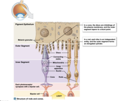
Why is the retina "inside out"? (i.e. why are the photoreceptors on the inside
discs from photoreceptors are shed and phagocytosed to PE cells
What is the name of the experiment that proved the function of PE cells? Describe it
pulse-chase experiment
injected methionine that contained radioactive tritium (3H) into retina, amino acids taken up by cells and used in proteins
over time, the marked proteins moved down the retina and eventually entered PE cells
What is the potential of a photoreceptor in the dark? What happens to the potential in response to light?
dark: -40 mV (depolarized)
light: hyperpolarizes down towards -65 mV, depending on the intensity of light
What is "dark current" and what are the mechanisms that cause it?
overall inward current of positive charge from the opening of a Na+/Ca++ ion channel in darkness
K+ leaves through leak channels
cGMP (cyclic guanosine monophosphate) acts as an intracellular ligand that keeps the Na+/Ca++ channel open
What happens to photoreceptors when exposed to light?
cGMP is degraded in the presence of light, causing the Na+/Ca++ channel to close
Less Na+ & Ca++ enters but K+ still leaves, resulting in hyperpolarization
When photoreceptors are in the dark, what neurotransmitter is released and why? What happens when the cell is exposed to light?
glutamate: since the cell is depolarized in the dark, voltage-gated Ca++ channels open and release glutamate
In light: cell hyperpolarizes and Ca++ voltage-gated channels close, less glutamate is released (note: how much less depends on how bright the light is)
What are opsin and retinal?
Opsin is a protein found in the discs of photoreceptors that contains retinal, a chromophore (molecule that absorbs light) that is a type of vitamin A
Photoisomerization in photoreceptors
when 11-cis retinal transofrms into all-trans retinal after being hit by light
What is PDE? What is its function dependent on?
Phosphodiesterase
an enzyme that degrades cGMP - activated by transducin which is activated by the photoisomerization of retinal due to a light stimulus
Describe what goes on in photoreceptors during the process of phototransduction
When light hits the retina, photoisomerization of retinal activates the enzyme transducin
transducin activates phosphodiesterase (PDE)
PDE degrades cGMP, cGMP channels close and cell hyperpolarizes (Na+ & Ca++ not entering, but K+ leaving)
What is guanylate cyclase? What is its function dependent on?
An enzyme within photoreceptors that produces cGMP
activity is inhibited by Ca++
What is happening in a photoreceptor during the process of photoadaption?
Since Ca++ inhibits guanylate cyclase, the depletion of intracellular Ca++ due to continued light exposure allows guanylate cyclase to produce cGMP
Rise in cGMP opens cGMP-gated channels channels, resulting in depolarization of the photoreceptor (return to -40mV baseline = adaptation)
True or False: the brain infers all colours in the visible light spectrum based on the activation of the cone type associated with that wavelength
False
brain uses information from the relative activation of cone types - the range of wavelengths covered by each cone type overlaps
What are the types of cones and their generic corresponding colours?
Short-wavelength: blue
Medium-wavelength: green
Long-wavelength: red
What wavelenths of light are rods most sensitive to?
Green/blue wavelengths, but they do not detect colour (only intensity)
What is the photopigment in rods? How many kinds of rods are there?
Rhodopsin
one type
Why does the system for rods have greater sensitivity than the system for cones? (3 reasons)
1. rods are longer (contain more photopigment), so there is a higher chance for dim light to hit
2. more rods (15-30) converge onto the same bipolar cell - higher chance for signalling to occur
3. greater amplification (more Na+ opened for the same stimulus strength), 1 photon = 800 transducin activated = 800 PDE activated = 4800 cGMP degrade = 200 closed ion channels = 1mV hyperpolarization (cones need to abssorb x~100 more photons to produce the same response)
Why does the system for cones have greater sensitivity than the system for rods? (2 reasons)
1. Cones are more highly concentrated on the fovea (region without overlying axons and blood vessels, i.e. reduced light scattering, looks like a pit in the retina)
2. Much less convergence (1 cone per 1 bipolar cell)
Where are rods and cones located on the retina? How many rods and cones are there per eye?
Rods: located on eccentric area of retina (outer regions), 90 million per eye
Cones: located in the central region of retina, 4.5 million per eye
What are off-center vs on-center ganglion cells?
On-center ganglion: fire when light stimulus falls on the center of the receptive field
Off-center ganglion: fire when light stimulus falls on the outer ring of the receptive field
What are the morphological differences and similarities between on-center and off-center ganglion cells?
No difference (physiologically the same), only differ by the type of bipolar cells they synapse with (on-center or off-center bipolar cells)
What is the difference between off-center bipolar cells and on-center bipolar cells?
Off-center: Have the normal glutamate receptors (AKA ionotropic glutamate receptors, AKA AMPA R)
On-center: Have metabotropic glutamate receptors (mGluR6)
How do the standard glutamate receptors differ from the ones found in on-center bipolar cells?
In response to glutamate, ionotropic glutamate receptors directly open ion channels within the receptor
In response to glutamate, mGluR6 receptors indirectly close external Na+ channels (not within the receptor)
Describe the cascade of events that happens when light hits the center of the receptive field
light causes photoreceptor to hyperpolarize, so it releases LESS glutamate onto biopolar cells
In off-center bipolar cells, ionotropic glutamate receptors' Na+ channels close, leading to hyperpolarization. Thus, off-center ganglion cells get no glutamate and do not fire
In on-center bipolar cells, mGLuR6 opens Na+ channels, so more glutamate is released onto the ganglion cell, causing it to fire more APs
Describe the cascade of events that happens when the center of the receptive field is made darker
absence of light stimulus causes photoreceptor to depolarize, and it releases glutamate onto biopolar cells
Off-center bipolar cells: ionotropic glutamate receptors' Na+ channels open, depolarizating the bipolar cell and causing it to release glutamate. Thus, off-center ganglion cells' firing rate increases
On-center bipolar cells, mGLuR6 closes Na+ channels (hyperpolarization), and less glutamate is released onto the ganglion cell, lowering its firing rate
What are ionotropic receptors? What receptors fall under this category?
AKA Ligand-gated receptors: open to allow ion flow when neurotransmitter binds
e.g. glutamate (AMPA) receptor, GABA (A) receptor
What are metabotropic receptors? What receptors fall under this category?
AKA G-Protein Coupled Receptors: When neurotransmitter binds, receptor activates G-protein within cell whose subunits modulate ion channels
e.g. mGluR6 Glutamate Receptor, Enkephalin (opiod) Receptor, Beta2 Adrenergic Receptor (on A-beta axons, binds norepinephrine released by Merkel cell)