Cardiac physiology
 Call Kai
 Learn
Learn Practice Test
 Spaced Repetition
 Match
Match Flashcards
 Knowt Play
1/8

There's no tags or description
Looks like no tags are added yet.
Name | Mastery | Learn | Test | Matching | Spaced | Call with Kai | Chat |
|---|
No analytics yet
Send a link to your students to track their progress
9 Terms
Describe the anatomy of the heart.
The heart has four chambers: left and right atria, and left and right ventricles, divided by the septum. The atria receive blood from veins, and valves ensure one-way blood flow.

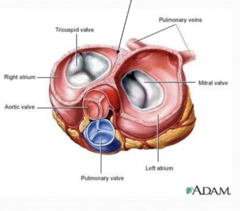
Describe the valves in the heart
- There are 2 AV valves, Bicuspid/mitral (left) and Tricuspid (right).
- Also the pulmonary and aortic valves.
- Valves are supported by chrodae tendineae.
- Papillary muscles contract with ventricles to prevent back-flow.

Describe the pulmonary and aortic valves.
- Allow movement of blood from ventricles to arteries.
- Made up of three cusps
- Can't be turned inside out by upturned nature of cusps.
- Close under back pressure
Describe diastole of the heart.
- Heart is resting and filling with blood from veins.
- All valves are closed
- As more blood fills, AV valves open allowing ventricles to begin filling.
Describe systole of the heart.
- AV valves close to prevent back flow of blood from ventricles to atria.
- As ventricles contract aortic and pulmonary valves open allowing blood to be forced into arteries.
What is stroke volume and how is it calculated?
The amount of blood ejected by the heart in any one contraction.
Stroke volume = end diastolic volume - end systolic volume
How are cardiocytes connected to one another?
- Desmosomes physically attach them together.
- Also a gap junction which allows action potential to cross between them.
- This produces intercalated discs between cardiocytes.
What is the pericaridal sac?
A protective, fluid-filled sac that surrounds your heart and helps it function properly.
Describe the pericardial sac.
- A tough double walled sac.
- Anchors the heart and secrets pericardial fluid which lubricates the heart as it rubs against rib cage.
- Can become infected and inflamed which can be painful (pericarditis).