Chapter 11
 Call Kai
 Learn
 Practice Test
 Spaced Repetition
 Match
 Flashcards
 Knowt Play
1/238

Earn XP
Description and Tags
A&P 1 Lecture
239 Terms
Universal characteristics of muscle
Excitability (responsiveness), conductivity, contractibility, extensibility, elasticity
Excitability (responsiveness)
To chemical signals, stretch, and electrical changes across plasma membrane
Conductivity
Local change triggers a wave of excitation that travels along the muscle fiber
Contractability
Shortens when stimulated
extensibility
capable of being stretched between contractions
Elasticity
Returns to original resting length after being stretched
Skeletal muscle
Voluntary, striated muscle attaches to one or more bones
Striations
Alternating light and dark transverse bands
Voluntary
Usually subject to conscious control
Skeletal muscle cell
Muscle fiber or myofiber
muscle fiber or myofiber
skeletal muscle cell
multinucleated
nucleus in skeletal muscle?
Endomysium, perimysium, epimysium
Skeletal muscle contains fibrous connective tissues:
fibrous connective tissues
the endomysium, perimysium, epimysium are what
Endomysium
Surrounds each muscle fiber
Perimysium
Bundles muscle fibers into fascicles
Epimysium
Surrounds entire muscle
Collagen
Stretches slightly under tension and recoils when released and resists excessive stretching
collagen
protects muscle from injury and returns muscle to its resting length
collagen
contributes to power output and muscle efficiency
extensible and elastic
collagen is somewhat_____

Sarcolemma
Plasma membrane

Sarcoplasm
Cytoplasm
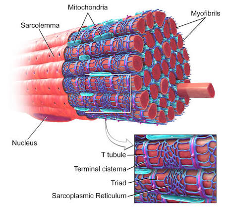
Myofibrils
Long protein bundles occupying most of sarcoplasm
Multiple nuclei
Flattened and pressed against inside of sarcolemma
Mitochondria
Packed into spaces between myofibrils

Sarcoplasmic reticulum (SR)
Smooth ER that forms a network around each myofibril; calcium reservoir
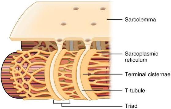
Terminal cisternae
Dilated end-sacs of SR; cross muscle fiber from one side to another

T tubules
Tubular infolding of the sarcolemma which penetrate through the cell and emerge on other side
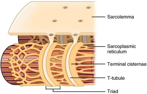
T tubules
signal sarcoplasmic reticulum (SR) wehn to release calcium which causes muscle contraction

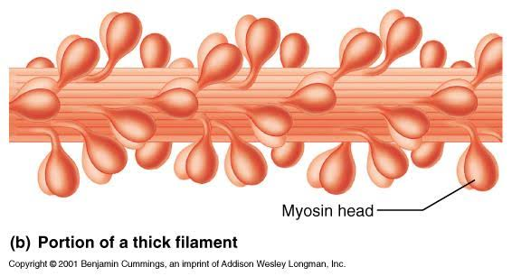
Thick filaments
Made of several hundred myosin molecules
Thick filaments
Shaped like golf club, 2 chains interwinds to form a shaft-like tail, double globular myosin head
Thick filament heads
Directed outwards in a helical array (spiral) around bundle
thick filament heads
heads on one half of the thick filament angle to the left and the other half angle to the right, bare zone with no heads in the middle
Thin filaments
Fibrous actin, globular actin subunits, tropomyosin molecules, troponin molecule

Fibrous actin
2 intertwined strands

fibrous (F) actin
string of globular (G) actin subunits each with an active site that can bind to head of myosin molecules

Tropomyosin molecules
Blocks active site on G actin when muscle is relaxed; prevents myosin from binding
Troponin molecule
Small, calcium-binding protein (attaches to calcium) on each tropomyosin molecule
regulatory proteins
determine when the muscle fiber can and cannot contract: tropomyosin and troponin
contractile protiens
do the actual work of shortening the muscle: myosin (head) and actin (primary protein of thin filament
Elastic filaments
run through core of thick filament; anchor it to Z disc and M line

elastic filaments
help stabilize and position thick filament
elastic filaments
prevent overstretching and provide recoil
Titan
Huge, springy protein
titan
elastic filaments contain what protein
Dystrophin
Links actin in outermost myofilaments to membrane proteins that link to endomysium

dystrophin
transfers forces of muscle contraction to CT ultimately leading to tendon

dystrophin
genetic defects in ______ produce disabling disease muscular dystrophy (muslces loses the ability to contract)

Striations
Organized in a precise way in skeletal and cardiac muscle

A band
Dark, thick (myosin) filaments

H band
Lighter region in middle of A band; thick filaments only
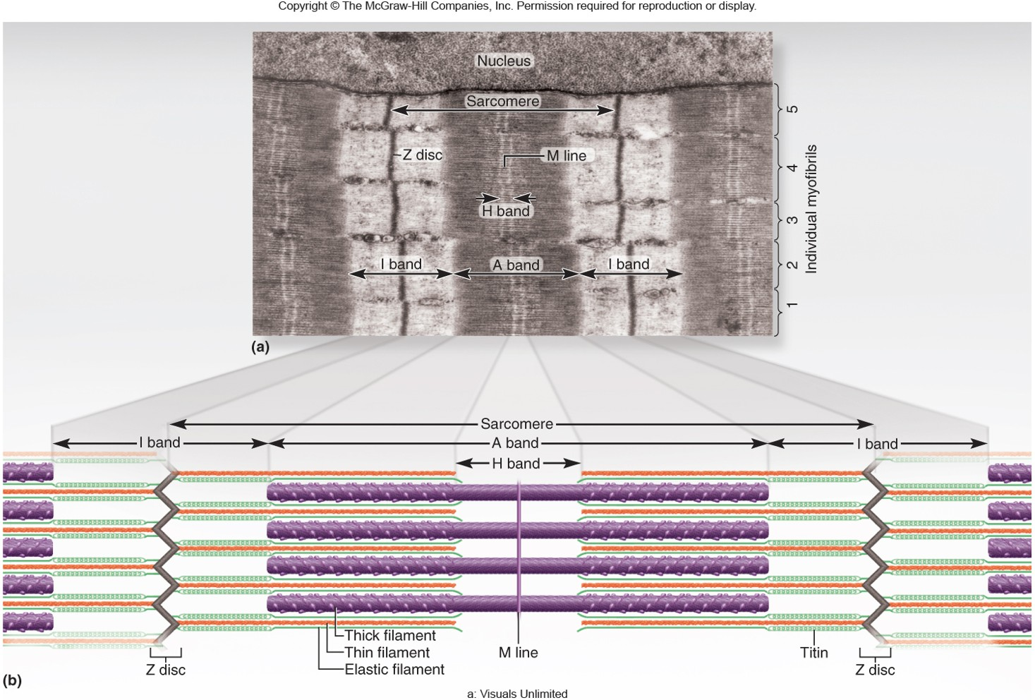
M line
Middle of H hand; transverse protein complex that links thick filaments

I band
light, thin (actin) filaments

Z disc (Z-line)
Provides anchorage for thin and elastic filaments

Sarcomere
Segment of a myofibril from Z disc to Z disc
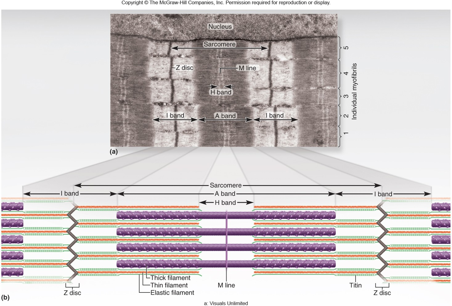
sarcomere
functional contractile unit of muscle fiber
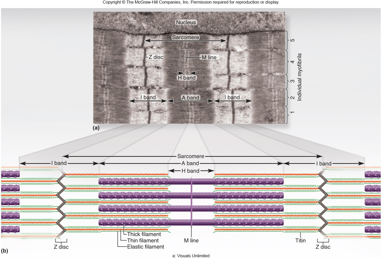
Their individual sarcomeres shorten
Muscle cells shorten because:
Thick and thin filaments slide past each other
Z disc (Z lines) are pulled closer together as:
Length during shortening; only amount of overlap changes
Neither thick nor thin filaments change in:
Dystrophin and linking proteins also pull on extracellular proteins which transfers pull to extracellular tissue
During muscle shortening

Nerve-muscle relationship
Skeletal muscle cannot contract unless stimulated by a nerve: what is this
Somatic motor neurons
Nerve cells with cell bodies in brainstem and spinal cord; serve skeletal muscles
Somatic motor fibers
Axons (long extensions) of somatic motor neurons (nerve cells for contraction) that lead to skeletal muscle

Branches out to a number of muscle fibers
Each nerve fiber does what

Is supplied by only one motor neuron
Each muscle fiber:
Motor unit
One nerve fiber and all the muscle fibers innervated by it; behave as a functional unit
Muscle fibers of one motor unit
Dispersed throughout muscle (not clustered)
muscle fibers of one motor unit
contract in unison
muscle fibers of one motor unit
produce weak contraction over wide area
muscle fibers of one motor unit
motor units take turns contracting to provide long-term contraction (postural control)
Average motor unit
200 muscles fibers distributed across 100 fascicles

Small motor units
Fine degree of control and 3-6 muscles fibers per neuron

small motor units
eye and hand muscles are examples of what kind of moto unit
Large motor units
More strength than control

large motor units
powerful contractions supplied by _____ with hundreds of fibers

large motor units
quadriceps femoris and gastrocnemius controlled by motor units that may have 1,000 muscle fibers per neuron
Synapse
Point where a nerve fiber meets its target cell
Neuromuscular junction (NMJ)/ motor end plate
When target cell is a muscle fiber

Separate synapse with muscle fiber
Each terminal branch of the nerve fiber within NMJ forms:
Axon terminal/synaptic knob
Swollen end of nerve fiber; 1 per synapse, contains synaptic vesicles filled either acetylcholine (ACh)- part 1 on picture

Synaptic cleft
Narrow gap between axon terminal and sarcolemma- part 2 on image

Postsynaptic membrane/junctional folds of sarcolemma
Increase surface area holding ACh receptors- part 3 on image

Basal lamina
Thin layer separating muscle fiber and nerve ending from surrounding CT; contains acetylcholinesterase (AChE)- part 4 on image

Electrically excitable cells
Muscle fibers and neurons are what kind of cells
Plasma membranes exhibit voltage changes in response to stimulation
Why are muscle fibers and neurons electrically excitable (cell can respond to a stiumulus by producing an electrical signal) ?
Concentration differences in ions in intracellular fluid vs. extracellular fluid
Electrical activity depends on:
Voltage (electrical potential)
A difference in electrical charge from one side of the membrane to the other
Resting membrane potential
About -90 mV
about -90 mV
what is the resting membrane potential
resting membrane potential
maintained by sodium-potassium pump
unstimulated (resting) cell
More anions on the inside of the plasma membrane negatively charged in comparison to the outside
negatively charged in comparison to the outside
these more anions make the inside of the plasma membrane____ (resting cell)
Electrically polarized (charged) with a resting membrane potential (RMP)
The plasma membrane is: (resting cell)
Extracellular fluid (ECF)
where are there excess sodium ions (Na+) (resting cell)
Intracellular fluid (ICF)
where are there excess potassium ions (K+) (resting cell)
intracellular fluid (ICF)
in _____, there are also anions such as proteins, nucleic acids, and phosphates that cannot penetrate the plasma membrane, these anions make the inside of the plasma membrane negative charged in comparison to its outer surface (resting cell)
proteins, nucleic acids, phosphates
in the intracellular fluid, what anions cannt penetrate the plasma membrane (resting cell)
they make the inside of the plasma membrane negatively charged in compariosn to its outer surface
what do the anions proteins, nucleic acids, and phosphates do when they are in the ICF (resting cell)
Stimulated (active) muscle fiber or nerve cell
Na+ ion gates open in the plasma membrane and Na+ flows down its electrochemical gradient into the cell