lab practical #2 origins/insertions
 Call Kai
 Learn
 Practice Test
 Spaced Repetition
 Match
 Flashcards
 Knowt Play
1/64

Earn XP
Description and Tags
cwru biol346 (superficial head & neck, brain, cranial nerves, and special senses)
65 Terms

platysma (ORIGIN)
fascia of superior portions of pectoralis major and deltoid

platysma (INSERTION)
1.) inferior mandible at symphysis
2.) skin and fascia of the lower parts of the face

sternocleidomastoid (ORIGIN)
1.) medial head: manubrium of the sternum
2.) lateral head: middle 1/3 of the clavicle
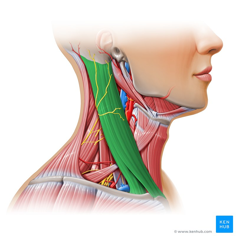
sternocleidomastoid (INSERTION)
mastoid process of the temporal bone
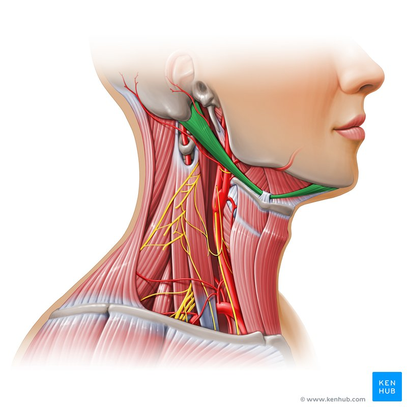
digastric (ATTACHMENTS)
note that the digastric does not have a formal origin/insertion.
1.) anterior belly: digastric fossa of internal mandible
2.) posterior belly: mastoid process of the temporal bone
both bellies meet and attach at the lateral aspect of the body of the hyoid bone by a pulley tendon

mylohyoid (ORIGIN)
inner surface of mandible

mylohyoid (INSERTION)
1.) body of hyoid
2.) along midline at the mylohyoid raphe
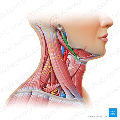
stylohyoid (ORIGIN)
styloid process of temporal bone

stylohyoid (INSERTION)
lateral margin of hyoid (near greater horn)
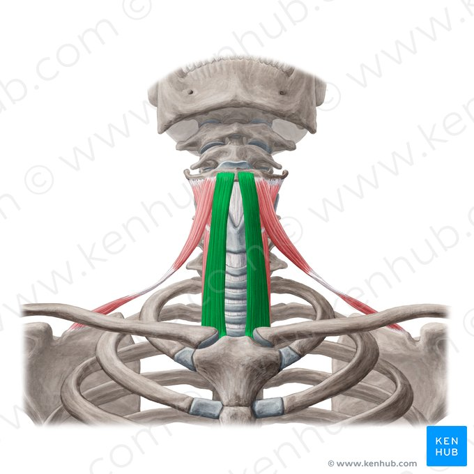
sternohyoid (ORIGIN)
1.) manubrium of sternum
2.) medial end of clavicle

sternohyoid (INSERTION)
body of hyoid bone

sternothyroid (ORIGIN)
manubrium of sternum

sternothyroid (INSERTION)
thyroid cartilage

thyrohyoid (ORIGIN)
thyroid cartilage of larynx

thyrohyoid (INSERTION)
greater horn of the hyoid bone

omohyoid (ORIGIN)
superior border of scapula

omohyoid (INSERTION)
body of the hyoid bone, lateral to the insertion of sternohyoid

frontalis (ORIGIN)
epicranial aponeurosis

frontalis (INSERTION)
skin above the eyebrows

masseter (ORIGIN)
1.) superficial portion: zygomatic process of maxilla
2.) deep portion: zygomatic arch

masseter (INSERTION)
1.) superficial portion: angle of the ramus of the mandible
2.) deep portion: upper portion of the ramus and coronoid process of mandible

temporalis (ORIGIN)
temporal fossa of the skull

temporalis (INSERTION)
coronoid process and ramus of the mandible

buccinator (ORIGIN)
1.) alveolar processes of the maxilla and mandible
2.) pterygomandibular raphe

buccinator (INSERTION)
fibers of orbicularis oris at the lateral aspect of the mouth
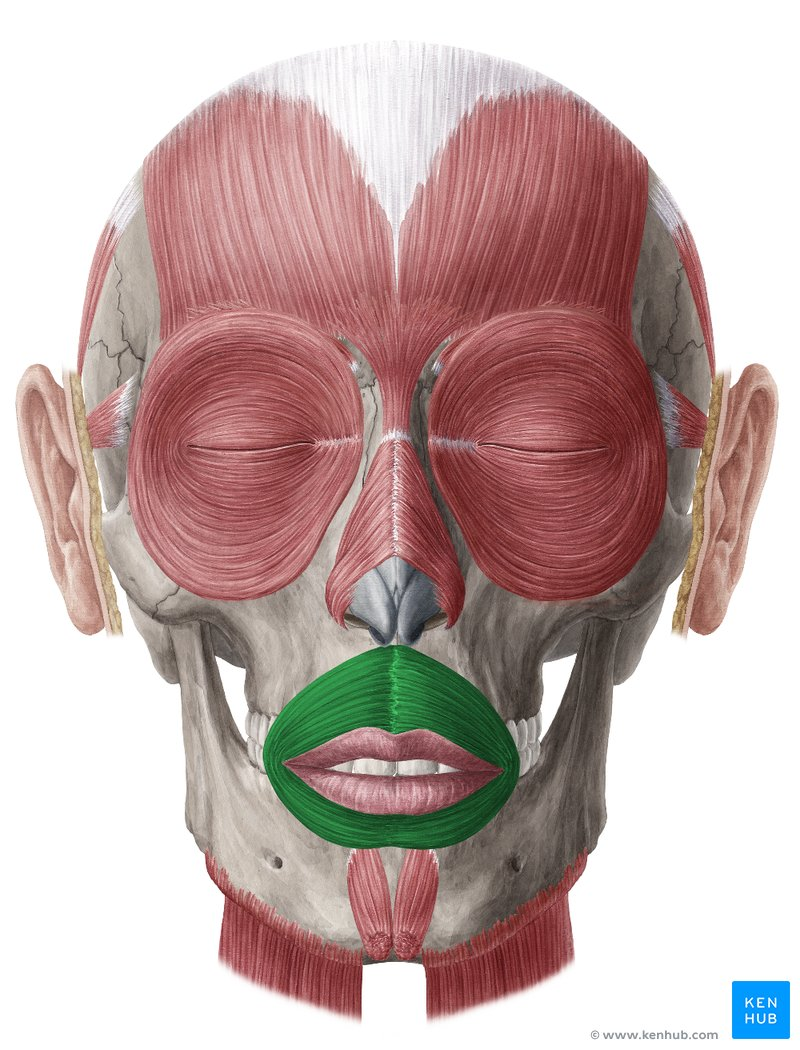
orbicularis oris (ORIGIN)
very complex; maxilla and mandible, in roughly 4 quadrants
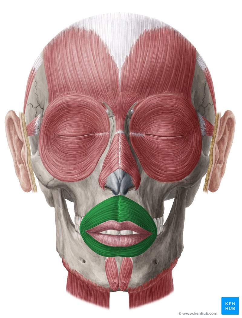
orbicularis oris (INSERTION)
skin around the lips

orbicularis oculi (ORIGIN)
medial portion of the orbit
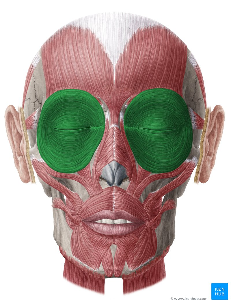
orbicularis oculi (INSERTION)
skin of the eyelid

levator palpebrae superioris (ORIGIN)
sphenoid bone
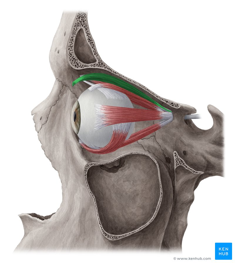
levator palpebrae superioris (INSERTION)
tarsal plate of upper eyelid

zygomaticus major (ORIGIN)
antero-lateral aspect of zygomatic bone

zygomaticus major (INSERTION)
connective tissue of the angle of the mouth

zygomaticus minor (ORIGIN)
antero-medial aspect of zygomatic bone

zygomaticus minor (INSERTION)
skin of the upper lip

levator labii superioris (ORIGIN)
medial infraorbital margin

levator labii superioris (INSERTION)
skin and muscle of the upper lip

levator labii superioris alaeque nasi (ORIGIN)
medial maxilla
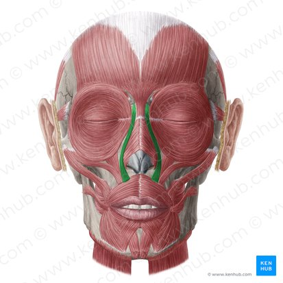
levator labii superioris alaeque nasi (INSERTION)
nostril and upper lip

risorius (ORIGIN)
fascia over parotid salivary gland
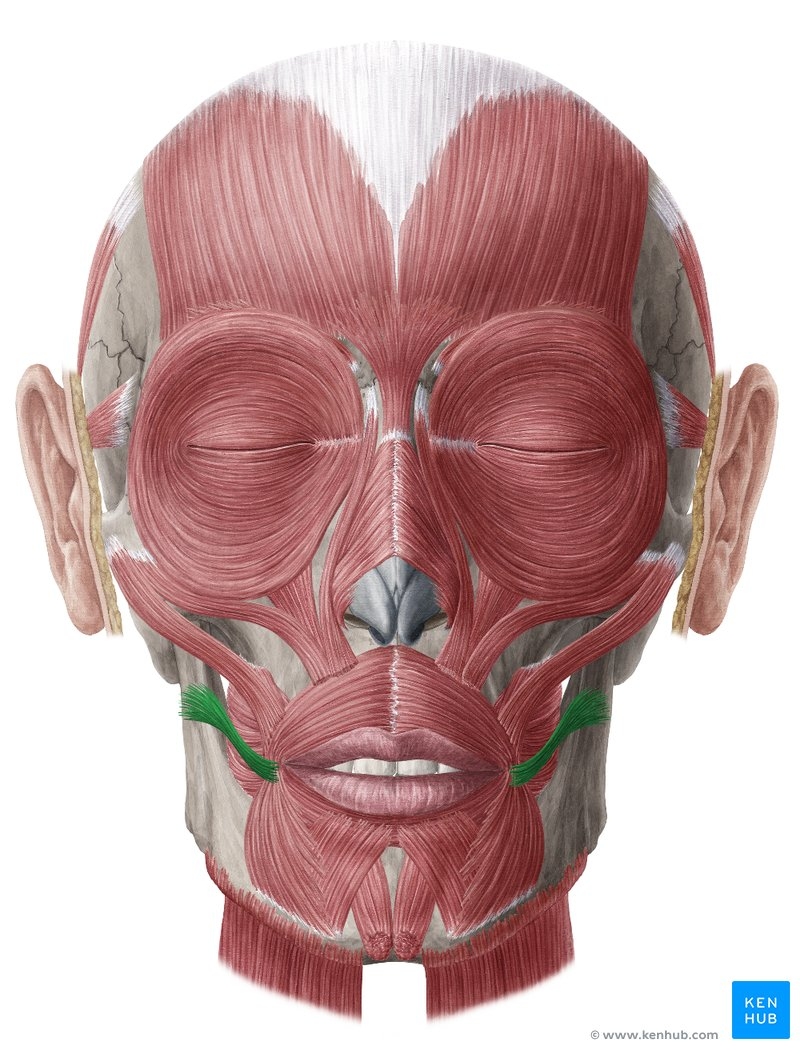
risorius (INSERTION)
connective tissue of the angle of the mouth
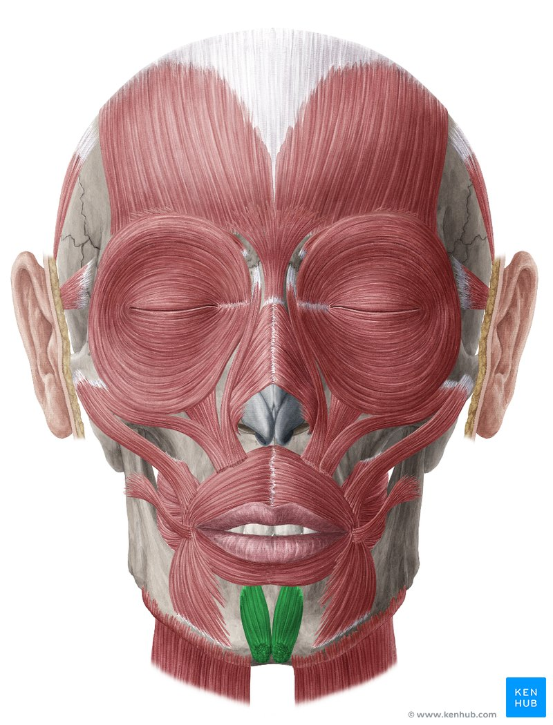
mentalis (ORIGIN)
anterior mandible
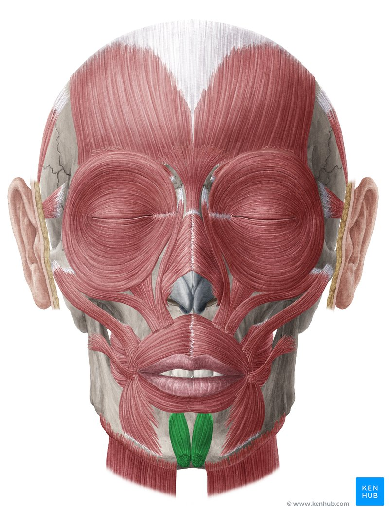
mentalis (INSERTION)
skin of the chin

depressor anguli oris (ORIGIN)
tubercle of mandible

depressor anguli oris (INSERTION)
connective tissue of the angle of the mouth
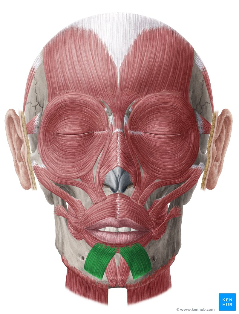
depressor labii inferioris (ORIGIN)
oblique line of mandible

depressor labii inferioris (INSERTION)
1.) skin of the lower lip
2.) fibers of orbicularis oris
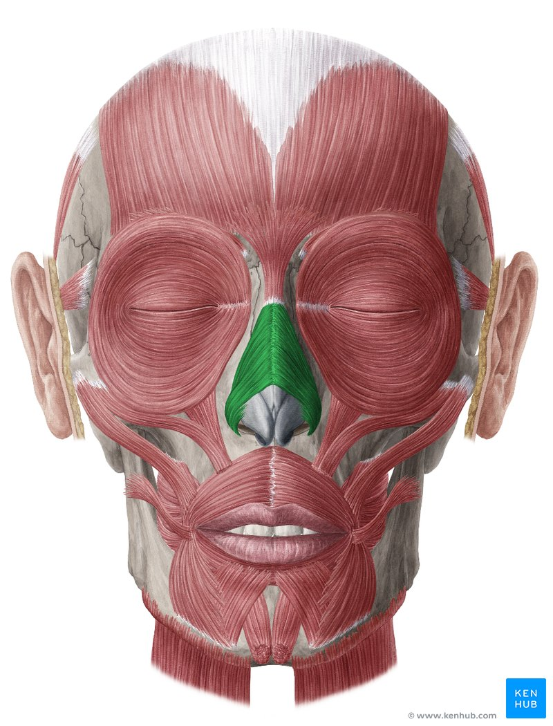
nasalis (ORIGIN)
1.) transverse portion: maxilla lateral to the nose
2.) alar portion: maxilla over the lateral incisor

nasalis (INSERTION)
1.) transverse portion: dorsum of the nose
2.) alar cartilage of the nose

corrugator supercilii (ORIGIN)
superciliary arch (brow ridge) of the frontal bone
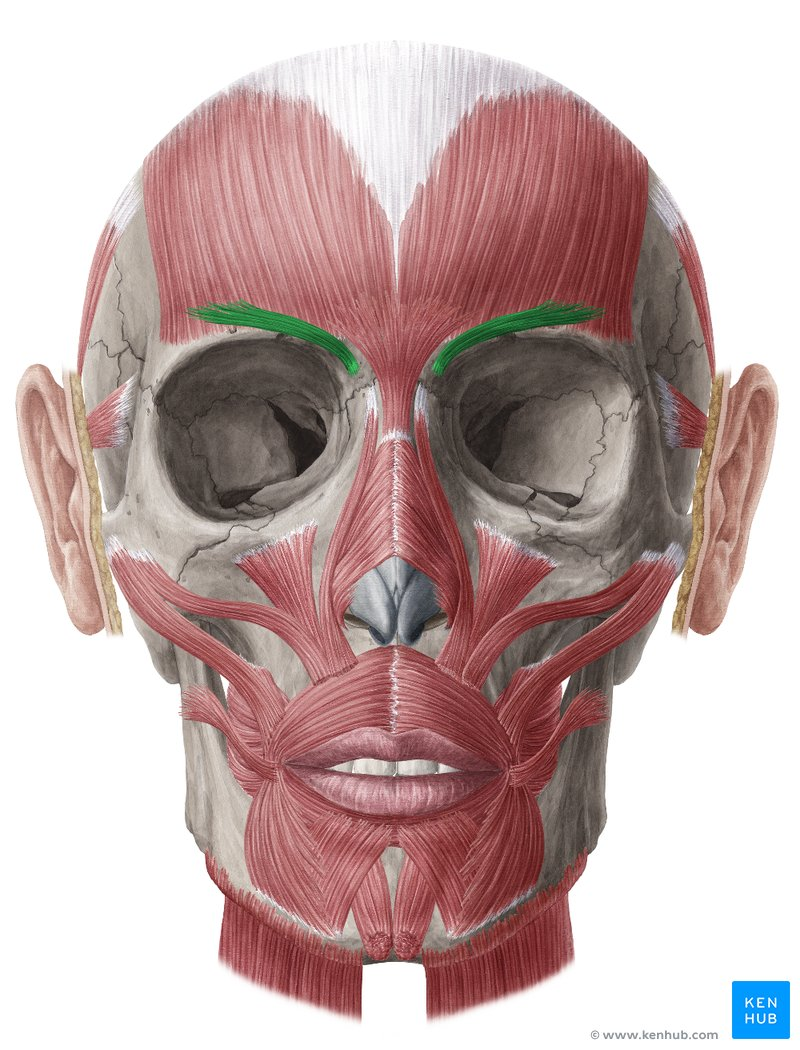
corrugator supercilii (INSERTION)
skin of forehead near eyebrow
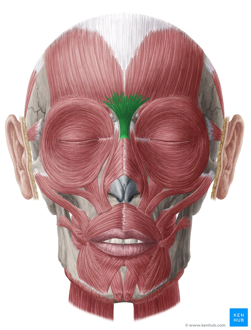
procerus (ORIGIN)
fascia over the lower half of the nasal bone

procerus (INSERTION)
skin of the forehead, between the eyebrows
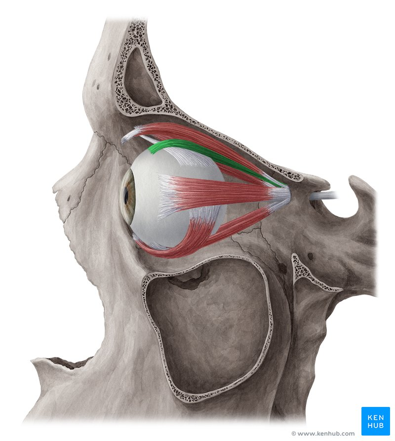
superior rectus (ORIGIN)
annular tendon at the apex of the orbit
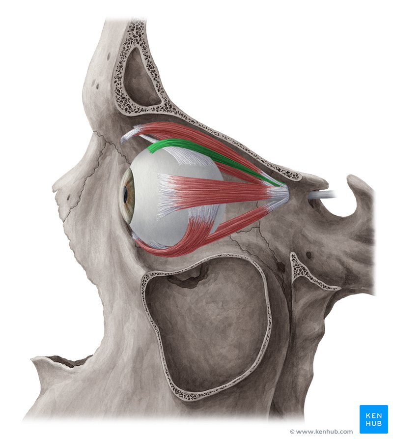
superior rectus (INSERTION)
superior aspect of sclera, posterior to the corneal limbus (where the cornea meets the sclera)

inferior rectus (ORIGIN)
annular tendon at the apex of the orbit

inferior rectus (INSERTION)
inferior aspect of sclera, posterior to the corneal limbus
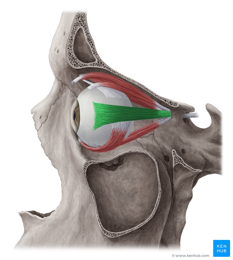
lateral rectus (ORIGIN)
annular tendon at the apex of the orbit

lateral rectus (INSERTION)
lateral aspect of the sclera, posterior to corneal limbus

medial rectus (ORIGIN)
annular tendon at the apex of the orbit

medial rectus (INSERTION)
medial aspect of sclera, posterior to corneal limbus

superior oblique (ORIGIN)
annular tendon at the apex of the orbit

superior oblique (INSERTION)
lateral posterior quadrant of the eyeball

inferior oblique (ORIGIN)
orbital surface of the maxilla, lateral to the lacrimal groove

inferior oblique (INSERTION)
lateral aspect of the eyeball, deep to the lateral rectus