Lab 2 practical (Intro to MLS) practice
 Call Kai
 Learn
 Practice Test
 Spaced Repetition
 Match
 Flashcards
 Knowt Play
1/40

There's no tags or description
Looks like no tags are added yet.
41 Terms

What is this crystal structure in a urine sample?
When is it found, and in what conditions?
Uric acid
Found in acidic urine (pH less than 7)

What is the arrow pointing to in this photo?
When is this found in urine?
Hayaline cast
This type of cast is usually found in patients with:
Prior exercise
Fever
Diuretics
These observed in urine can be normal.
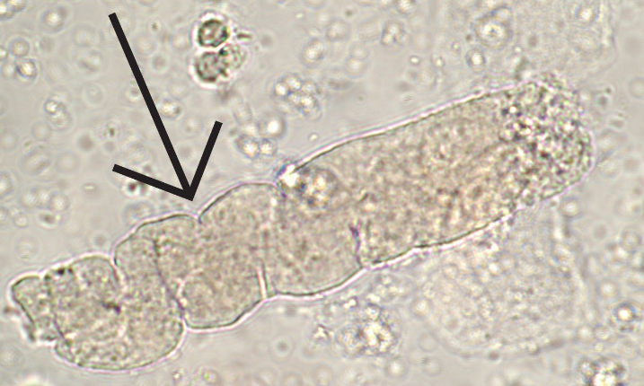
What type of cast is illustrated in the image?
When is it found?
Waxy cast
It usually indicates chronic renal disease when present.

What cell is being pointed to?
White blood cell
They are usually present as Leukocyte esterase in urine and is commonly present in association with elevated Nitrite levels (usually seen in UTI or inflammation).
Usually appear granular and sparkly and are larger than RBCs.

Identify the cell illustrated? Under what conditions is it present in urine?
Red Blood Cell
They are usually common in women during menstruation, but are often found in cases of trauma, hematuria, and glomerular disease.
Three types of observed RBCs in urine:
1. Crenated RBCs (spiky appearance)
2. Ghost cells (only membrane remaining)
3. Normal red blood cells (oval and concave)

Identify cell observed in this urine sample.
RBC
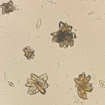
What is present in this image? When and why is it present in urine?
Uric acid (Rosette form)
Commonly present in acidic urine (pH less than 7).
There are 3 types of Uric acid crystals:
1. Barrel
2. Diamond
3. Rosettes

What cell is in this image ?
RBC

What cell is being pointed to in this image?
WBC

What type of cell is the arrow pointing to? In what instances can this be found in urine?
Squamous Epithelial cell
Usually present during contamination (clean catch test) or common in women (1-5 normal range) due to uterus.
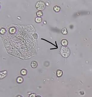
What is the arrow pointing to in this image? Why is this present in urine?
Trichomonas
Important in identifying as it is associated with STI.
Characteristics:
Pear-shaped
Motile
Flagella

What is observed in this image? Why is it found in urine?
Amorphous crystals
They are considered junk crystals. Getting rid of them depends on pH, in order to properly dissolve without interfering with crystals, RBCs, or WBCs.

What are the circled portions indicating in this image? When is this commonly present in urine samples?
Bacteria
Usually rod-shaped and present with a UTI.
Corresponding dipstick results:
WBCs present in urine
Positive nitrites
Positive leukocyte esterase

What is the arrow pointing to in this image? It’s significance in urine?
Yeast
Usually associated with a UTI with the most common form being “Candida albicans”.
Characteristics:
Budding cells
Pseudohyphae

What is in this image? What is a reason it could be present in urine?
Bilirubin crystal
Should not be present in urine, but usually indicates liver disease or biliary obstruction.

Whats in this image? When is it likely present in a urine sample?
Triple phosphate
It is usually coffin lid/ enveloped shaped and is commonly found in alkaline urine (pH greater than 7).

What is present in this image? Why is it found in urine?
Calcium Oxalate
Usually appears as a small square with an X on it and is found in acidic urine (pH greater than 7).

What is present in this image?
Uric acid (barrel shaped)

Identify this item in the urine slide.
Triple Phosphate

What type of cast is this? Why is it usually found in urine?
Granular cast
Usually considered abnormal when present in urine and indicates an issue in kidney function.

What is the arrow pointing to in this photo? When should it be reported?
Sperm
It is common in adults 18+ (just state that it is present); anyone under 18 must be reported.

What type of WBC is this? When is it present in blood?
Lymphocyte
Commonly associated when a viral infection is present.
Compared to monocytes, they contain a large round nucleus.

What type of RBC is in this image?
Polychromatic RBC (reticulocytes)
Usually Bluish/ purple color, and they are the last stage in RBC maturation.

What type of cell is this?
Sickle cell (RBC)
They are usually crescent-shaped and associated with sickle cell disease.

what type of WBC is in this image?
Hypersegmented Neutrophil
Commonly present when there is more than 5 lobes in the nucleus.
Hyposegmented Neutrophil → less than 2 lobes.

What type of WBC is this? When is it present in blood?
Eosinophil
Usually contains orange granules and a purple nucleus.
They are present in instances of:
Parasitic infection
Allergic reactions
asthma

What is in this image?
Platelet (Tiny cells)
They are anucleated and usually stain a bluish-purple color.
There are 3 types of platelets:
Large platelets → Same size as RBCs
Giant Platelets → Bigger than RBCs
Platelet clumping → clotting occurred or micro-clotting present in sample.

What type of WBC is this?
Reactive lymphocyte
Large lymphocyte: note the dark cytoplasmic edges next to the RBCs. These are the ones seen in infectious mononucleosis.

What is this cell shown? When is it found in the blood?
Megakaryocyte
Megakaryocytes are typically found in the bone marrow. Platelets break off from this cell (HUGE platelet-producing machine).
This is one of my favorite cells. I call it "The Mothership" because it is like a spaceship, and the little platelets that come off it are like fighter jets.

What type of RBC is this?
Target cell
As the name suggests, the cell looks like a target sign.

What type of RBC is this?
Normal RBC
The RBC size is similar to the nucleus of the Lymphocyte seen on the bottom left of the cell.

What type of cell is this?
Giant Platelet
Usually bigger than RBCs.

What cell is present in this photo?
Basophil
It has a dark blue/ purple granulated appearance. When present, it is usually associated with an allergic reaction or chronic myeloproliferative disorders.

What type of RBC is this ?
Teardrop cell
Note: If the tip of the teardrop cells are all facing the same direction the it was an error in technique.
However, if they are facing different directions, then it is a morphological feature.
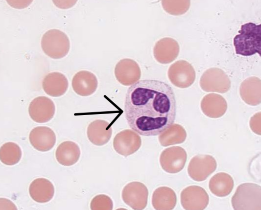
What type of cell is this? Why is it usually present in the blood?
Neutrophil
When elevated, they are usually associated with Bacterial infection and acute inflammation.

What type of cell is this?
Monocyte
visible vacuoles and irregularity in nucleus shape.

What type of cell is this? When is it usually present in the blood?
Monocyte
They are similar in appearance to lymphocytes, but contain an irregular shaped nucleus.
They are usually associated with chronic inflammation and recovery from infection.
Note: They are the largest WBC.

What is the arrow pointing to?
Clumped Platelets
Usually signifies clotting occurred or micro-clotting present in sample.

What type of RBC is this?
nRBC (nucleated Red Blood Cell)
This is the last step before the nucleus is ejected from RBC.
Increased nRBCs often indicate intense bone marrow stimulation and results in illnesses such as:
Acute blood loss
anemia
blood disorders

What kind of cell is this?
Smudge cell (Ruptured WBC)
We don't count these toward the 100-cell differential because we don't know what they were.

What type of RBC is this?
Schistocyte (RBC fragment)
This occurs when the cell breaks into fragments and is often associated with bad preparation.