MSK ANATOMY PART 2
 Call Kai
 Learn
Learn Practice Test
 Spaced Repetition
 Match
Match Flashcards
 Knowt Play
1/11

Earn XP
Description and Tags
PART 2
Name | Mastery | Learn | Test | Matching | Spaced | Call with Kai | Chat |
|---|
No analytics yet
Send a link to your students to track their progress
12 Terms
What are the boundaries of the axilla?(BRACHILLE LECTURE)
Apex/inlet:
Floor:
Anterior wall:
Posterior wall:
Medial wall:
Lateral wall:
The anterior axillary fold =.
The posterior axillary fold =what surrounds the axillary artery, vein, and brachial plexus
How is the brachial plexus formed, and what are its 5 organizational levels?
What are prefixed and postfixed brachial plexuses?
Clinical relevance of postfixed plexus:
How are the brachial plexus cords related to the axillary artery?
The names lateral, medial, posterior cords are based on their relationship to the…..part of the axillary artery.
What are the branches of the roots of the brachial plexus?
C5 also contributes to the phrenic nerve BUT WHATS THE MAIN POINT
What branches arise from the upper trunk and lateral cord?
C5–C7 + anterior arm muscles,WHICH NERVE?
What are the branches of the medial cord?
The median nerve is formed by:
What are the branches of the posterior cord?
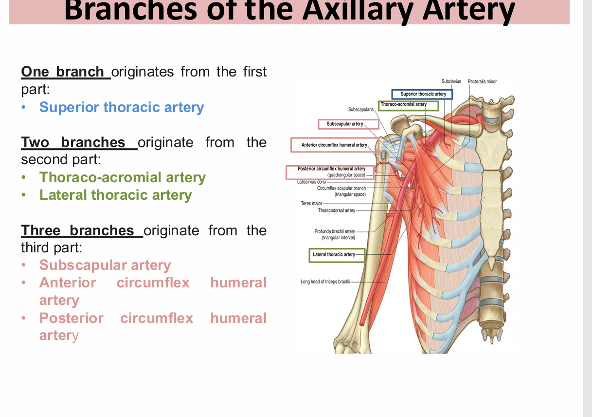
What are the origin, termination, parts, and branches of the axillary artery?
Apex/inlet: 1st rib, clavicle, superior border of scapula
Floor: skin + fascia stretched between anterior and posterior walls
Anterior wall: pectoralis major, pectoralis minor, clavipectoral fascia
Posterior wall: subscapularis, teres major, latissimus dorsi
Medial wall: serratus anterior + upper 4–5 ribs + intercostal muscles
Lateral wall: intertubercular groove of humerus
The anterior axillary fold = pectoralis major.
The posterior axillary fold = latissimus dorsi + teres major.AXILLARY SHEATH
Formation: ventral rami of C5–T1 Roots + trunks → posterior triangle of neck -Divisions → behind clavicle -Cords + branches → axilla
Prefixed:
Superior root = C4
Inferior root = C8
Postfixed:
Superior root = C6
Inferior root = T2
The inferior trunk may be compressed by the 1st rib, producing neurovascular symptoms in the upper limb.
In the 1st part, the Medial Cord is behind the artery, and Lateral/Posterior cords are lateral to it BUT IN THE SECOND PART ITS Lateral Cord: Lateral to artery Medial Cord: Medial to artery Posterior Cord: Posterior to artery
2ND PART
C5 → dorsal scapular nerve
Levator scapulae
Rhomboid major
Rhomboid minor
C5–C7 → long thoracic nerve
Serratus anterior
Clinical: injury to the long thoracic nerve → winged scapula.
C5 also contributes to the phrenic nerve, but the phrenic nerve is considered a branch of the cervical plexus, not the brachial plexus.
Upper trunk:
Nerve to subclavius → C5–C6
Suprascapular nerve → C5–C6
Lateral cord:
Lateral pectoral nerve → C5–C7
Musculocutaneous nerve → C5–C7
Lateral root of median nerve → C5–C7
think muesculocutaneous nerv.
Medial pectoral nerve → C8–T1 Medial cutaneous nerve of arm → C8–T1 Medial cutaneous nerve of forearm → C8–T1 Medial root of median nerve → C8–T1 Ulnar nerve → C7–T1
LATERAL ROOT+ MEDIAL ROOT
Upper subscapular nerve → C5–C6 Lower subscapular nerve → C5–C6 Thoracodorsal nerve → C6–C8 Axillary nerve → C5–C6 Radial nerve → C5–T1
Origin: continuation of subclavian artery at lateral border of rib 1
Termination: becomes brachial artery at inferior border of teres major
Divided by: pectoralis minor
What is Erb-Duchenne palsy, and what deformity does it produce?
A newborn has an arm hanging by the side, medially rotated, with a pronated forearm
What is Klumpke palsy, and what deformity does it produce?
What happens to the axillary vein at the 1st rib, and what is clinically important about its relationship to the axillary artery and brachial plexus?
Which two nerves are particularly at risk during axillary lymph node dissection, and what happens if each is injured?
these vessels form collateral circulation around the scapula/shoulder.
traction injury to C5–C6, particularly the upper trunk.
Affected functions:
Shoulder abduction
Shoulder lateral rotation
Forearm supination
Resulting position — "policeman's tip":
Arm adducted
Arm medially rotated
Forearm extended
Forearm pronated
Erb palsy (C5–C6).
Cause: injury to the lower roots C8–T1.
Possible causes:
Cervical rib
Birth injury
Excessive abduction of arm
Mainly affected: intrinsic/small muscles of the hand.
Deformity: claw hand
Hyperextension of MCP joints
Flexion of IP joints
During "subclavian" vein puncture, the actual point of entry may be into the terminal axillary vein, with the needle tip quickly entering the subclavian vein.Axillary vein lies superficial to the axillary artery and brachial plexus.This makes the vein relatively accessible for venous procedure
Long thoracic injury = winged scapula
Thoracodorsal injury = weakened latissimus function without characteristic deformityImportant contributors include:
Suprascapular artery
Transverse cervical artery
Subscapular artery
Circumflex scapular artery
Branches of subclavian/axillary systems
Axillary artery VEINS branches
Internal thoracic artery AND VEINS
Intercostal arteries AND VEINS

(MUSCLES OF BACK LECTURE)
How are the back muscles divided, and how do the groups differ in nerve supply, function, and examples?
Give the insertion, actions, and nerve supply of trapezius. What happens if its nerve is injured?
Patient cannot shrug shoulder against resistance →NERVE
Give the attachments, actions, and nerve supply of latissimus dorsi. What is the clinical consequence of injury to its nerve?
What are the attachments, actions, and innervation of levator scapulae and the rhomboids?
Which group is supplied by posterior rami
Compare the actions of erector spinae and transversospinales.
What are the attachments, nerve supply, and actions of serratus anterior? What happens when its nerve is injured?
Give the origin, insertion, nerve supply, and actions of pectoralis major.
Compare pectoralis minor, subclavius,
Extrinsic = movement of upper limb/rib
Intrinsic = vertebral column/posture
Intrinsic muscles = posterior rami
nsertion:
Lateral 1/3 of clavicle
Acromion
Spine of scapula
Actions:
Upper fibers → elevate scapula
Middle fibers → retract scapula
Lower fibers → depress scapula
Upper + lower fibers → rotate scapula to help raise arm above head
Nerve: Spinal accessory nerve (CN XI)
Lesion →
Drooping shoulder
Weak shoulder shrug
Difficulty raising arm above head
think CN XI → trapezius.Clavicle - lateral 1/3- Scapula - acromion and spinous proces
Insertion:
Bicipital groove of humerus
Actions:
Extension
Adduction
Medial rotation of arm
Nerve: Thoracodorsal nerve (C6–C8)
Injury → weakness pulling body upward, e.g.:
Climbing
Pull-ups
RHOMBOIS MAJOR+ MINOR ——-.>Insertion → medial border of scapula Action → retraction Nerve → dorsal scapular nerve Levator scapulae
Insertion → medial border of scapula
Action → elevates + medially rotates scapula
Nerve → dorsal scapular nerve (C5)
Intrinsic/deep back muscles.
Erector spinae
Bilaterally:
Extend vertebral column
Pull head posteriorly
Unilaterally:
Laterally flex vertebral column
Bend head toward contracting side
Transversospinales
Bilaterally:
Extend the back
Unilaterally:
Rotate trunk/head toward the opposite side
Insertion: ventral surface of medial border + inferior angle of scapula
Nerve: long thoracic nerve
Actions:
Protracts scapula
Holds medial border/inferior angle against thoracic wall
Important for movements such as punching/boxing
Long thoracic nerve injury → serratus anterior paralysis → winged scapula.
Insertion:
Lateral lip of bicipital groove
Nerve supply:
Medial pectoral nerve
Lateral pectoral nerve
Actions:
Adduction
Medial rotation
Flexion of arm
Pectoralis minor
Origin → ribs 3–5
Insertion → coracoid process
Nerve → medial pectoral nerve
Action → depresses shoulder
Subclavius
Origin → 1st rib/costochondral junction
Insertion → subclavian groove on inferior clavicle
Nerve → nerve to subclavius
Action → steadies clavicle
What are the boundaries and clinical significance of the auscultatory triangle and lumbar triangle?
What are the boundaries and contents of the quadrangular space?
Compare the triangular space and triangular interval: boundaries and structures passing through each.
What are the four rotator cuff muscles, their nerves, actions, and insertions?
Stabilize the humeral head within the glenoid cavity while preserving movement.AND Abducts 0–15°
what are the actions and innervation of deltoid?
WHAT IS Important sequence for abduction:
WHAT IS Clavipectoral fascia AND WHERE IS IT PIERCED BY?
Also remember that initial shoulder abduction (0–15°) is driven by supraspinatus (suprascapular nerve), whereas the deltoid takes over from 15–90°.
Auscultatory triangle
Boundaries:
Medial border of scapula
Trapezius
Latissimus dorsi
Clinical significance:
Area where breath sounds are most easily heard with a stethoscopE
Lumbar triangle / Triangle of Petit
Boundaries:
Latissimus dorsi
Posterior border of external oblique
Iliac crest
Clinical significance:
Potential site of abdominal hernia
Pus may emerge through the abdominal wall.
oundaries:
Superior → teres minor
Inferior → teres major
Medial → long head of triceps
Lateral → surgical neck of humerus
Contents:
Axillary nerve
Posterior circumflex humeral artery + vein Clinical relevance: injury around the surgical neck of humerus can endanger the axillary nerve.
Triangular space
Boundaries:
Superior → teres minor
Inferior → teres major
Lateral → long head of triceps
Contains:
Circumflex scapular artery + vein
Triangular interval
Boundaries:
Superior → teres major
Medial → long head of triceps
Lateral → shaft of humerus
Contains:
Radial nerve
Profunda brachii artery
ROTATOR CUFF MUSCLES
Insertion:
Deltoid tuberosity of humerus
Nerve:
Axillary nerve
Actions by fibers:
Anterior → flexion + medial rotation
Middle → abduction 15°–90°
Posterior → extension + lateral rotation
Supraspinatus: 0–15°
Deltoid: 15–90°Clavipectoral fascia
Located between:
Subclavius + pectoralis minor
Pierced by:
Lateral pectoral nerve
Thoracoacromial artery
Cephalic vein
Lymphatic vessels


LEG AND FOOT
What are the muscles of the posterior compartment of the leg, and how are they divided?
Which superficial posterior-compartment muscles plantar-flex the ankle, and which also flex the knee?
What structures pass deep to the flexor retinaculum from posterior → anterior?
If asked which nerve passes under the flexor retinaculUM?
A muscle initiates flexion of an extended knee by laterally rotating the femur on a fixed tibia. Which muscle?
What is its nerve supply?
Which muscle flexes the MTP and IP joints of the lateral four toes?
Does it assist ankle plantar flexion?
Which muscle flexes the MTP and IP joints of the great toe?
What other action does it have?
Which deep posterior muscle plantar-flexes AND inverts the foot?
Where does it insert mainly?
What muscles are found in each layer of the sole?
Which muscles of the sole are supplied by the medial plantar nerve?
What about the lateral plantar nerve?
What are the muscles of the anterior compartment?
What are the actions of tibialis anterior?
Dorsiflexion + inversion →
Which anterior-compartment muscle extends the lateral four toes?
Which extends the great toe?
Which dorsiflexes + everts the foot?
All are supplied by the??
What are the muscles of the lateral compartment, their nerve supply, and their main actions?
Eversion =??
A patient has numbness specifically in the skin between the great toe and second toe. Which nerve is involved?
A patient develops foot drop after an injury near the neck of the fibula. Which nerve is most likely injured?
Why does this cause foot drop?
What sensory deficit may occur?
tibial nerve 2 branches?
gastrocenamous important point
soleus important feature
The tendon penetrates the fibrous membrane of the joint capsule of the knee. • The tendon passes between the lateral meniscus and the fibrous membrane • The it attaches to and originates from a depression at the anterior end of the groove on the lateral femoral condyle???
In the sole, it crosses inferior to the tendon of the flexor hallucis longus muscle
Flexor digitorum brevis and abductor hallucis supplied by?
The Abductor digiti minimi supplied by the
Adductor hallucis and flexor digiti minimi brevis are supplied by
Flexor hallucis brevis is supplied by
interossi intervatd by
tibialis anterior insertionn is?
Insertion: Base of 5th, metatarsal bone?
extensor hoods?
dorsalis pedis artery?
A vascular surgeon evaluates a patient with suspected peripheral artery disease by palpating the dorsalis pedis pulse. To locate this pulse accurately, where should the physician place their fingers on the dorsum of the foot?
A.
Lateral to the tendon of extensor hallucis longus
Correct answer
The dorsalis pedis artery (continuation of the anterior tibial artery) is best palpated on the dorsum of the foot just lateral to the prominent tendon of the extensor hallucis longus.
dorsal interossi abducts toes (dab)
pad
Superficial: Gastrocnemius, Soleus, Plantaris
Deep: Popliteus, Flexor digitorum longus, Flexor hallucis longus, Tibialis posterior.
Nerve: Tibial nerve.
Artery: Posterior tibial artery.Gastrocnemius, Soleus and Plantaris → plantar-flex ankle.
Gastrocnemius + Plantaris → also flex knee.
All are supplied by the tibial nerve. G ANATOMY LEG AND FOOTTibialis posterior → FDL → posterior tibial vessels → tibial nerve → FHL
Mnemonic: Tom Does Very Nice Hats. G ANATOMY LEG AND FOOT
→ tibial nerve.
: Popliteus.
Tibial nerve.
Flexor digitorum longus (FDL).
YES
Flexor hallucis longus (FHL).
Assists plantar flexion of the ankle.
Tibialis posterior.
Tuberosity of navicular + adjacent medial cuneiform.
1st layer:
Abductor hallucis
Flexor digitorum brevis
Abductor digiti minimi
2nd layer:
Quadratus plantae
Lumbricals
3rd layer:
Flexor hallucis brevis
Adductor hallucis
Flexor digiti minimi brevis
4th layer:
3 plantar interossei
4 dorsal interosse
A:
Abductor hallucis
Flexor digitorum brevis
Flexor hallucis brevis
1st lumbrical. G ANATOMY LEG AND FOOT
Supplies most of the remaining intrinsic muscles, including the interossei, adductor hallucis, flexor digiti minimi brevis, quadratus plantae and lumbricals except the 1st.
A:
Tibialis anterior
Extensor digitorum longus
Extensor hallucis longus
Fibularis tertius
Nerve: Deep fibular nerve
Artery: Anterior tibial arteryDorsiflexion + inversion of the foot.
Also helps maintain the medial longitudinal arch.Nerve: Deep fibular nerve
Tibialis anterior.
Extensor digitorum longus.
Extensor hallucis longus.
Fibularis tertius.
Deep fibular nerve.
A:
Muscles: Fibularis longus + Fibularis brevis
Nerve: Superficial fibular nerve
Actions: Plantar flexion + eversionthink fibularis muscles.
Deep fibular nerve.
It supplies the 1st interdigital web space, the only cutaneous area supplied by this nerve. G ANATOMY LEG AND FOOT
Clinical variation: Deep fibular nerve entrapment can cause pain radiating to this same web space (“ski boot syndrome”).
Common fibular nerve.
Injury causes loss of dorsiflexion, producing foot drop.
Variable sensory loss over the anterolateral leg and dorsum of the foot.
Tibial nerve (branch of sciatic nerve). • It has two cutaneous branches, the sural nerve and the medial calcaneal nerve. • The sural nerve supplies skin on the lower posterolateral surface of the leg and the lateral side of the foot and little toe
Insertion : tendon). > the calcaneal tendon (Achilles Special Features : The lateral head may contain a sesamoid bone called fabella
The muscle contains a rich plexus of veins, which is connected to the great saphenous vein by a series of perforating veins
poplitius
flexor digitorum longus
medial planter N.
lateral planatar nerve
lateral planatar
medial planatar
lateral planter N
Insertion: Medial cuneiform and base of 1st, metatarsal bone
fibularis tertius
The tendons of the extensor digitorum longus, extensor digitorum brevis, and extensor hallucis longus pass into the dorsal aspect of the digits and expand over the proximal phalanges to form dorsal digital expansions, forming extensor hoods
he dorsalis pedis artery (continuation of the anterior tibial artery) begins as the anterior tibial artery crosses the ankle joint. It passes inferiorly, as the deep plantar artery, between the two heads of the first dorsal interosseous muscle to join the deep plantar arch in the sole (Its pulse of the dorsalis pedis artery on the dorsal surface of the foot can be fel

BACK OF THIGH
Which muscles make up the hamstrings, and where are they positioned?
What are the origins of the 3 hamstrings?
Give the origin, insertion, nerve supply, and actions of biceps femoris
A lesion affects the common fibular division of the sciatic nerve. Which hamstring component is denervated?
Give the origin, insertion, nerve supply, and actions of semitendinosus.
Give the origin, insertion, nerve supply, and actions of semimembranosus.
Which hamstrings flex the knee, extend the hip, and medially rotate?
Which hamstring contributes to pes anserinus, and where does it insert?
What forms the four muscular boundaries of the popliteal fossa?
What form the floor and roof of the popliteal fossa?
What are the major contents of the popliteal fossa?
Describe the course and termination of the popliteal artery.
How do the tibial and common fibular nerves course through the popliteal fossa?
Where is the popliteal vein relative to the artery, and what does it become?
A patient has a lesion involving the medial hamstrings. Which actions are weakened, and which nerve division is affected?
A:
Biceps femoris → lateral
Semitendinosus → medial
Semimembranosus → medial
Exam trap: Biceps femoris is the lateral hamstring; the other two are medial.
A:
Biceps femoris long head → inferomedial upper ½ ischial tuberosity
Semitendinosus → same area
Semimembranosus → superolateral upper ½ ischial tuberosity
High-yield: Long head BF + semitendinosus share the origin.
A:
Long head: ischial tuberosity → tibial division
Short head: linea aspera → common fibular division
Both insert → head of fibula
Hip: extends + laterally rotates
Knee: flexes + laterally rotates
A: Short head of biceps femoris.
Why:
Long head → tibial division
Short head → common fibular division.This is a classic distinction.
A:
Origin → ischial tuberosity
Insertion → medial proximal tibia via pes anserinus
Nerve → tibial division of sciatic
Hip → extension + medial rotation
Knee → flexion + medial rotation
A:
Origin → superolateral upper ½ ischial tuberosity
Insertion → medial/posterior medial tibial condyle
Nerve → tibial division of sciatic
Hip → extension + medial rotation
Knee → flexion + medial rotation
A: Semitendinosus + semimembranosus.
Contrast:
Biceps femoris → lateral rotation.A: Semitendinosus contributes to pes anserinus.
It inserts on the upper medial surface of the tibia, behind sartorius and gracilis.
S.G.S. = sartorius, gracilis, semitendinosus
A:
Superolateral: biceps femoris
Superomedial: semitendinosus + semimembranosus
Inferolateral: lateral head gastrocnemius + plantaris
Inferomedial: medial head gastrocnemiusShape: diamond-shaped.
A:
Floor: knee joint capsule, adjacent femur/tibia, popliteus.
Roof: skin + superficial fascia + deep fascia.Superficial fascia contains small saphenous vein + posterior cutaneous nerve of thigh.
A:
Tibial nerve
Common fibular nerve
Popliteal vein
Popliteal artery
Superficial → deep:
Nerves → vein → arteryContinuation of femoral artery.
It is the deepest major structure in the fossa.
Passes obliquely downward.
Ends by dividing into anterior + posterior tibial arteries.Both are branches of sciatic and enter beneath biceps femoris.
Tibial: descends vertically.
Common fibular: follows medial border of biceps femoris.Both are more superficial than the popliteal vessels.
A:
It lies superficial to the popliteal artery and travels with it.Superiorly, it continues as the femoral vein.
A:
Weak knee flexion, hip extension, and medial rotation.Affected muscles: semitendinosus + semimembranosus.
Nerve: tibial division of sciatic nerve.
Contrast: biceps femoris → lateral rotation.
GLUTEAL
What defines the gluteal region, and what are the functions of its 2 major ligaments?
What structures pass through the greater sciatic foramen superior to piriformis?
What structures pass through the greater sciatic foramen inferior to piriformis?
What enters and exits the lesser sciatic foramen?
Give the origin, insertion, nerve supply and actions of gluteus maximus.
Why is gluteus maximus the chief antigravity muscle of the hip?
What are the actions and nerve supply of gluteus medius and minimus?
A patient has superior gluteal nerve injury. What happens and why?
Give the origin, insertion, nerve supply, action and key importance of piriformis.
Give the origin, insertion, nerve supply and action of obturator internus.
Gluteal region: behind pelvis, iliac crest → gluteal fold.
Ligaments: sacrospinous + sacrotuberous.
They convert sciatic notches → greater/lesser sciatic foramina and stabilize sacrum/prevent posterior rotation.Superior gluteal nerve + superior gluteal vessels.
TRAP: Piriformis divides the greater sciatic foramen into above vs below.
A:
Inferior gluteal nerve/vessels
Sciatic nerve
Posterior cutaneous nerve of thigh
Pudendal nerve + internal pudendal vessels
Nerve to obturator internus
Nerve to quadratus femoris
High-yield: Most structures pass below piriformis.
Enters: pudendal nerve + internal pudendal vessels.
Exits: tendon of obturator internus.Think: Pudendal nerve leaves pelvis → loops around → re-enters through lesser sciatic foramen.
Origin: ilium, sacrum/coccyx, sacrotuberous ligament.
Insertion: ¾ IT tract; deep fibers → gluteal tuberosity.
Nerve: inferior gluteal (L5–S2).
Action: hip extension + lateral rotation.Powerful hip extension raises the trunk against gravity.
Important activities:
Standing from sitting
Running
Climbing stairs
Also stabilizes hip/knee through the iliotibial tract.
Both:
Abduct thigh
Medially rotate thigh
Stabilize pelvis during walking
Nerve: superior gluteal nerve (L4–S1).
Paralysis/weakness of gluteus medius + minimus → inability to stabilize pelvis during single-leg stance.
The pelvis drops on the unsupported/swing side → Trendelenburg sign and waddling gait.
Origin: anterior sacrum.
Insertion: upper greater trochanter.
Nerve: nerve to piriformis (S1–S2).
Action: lateral rotation + abduction of femur.
Importance: landmark dividing greater sciatic foramen into superior/inferior parts.Origin: medial obturator membrane + adjacent bone.
Insertion: greater trochanter.
Nerve: nerve to obturator internus (S1–S2).
Action: lateral rotation + abduction of femur.Key anatomical fact: Its tendon bends ~90° through the lesser sciatic foramen
What are the attachments, actions and nerve supplies of the superior and inferior gemelli?
: Give the origin, insertion, nerve supply and action of quadratus femoris.
Match the major gluteal nerves to the muscles they supply.
Compare the superior and inferior gluteal arteries.
Where is the safe site for gluteal IM injection, and what structure is being protected?
Both attach to the obturator internus tendon and assist its action.
Superior: ischial spine → nerve to obturator internus.
Inferior: ischial tuberosity → nerve to quadratus femoris.Action: lateral rotation + abduction.
Origin: anterior to ischial tuberosity.
Insertion: quadrate tubercle/intertrochanteric crest.
Nerve: nerve to quadratus femoris.
Action: lateral rotation of femur.Superior gluteal → gluteus medius + minimus.
Inferior gluteal → gluteus maximus.
Nerve to piriformis → piriformis.
Nerve to obturator internus → obturator internus + superior gemellus.
Nerve to quadratus femoris → quadratus femoris + inferior gemellus.Both arise from internal iliac artery.
Superior gluteal: exits greater sciatic foramen above piriformis → accompanies superior gluteal nerve.
Inferior gluteal: exits below piriformis → accompanies inferior gluteal nerve; mainly supplies gluteus maximus.
Upper outer quadrant of buttock.
It minimizes risk of injuring the sciatic nerve, which passes through the gluteal region.
The things I REALLY want you to know
If you can retrieve these without looking, you're in a much stronger position:
Piriformis = landmark
Above piriformis:
→ superior gluteal nerve + vessels
Below piriformis:
→ sciatic nerve
→ inferior gluteal nerve + vessels
→ posterior cutaneous nerve of thigh
→ pudendal nerve + internal pudendal vessels
→ nerves to obturator internus + quadratus femoris
Superior gluteal nerve
→ gluteus medius + minimus
→ Trendelenburg
Inferior gluteal nerve
→ gluteus maximus
Gluteus maximus
→ extension + lateral rotation
Gluteus medius/minimus
→ abduction + medial rotation
Deep lateral rotators
→ piriformis, obturator internus, gemelli, quadratus femoris
Safe IM injection
→ upper outer quadrant → avoid sciatic nerve
SUMMARY
Rapid-Fire Self-Test
What nerve innervates Gluteus Maximus?
.
If the left superior gluteal nerve is cut, which side of the pelvis drops during walking?
What is the safe quadrant for gluteal injections?
Which muscle exits the pelvis through the greater sciatic foramen?
Which muscle tendon exits through the lesser sciatic foramen?
Answer: Inferior Gluteal Nerve
Answer: The Right side (the unsupported swing limb).
Answer: Outer upper (superolateral) quadrant.
Answer: Piriformis.
Answer: Obturator Internus.
: What is fascia lata, what does its lateral thickening form, and what opening does it contain?
What are the key features of fascia lata?
Give the origin, insertion, nerve supply and actions of sartorius.
Name the 4 quadriceps heads, their common insertion/nerve, and main action.
What distinguishes rectus femoris from the three vasti?
Compare iliacus and psoas major by origin, innervation and action.
What are the boundaries, floor and roof of the femoral triangle?
What structures lie in the femoral triangle from lateral → medial?
Describe the origin, course, termination and major branch of the femoral artery.
A:
Anterior: iliopsoas, sartorius, quadriceps → femoral N
Medial: adductors, gracilis, pectineus, obturator externus → mainly obturator N
Posterior: hamstrings → sciatic N
Trap: Pectineus can receive femoral innervation.
Thick deep fascia surrounding thigh.
Laterally thickens → iliotibial tract.
Contains saphenous opening, through which great saphenous vein enters femoral vein.A:
O: ASIS
I: upper medial tibia
N: femoral N
Actions: hip flexion + abduction + lateral rotation; knee flexion.Memory: “tailor's muscle.”
Rectus femoris, vastus lateralis, medialis, intermedius.
All → quadriceps tendon → patella → ligamentum patellae → tibial tuberosity.
Nerve → femoral N.
Action → knee extension.
RF is the only head crossing the hip → also flexes hip.
Rectus femoris: crosses hip + knee → hip flexion + knee extension.
Vasti: cross only knee → knee extension.
RF originates from AIIS + ilium above acetabulum.
Iliacus: iliac fossa → femoral N → hip flexion.
Psoas major: T12–L5 vertebral bodies/discs + transverse processes → anterior rami L1–L3 → hip flexion.
Both insert at lesser trochanter and act as hip flexors.
Base: inguinal ligament
Lateral: sartorius
Medial: adductor longus
Apex: continues into adductor canalFloor medial → lateral: adductor longus → pectineus → iliopsoas.
Roof: fascia lata + saphenous opening.
Nerve → artery → vein → lymphatics
= NAVEL
Femoral nerve lies outside the femoral sheath.
Femoral artery, vein and lymphatics are enclosed by the femoral sheath.
Continuation of external iliac artery after passing beneath inguinal ligament.
→ femoral triangle → adductor canal → adductor hiatus.
After hiatus → popliteal artery.
Major branch: profunda femoris, the major blood supply of thigh.
Describe the femoral vein and distinguish the great vs small saphenous veins.
What are the course, major motor branches and cutaneous territories of the femoral nerve?
What are the major medial thigh muscles and their principal actions?
: Why is adductor magnus unusual? Give its 2 parts, innervation and key anatomical feature.
Which medial thigh muscles have notable nerve-supply exceptions?
What is a femoral hernia, where does it occur, and why can it strangulate?
Femoral vein = continuation of popliteal vein → becomes external iliac vein above inguinal ligament.
Tributaries include great saphenous + profunda femoris veins.
Great saphenous: medial dorsal venous arch → femoral vein.
Small saphenous: lateral dorsal venous arch → popliteal vein.
From lumbar plexus → emerges lateral to psoas → passes posterior to inguinal ligament → enters femoral triangle outside femoral sheath.
Motor: iliacus, pectineus, sartorius, quadriceps.
Cutaneous:
medial/intermediate cutaneous nerves → anterior thigh
saphenous nerve → medial leg
Pectineus: flexes + adducts thigh
Gracilis: adducts thigh + flexes knee
Adductor longus: adduction + medial rotation
Adductor brevis: adduction + medial rotation
Adductor magnus: powerful adduction + medial rotation
Obturator externus: lateral rotationMost are supplied by obturator N.
A:
Pubic part: obturator N → adduction + medial rotation.Ischial part: sciatic N → adduction.
Ischial part inserts on adductor tubercle.
A gap between its parts = adductor hiatus, transmitting femoral vessels → popliteal vessels.
Major trap: Adductor magnus has dual innervation.
A:
Pectineus → femoral N (classically; may receive obturator/accessory obturator)
Adductor magnus → obturator + sciatic
Obturator externus → obturator N
Gracilis → obturator N
Exam focus: Don't assume every medial muscle is simply obturator nerve.
Abdominal viscera protrude through the femoral ring → femoral canal.
Presents as a mass inferolateral to pubic tubercle.
May enlarge toward the saphenous opening.
Strangulation risk: rigid/sharp lacunar ligament can constrict the hernia.
HE EXAM TRAPS I WANT YOU TO KNOW COLD
These are the ones I'd actively test yourself on:
NAVEL
Femoral Nerve → Artery → Vein → Empty space/Lymphatics
Femoral nerve = OUTSIDE femoral sheath.
Femoral artery
External iliac → femoral → adductor hiatus → popliteal
Femoral vein
Popliteal → femoral → external iliac
Great saphenous
medial dorsal venous arch → femoral vein
Small saphenous
lateral dorsal venous arch → popliteal vein
Quadriceps
4 heads → knee extension
Rectus femoris alone crosses hip
Iliopsoas
major hip flexor
Adductor magnus
obturator + sciatic
adductor hiatus → femoral vessels
Femoral hernia
femoral ring/canal → inferolateral to pubic tubercle
SUMMARY
Your lecture says the adductor hiatus gives passage for “femoral vessels.” More precisely, the femoral artery and vein pass through the hiatus and become the popliteal artery and vein posterior to the knee.
And one other important nuance: the lecture's “femoral nerve supplies pectineus” is a useful exam answer, but pectineus can have variable/accessory innervation, so don't let that confuse you if you encounter it in a more detailed source