Dentals
 Note
Note Call Kai
 Learn
Learn Practice Test
 Spaced Repetition
 Match
Match Flashcards
 Knowt Play
1/45

There's no tags or description
Looks like no tags are added yet.
Name | Mastery | Learn | Test | Matching | Spaced | Call with Kai | Chat |
|---|
No analytics yet
Send a link to your students to track their progress
46 Terms
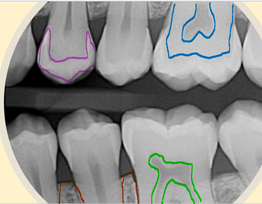
what is the structure highlighted in pink?
enamel
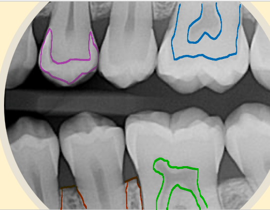
what is the structure highlighted in blue?
dentin
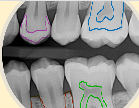
what is the structure highlighted in red?
bone
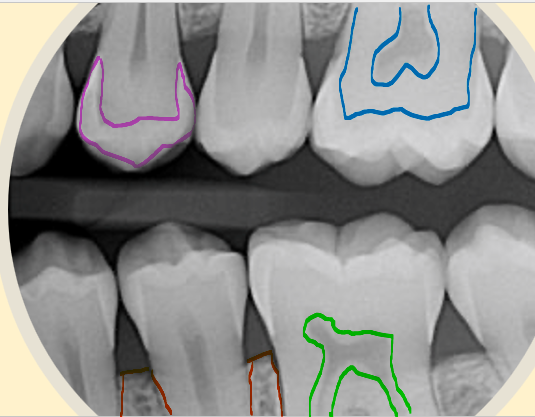
what is the structure highlighted in green?
pulp
what are the four quadrants in palmer’s notation?
upper left, upper right, lower left, lower right
which are the maxillary teeth?
upper teeth
which are the mandibular teeth?
lower teeth
what does 1 represent in palmer’s notation?
central incisor
what does 2 represent in palmer’s notation?
lateral incisor
what does 3 represent in palmer’s notation?
canine
what does 4 represent in palmer’s notation?
first premolar
what does 5 represent in palmer’s notation?
second premolar
what does 6 represent in palmer’s notation?
first molar
what does 7 represent in palmer’s notation?
second molar
what does 8 represent in palmer’s notation?
third molar
what are the two types of dental x-rays?
intraoral and extraoral
what are the different types of intraoral techniques?
periapical, bitewing, and occlusal
what are the two different periapical techniques?
paralleling and bisecting angle techniques.
what are the different types of extraoral techniques?
lateral cephalometry, OPG, lateral oblique mandible, PA mandible
what does OPG stand for?
Orthopantomogram
what is a periapical dental x-ray?
captures the entire tooth structure and surrounding alveolar bone, to assess the 2nd to 4th teeth
what are the clinical indications for a periapical x-ray?
apical infection/cyst, impacted tooth, root morphology prior to surgery, evaluate implants
what is the parallel technique for a periapical x-ray?
the x-ray film is parallel to the long axis of the teeth with the central beam perpendicular to the film/tooth
what are the benefits of the parallel periapical technique?
reduces distortion, has true anatomical relation, reduces magnification, reproducible
what are the disadvantages to a parallel periapical x-ray?
requires a film holder which can be hard to place and is uncomfortable to patient
what is the bisecting technique for periapical x-rays?
film is positioned along the tooth and the central ray is directed perpendicular to an imaginary line that bisects the angle formed by the long axis of the tooth and the film
what are the advantages to the bisecting periapical x-ray?
no need for film holder, more comfortable to the patient, quicker
what are the disadvantages to the bisecting periapical x-ray?
higher chance of magnification and super imposition, not reproducible
what is a bitewing dental x-ray?
captures both maxillary and mandibular teeth, interproximal area and bone in a single film
what are the clinical indications of a bitewing dental x-ray?
detection of cavities, periodical review, evaluate alveolar bone level and pulp chamber
what is the postioning for a bitewing x-ray?
film is placed parallel to the crown of the teeth then is stabilized by the patient biting onto a tab, beam is perpendicular to the receptor
what is an occlusal dental x-ray?
technique which exposes all the maxilla or mandible
what are the clinical indications for an occlusal x-ray?
detection of presence of unerupted teeth, supernumeraries and odontomes, evaluate size and extent of lesions
what is a positioning for a maxillary occlusal technique?
patient is seated with IR on occlusal plane which is parallel to floor then patient bites down gently
tube positioned either above patient angled down at 65 degrees or below patient angled up at 65 degrees
what is an orthopantomogram?
a panoramic single image radiograph of the mandible, maxilla and teeth
what are the clinical indications of an OPG?
general dental health evaluation, trauma assessment for fractures, infection evaluation, temporomandibular joint assessment, growth monitoring of paediatric teeth
what are the advantages of OPGs?
broad anatomical region, lower dose, can view both sides of jaw, allows comparisons of condylar heads
what are the disadvantages of OPGs?
poor anatomical detail, soft tissue and air shadows can superimpose the structures
what is the positioning for an OPG?
MSP is perpendicular to floor, frankfort line is parallel to floor, patient is erect with their head straight, incisors are placed correctly on bite block
what happens to the appearance of the OPG if the patient’s chin is too high?
condyles may not be visible, maxillary incisors will appear blurred and magnified
what happens to the appearance of the OPG if the patient’s chin is too low?
condyles will appear higher on image and mandibular incisors will appear blurred and roots will look shorter
what happens to the appearance of the OPG if the patient sits too far forward?
anterior teeth will be narrowed and spine will be visible
what happens to the appearance of the OPG if the patient sits too far back?
anterior teeth will be magnified and ramus wont be entirely visible
what happens to the appearance of the OPG if the MSP isnt centred?
ramus and posterior teeth will appear unequally magnified - the side furthest from receptor will appear magnified
what is a lateral cephalogram?
evaluates the relationship between structures of the facial skeleton, primarily for orthodontic assessment
what are the clinical indications of a lateral cephalogram?
dental/skeletal development, analysis of dentak/skeletal relationships, orthodontic or maxillofacial treatment, planning and monitoring