2 - Family Fasciolidae, Superfamily Paramphistomoidea
 Call Kai
 Learn
Learn Practice Test
 Spaced Repetition
 Match
Match Flashcards
 Knowt Play
1/101

There's no tags or description
Looks like no tags are added yet.
Name | Mastery | Learn | Test | Matching | Spaced | Call with Kai | Chat |
|---|
No analytics yet
Send a link to your students to track their progress
102 Terms
Large flukes mostly in the bile ducts of ungulates
FAMILY FASCIOLIDAE
Ventral sucker close to oral sucker
FAMILY FASCIOLIDAE = Distomes
Ovary and testes branched and ceca plus excretory bladder usually
highly branched or dendritic
FAMILY FASCIOLIDAE
Usually in digestive tract of fishes, amphibians, reptiles, birds and mammals
SUPERFAMILY PARAMPHISTOMOIDEA
Usually thick, fleshy and circular in transverse
section, some are cone-shaped
SUPERFAMILY PARAMPHISTOMOIDEA
Ventral sucker at posterior end
SUPERFAMILY PARAMPHISTOMOIDEA = Amphistomes
eggs – large (130–150 μm long by 60–90 μm wide), thin-
shelled, operculate, unembryonated when pass with the
feces
Family Fasciolidae
IH of Family Fasciolidae
FW aquatic snails
Fasciolidae species that are zoonotic
Fasciola hepatice, gigantica
Fasciolopsis buski
common liver fluke
Fasciola hepatica
in the bile ducts of large and small
ruminants, pigs, hare, rabbits, beavers,
coypus, elephants, horses, dogs,
kangaroos, cat and man throughout the
world
Fasciola hepatica
Fasciola hepatica can also occur where except for the usual bile ducts
may also occur in the lungs, under the
skin, etc. of unusual hosts, e.g. man
and horse
causes liver rot or liver fluke disease
Fasciola hepatica or gigantica
eggs of Fasciola are color __ when _ and its yolk balls or vitelline cells are less distinct or more diffuse vs. amphistome eggs
yellow when fresh
Fasciola eggs embryonate in __ and hatch to into _ at what °C
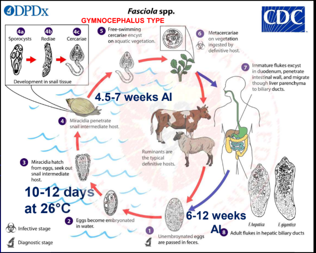
embryonate in water and hatch to miracidia in 10-12
days at 26°C
most important snail IH of Fasciola hepatica
Lymnaea truncatula
Stagnicola (Lymnaea) bulimoides
L. columella
L. tomentosa
L. viator
L. cubensis
explain life cycle of Fasciola hepatica (also for others in the family fasciolidae and Superfamily Paramphistomoidea = same only)
daughter rediae are produced under unfavourable
condition
cercariae – settle within few to 2 h on blades of grass or
other plants just below water level or on water surface,
cast off tail and form cyst to become metacercariae
(size: 280 μm or 0.28 mm in diameter)
after ingestion by DH
- metacercariae excyst in the duodenum; young flukes
emerge after being elicited by a bile component,
glycocholic acid
young flukes intestinal wall most are in the
abdominal cavity within 24 h have penetrated the liver
capsule by 4-6 d AI migrate in liver parenchyma where
they feed for 4-7 weeks
finally, they enter bile ducts and mature
1 egg of fasciola is equivalent to how many metacercariae and is due to
1 egg = ~600 metacercariae because of paedogenesis
or polyembryony
Pre-patent period and longevity of fasciola heaptica
6-12 weeks
11 years
Pathogenesis of fasciola hepatica - penetrates _
intestine and Glisson’s capsule – little damage
liver effects of fasciola hepatica are due to 3
a) Mechanical destruction
b) Toxic excretory and secretory (ES) products
c) Loss of blood
migrating flukes and irritation
of bile ducts by the adults
Mechanical destruction
inflammation, edema, and fibrosis of the bile ducts
(pipestem fibrosis or liver); consequence of the latter
is atrophy of the liver due to back pressure, cirrhosis
and possibly jaundice
Toxic excretory and secretory (ES) products
due to liver hemorrhage (acute form)
and hematophagous feeding of the adults anemia is
perhaps caused by parasite proline; proline also induces
bile duct collagen deposition
Loss of blood
more prone to the pathogenic effects of
infection of fasciola hepatica; no age resistance
young hosts
for sheep infection of fasciola hepatica, what constitutes as moderate infection
less than 30 adults
for sheep infection of fasciola hepatica, what constitutes as medium infection
30-200 adults
for sheep infection of fasciola hepatica, what constitutes as severe infection
more than 200 adults
clinical forms of fasciolosis
acute
subacute
chronic
subclinical
occurs after sudden massive ingestion of metacercariae occurs after a few days
Acute fasciolosis
Acute fasciolosis occurs with acute, traumatic and hemorrhagic hepatitis with
appearance of enzymes like
aspartate transferase (AST),
glutamate dehydrogenase (GDH), and sorbitol
dehydrogenase (SDH), in plasma from 2nd week AI
after longer massive ingestion of metacercariae in the course of 4-8 weeks
Subacute fasciolosis
subacute, traumatic and hemorrhagic hepatitis, peritonitis,
enlarged liver, cholangitis, liver fibrosis, loss of appetite
and weight, progressive hemorrhagic anemia, liver failure,
death
subacute fasciolosis
following prolonged moderate intake of metacercariae, occurs for several months
Chronic fasciolosis
lowered appetite, slowly developing
hypoproliferative anemia, emaciation, edema under
the jaw (bottle jaw), hypoalbuminemia, appearance
of gamma glutamyl transferase or transpeptidase
(GGT) in the plasma indicates blockade or chronic
damage of bile ducts
Chronic fasciolosis
chronic fasciolosis can be determined by the appearance of what enzymes in the plasma which indicates
appearance
of gamma glutamyl transferase or transpeptidase
(GGT) in the plasma indicates blockade or chronic
damage of bile ducts
most common form of fasciolosis, in very low level
and prolonged intake of metacercariae
subclinical form
CS of subclinical fasciolosis
moderate cholangitis
how are antibodies of fasciola hepatica detected
specific antibodies can be detected from 3rd week AI by
ELISA, IHA, etc.
cattle and sheep condition of fasciolosis
no evidence of acquired resistance in sheep and cattle
cattle can recover spontaneously and partially resist
reinfection
how is fasciolosis diagnosed
grazing history
clinical signs
fecal examination (sedimentation (NOT FLOTATION), Stoll egg counting
technique)
serology – ELISA for blood and milk samples (BEST method to detect Abs early at 3wks AI at greatest damage on migratory period to liver parenchyma)
Liver enzymes detection in plasma
flukecide treatment for 1-6 days of fasciolosis
Diamphenetide
currently the drug of choice for fasciolosis + the other one
Triclabendazole
Bithionol
flukecide treatment for 6wks and older of fasciolosis
Rafoxanide, Closantel, Nitroxynil
flukecide treatment for >12 wks r of fasciolosis
Albendazole, Brotianide, Clorsulon, Niclofolan, Netobimin, Oxyclozanide
ABCNNO
how can fasciolosis be prevented
farm management and grazing practices – zero grazing,
avoidance or fence off snail-infested areas, drain wet
areas or improve water flow
control snail – ducks, by chemicals only when negative
environmental impact is minimal (niclosamide, sodium
pentachlor phenate, copper sulfate)
chemicals to use when environmental impact of fasciola is minimal
niclosamide, sodium pentachlor phenate, copper sulfate
large liver fluke
F. gigantica
more common in Africa, Asia (including the Phils.),
Hawaii and tropical and subtropical regions
F. gigantica
DH of F. gigantica that is relatively resistant
Indonesian Thin Tail sheep
most important helminth of livestock in PH
F. gigantica
IH of F. gigantica
Radix rubiginosa (formerly Lymnaea auricularia rubiginosa SEA, PH)
L. natalensis (Africa)
L. viridis (China, Hawaii, Japan)
L. rufescens (Indian subcontinent),
L. acuminata (Malaysia)
metacercariae of F. gigantica can survive up to how many months
survive up to 4 months on stored straw
PPP of F. gigantica in host
12 weeks or more (longer than hepatica)
difference in the pathogenesis of F. hepatica and gigantica
12 weeks or more
large American liver fluke, giant deer fluke
Fascioloides magna
in bile ducts and or liver tissue
of white-tailed deer, other
Cervidae (roe deer, moose,
elk, fallow deer) wapiti, cattle,
bison, sheep, goat, pig, horse,
etc. of North America, central
Europe (invasive species),
Mexico and South America
Fascioloides magna
eggs – single-cell stage when
passed with the feces
Fascioloides magna
Fasciolidae member where its eggs cannot be sen on fecalysis and does not go out into the liver and causes patent infection to bovids
Fascioloides magna
pathogenesis of fascioloides magna in deer
deer – found in thin-walled, loose vascular
connective tissue cysts with bile duct connections;
associated with reduced body size and lowered
number of antler points (less “pogi”!)
pathogenesis of fascioloides magna in bovids
enclosed in thick-
walled or calcareous cysts
preventing discharge of
eggs, cause patent infection
pathogenesis of fascioloides magna in sheep and goats
sheep and goats – migration in the liver is
unrestricted due to absence of cyst formation = Non encapsulated
more severe infection than bovids or deer
effect of Fascioloides magna on the excretory system of DH
produces large amount of melanin
(black iron porphyrin pigment); the pigment accumulates in
liver, omentum, kidneys and other internal organs of hosts
causes anemia, loss of weight,
hypoproteinemia, submandibular edema and
sometimes death in Asian elephants
F. (=Fasciola) jacksoni
large intestinal fluke
Fasciolopsis buski
in small intestine, rarely the stomach and colon, of humans, pigs and dogs in
the Orient (South and East Asia such as China, Indonesia, Vietnam, India
and Thailand)
large (3.0-7.5 by 0.8-2.0
cm), thick-set fluke,
without shoulders
ceca not branched
Fasciolopsis buski
IH of Fasciolopsis buski
flat, spiralled freshwater snails
(Planorbis, Hippeutis and Segmentina)
metacercariae of Fasciolopsis buski are formed where and in what plants
underwater vegetation
- water chestnut Eliocharis tuberosa)
- water caltrop (Trapa natans, T. bicornis)
- lotus and water bamboo
how do DH of Fasciolopsis buski get infected
DH – eating of contaminated vegetation; the tubers
are eaten raw and often, children remove the outer
shell by their teeth; matures in about 3 months
heavy infection of Fasciolopsis buski causes what
diarrhea, ulceration and abscessation
of intestinal mucosa, anemia, and hypersensitization in
humans
treatment for fasciolopsis buski infection
albendazole, praziquantel
prevention for fasciolopsis buski infection
improved hygiene in vegetable production and consumption
in gall bladder and digestive tract of elks and wild goats
in the former USSR and Poland
Parafasciolopsis fasciolaemorpha
IH of Parafasciolopsis fasciolaemorpha
Planorbis corneus that inhabits swampy areas
family members of Superfamily Paramphistomoidea (amphistomes)
Family Paramphistomidae
Family Gastrothylacidae
Family Gastrodiscidae
in mammals, especially herbivores
body is “undivided” without a ventral pouch
Family Paramphistomidae
body is “undivided” with a ventral pouch
Family Gastrothylacidae
body is “divided” without a ventral pouch
Family Gastrodiscidae
in rumen of domestic
animals throughout the
world, conical and pink
when fresh
Paramphistomum cervi
Paramphistomid in European cattle
Paramphistomum daubneyi
Paramphistomid in Australasia
Paramphistomum ichikawa
Paramphistomid in rumen and reticulum of ruminants
Calicophoron
Orthocoelium
Cotylophoron spp.
Paramphistomid in rumen and abomasum of ruminants
Gigantocotyle formosanum
Paramphistomid in bile ducts and liver
Explanatum (=Gigantocotyle)
Paramphistomid in in stomach of beavers
Stichorchis subtriquetrus
Family Gastrothylacidae sp in in rumen and reticulum, testes – horizontal
Gastrothylax
Family Gastrothylacidae sp with ventral pouch – very large, extending over the whole ventral surface to posterior sucker
Gastrothylax crumenifer
Family Gastrothylacidae sp in rumen of
bovids
testes – tandem
uterus – runs forward in
midline
Fischoederius
most common
amphistome in cattle and
water buffaloes in the
Philippines,
Fischoederius elongatus
IH in the Philippines of Fischoederius elongatus
Gyraulus convexiusculus
Lymnaea philippinensis
Carmyerius of Family Gastrothylacidae occurs in where
rumen
occurs in rumen of water buffaloes in the
Philippines (Family Gastrothylacidae)
Velasquezotrema brevisaccus
in LI, ceca, rectum, and bile ducts of cattle, water
buffaloes, and other ruminants
Homalogaster paloniae
in intestines of equids and pigs
Gastrodiscus aegyptiacus
cecum and colon of pigs, also in primates, man as
accidental host being considered the only common
amphistome of man
Gastrodiscoides hominis
IH of amphistomes
FW or amphobious smails (Planorbis, Lymnaea, Gyraulus)
discuss specific LC of amphistomes that differes from the similar LC of F. hepatica
after ingestion
- metacercariae excyst in duodenum mucosa
migrate anteriorly through the tissues abomasum
creep to the rumen, mature in 2-4 months while attached
to the villi
- for other species, migrate to the large intestine or bile
ducts
cercariae of amphostomes are
large, pigmented and have eyespots
metacercariae of amphistomes may survive up to
3 months in pasture
Pathogenesis of amphistomes forms
acute or intestinal amphistomosis - migrating juveniles
following excystation
chronic amphistomosis
hepatic amphistomosis