Disorder of Primary Hemostasis
 Call Kai
 Learn
Learn Practice Test
 Spaced Repetition
 Match
Match Flashcards
 Knowt Play
1/16

Earn XP
Description and Tags
Chapter 26
Name | Mastery | Learn | Test | Matching | Spaced | Call with Kai | Chat |
|---|
No analytics yet
Send a link to your students to track their progress
17 Terms
Platelet Satellitism
Platelets clump around neutrophils that leads to pseudothrombocytopenia (falsely low count)

Tests to Classify Bleeding Disorders
Automated platelet count (quantitative analysis)
Peripheral smear inspection: assess large/dysplastic platelets, clumping & platelet satellitism
PFA-100: automated function analyzer that screen for primary hemostasis. This replaces the traditional bleeding time test
PT & aPTT: should have normal result in primary hemostatic disorder
Thrombopoietin (TPO)
Growth factor produced primarily by the liver, some from spleen that tell BM to produce platelets
Thrombocytopenia {Causes}
Hallmark clinical sign is petechiae on the lower legs
Deficient production due to impaired megakaryopoiesis
Splenic sequestration, a large amount of blood gets trapped in the spleen, causing it to swell (splenomegaly). As a result, excessive circulating platelets are trapped at the spleen
Increased destruction from immune-compromised

Myelophthisic (Infiltrative)
A severe type of bone marrow failure where normal marrow space is crowded out and replaced by abnormal cells, fibrosis, or tumors
Leukemia, lymphoma, multiple myeloma
Metastatic carcinoma, myelofibrosis
Thrombocytopenia {Diseases}
Myelophthisic
leukemia, lymphoma, multiple myeloma, metastatic carcinoma, myelofibrosis
Aplasia
Aplastic anemia (Fanconi’s)
Amegakaryocytic thrombocytopenia
Radiation (chemotherapy), drug effect
Ineffective Hematopoiesis
Megaloblastic anemia (Pernicious)
Liver disease
Myelodysplastic syndrome (MDS)
Paroxysmal nocturnal hemoglobinuria (PNH)
Congenital Thrombocytopenia Disorders (Quantitative Category)
Alport syndrome
Chediak-Higashi syndrome
Hermansky-Pudlak syndrome
May-Hegglin anomaly
TAR syndrome
Wiskott-Aldrich syndrome

Hypersplenism {Disorders}
Normal spleen sequesters (traps) 1/3 of platelets. Conditions that cause enlarge spleen include
Liver cirrhosis
Hematologic malignancies
Portal vein thrombosis (clotting in the liver vein)
Hemangiomas (vascular tumors trap & destroy platelets
Immune Thrombocytopenic Purpura (ITP)
Autoantibodies attack own platelets, which causes isolated thrombocytopenia (normal RBC & WBC)
Diagnosis of exclusion (no single test diagnosis)
Symptoms include menorrhagia (heavy menstrual bleeding), epistaxis (nosebleeds), petechiae, easy bruising
Vaccine such as measles & COVID-19 can cause ITP

ITP Laboratory Diagnosis & Treatments
Diagnosis
Platelet count & blood smear to assess for large platelets and low count
Viral testing to exclude Hepatitis C & HIV
Antiplatelet antibody test, however it’s not specific
Treatments
Corticosteroids: first-line (initial) treatment that suppress immune response
IV IgG: plasma injection for IgG that block receptors of macrophages, extremely fast response
Splenectomy: long-term option
Rituximab: monoclonal antibody that target CD20 on B cells, option for recurring cases
Post-Transfusion Purpura (PTP)
Anamnestic immune response (secondary reaction) from prior exposure to platelet antigen from blood transfusion or pregnancy
Common in middle-age women who have had pregnancy, fetus has HPA-1a platelet antigen and the mother is negative. The antigens cross placenta and enter maternal circulation. Opposite can occurs and destroy fetus platelets as well (isoimmune neonatal thrombocytopenia)
Heparin-Induced Thrombocytopenia Thrombosis Syndrome (HITTS)
Thrombocytopenia and thrombosis occurs 5-10 days after heparin exposure
Heparin + PF4 (Not calcium/factor 4) → IgG antibodies → platelets activate → thrombosis → platelets consumption
Discontinue heparin and switch to alternative anticoagulant
4T pretest probability scoring: intermediate 4-5, high: 6-8

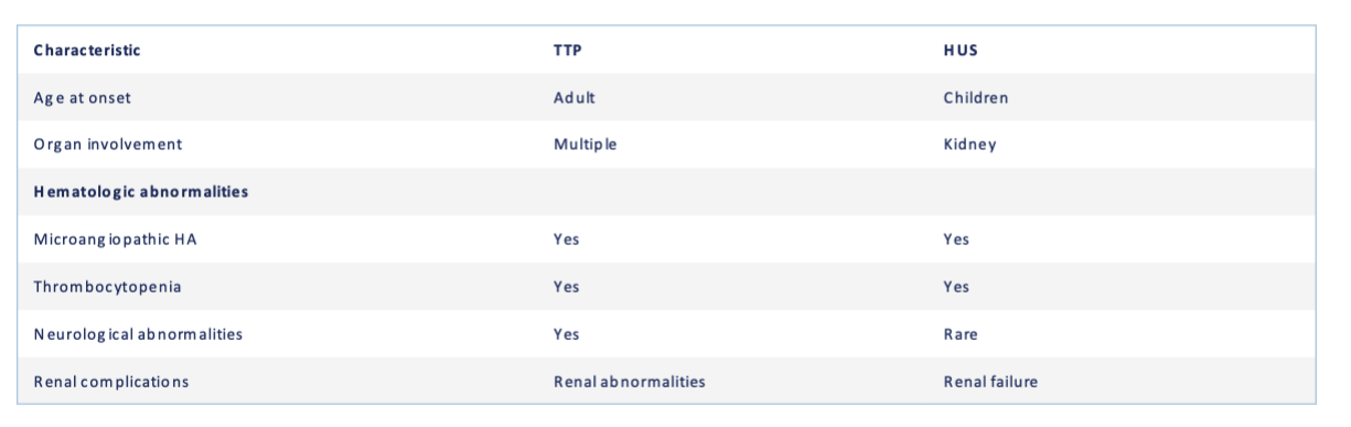
Thrombotic Thrombocytopenic Purpura (TTP) {Treatment}
Deficient in ADAMTS13 enzyme that is designed to cleave large vWF multimers → platelets aggregate & thrombi forms and obstruct small vessels
Associate with microangiopathic hemolytic anemia (increased schistocytes, reticulocytes, LDH & indirect bilirubin)
Treat by plasma exchange
Pentad signs/symptoms:
Fever
Microangiopathic hemolytic anemia
Thrombocytopenia
Neurological abnormalities
Renal dysfunction
Hemolytic Uremic Syndrome (HUS) {Treatment}
Share many of the same symptoms with TTP
Most common in children presents with diarrhea from E.coli/Shigella toxin
Atypical due to deficiency of complement regulators & proteins
Treat by fluid replacement, RBC & platelets transfusion
Disseminated Intravascular Coagulation (DIC)
A non-immune thrombocytopenia due to excessive consumption of platelets that caused by sepsis, severe trauma
Pregnancy-Associated Thrombocytopenia
Gestational thrombocytopenia: mild decrease of WBC in late pregnancy
Eclampsia: hypertension & proteinuria in late pregnancy
HELLP syndrome: hemolyze, elevated liver enzymes, low platelets. Associates with microangiopathic HA
Thrombocytosis {Primary vs Secondary}
Primary thrombocytosis: abnormal platelet morphology and nonfunctional. Due to myeloproliferative neoplasm (MPN) from abnormal stem cell that produce excessive RBC including megakaryocyte
Secondary thrombocytosis: normal & functional platelets, high platelets due to chronic inflammation/infection, or malignancy