Bone Tumor

0.0(0)
Studied by 1 person
call kaiCall Kai
learnLearn
examPractice Test
spaced repetitionSpaced Repetition
heart puzzleMatch
flashcardsFlashcards
GameKnowt Play
Card Sorting

1/53

encourage image

There's no tags or description

Looks like no tags are added yet.

54 Terms

1
New cards

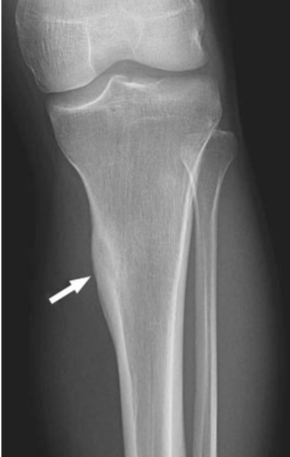
What periosteal reaction is BENIGN

Solid ONLY → rest are more likely to be malignant
2
New cards
<p>What is this</p>

What is this

SOLID periosteal reaction
3
New cards
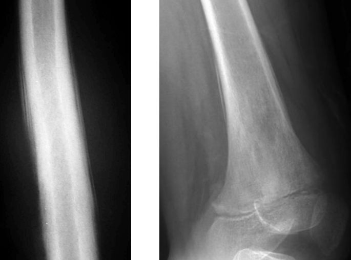
<p>What is this</p>

What is this

Laminated/onion peel periosteal reaction

4
New cards
<p>What is this</p>

What is this

Sunburst periosteal reaction

5
New cards
<p>What is this</p>

What is this

Codman triangle

6
New cards
<p>What is this</p>

What is this

Geographic bony destruction
7
New cards
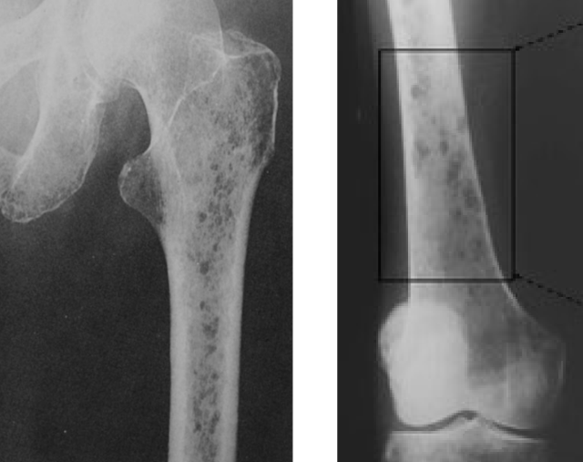
<p>What is this</p>

What is this

Moth eaten bony destruction

8
New cards
Where does a giant cell tumor (GCT) typically occur?

Eccentric epiphysis; occurs AFTER growth plate closure.

9
New cards
<p>Describe this</p>

Describe this

Geographic bony destruction + at epiphysis + growth plate is CLOSED → Giant cell tumor

10
New cards
What is the characteristic X-ray appearance of GCT?
Expansile, eccentric, bubbly lytic lesion with a non-sclerotic margin.
11
New cards
<p>What is this</p>

What is this

Geographic bony destruction + involve METAphysis → Bone CYST

12
New cards
What distinguishes GCT from a simple bone cyst?
GCT involves the epiphysis; simple bone cyst is central and metaphyseal.
13
New cards
Where does a simple bone cyst typically occur?
Central metaphysis of long bones, usually proximal humerus or femur.
14
New cards
What is the fallen fragment sign?
A bone fragment settling dependently within a fractured simple bone cyst.
15
New cards
<p>What is this</p>

What is this

Geographic lesion + eccentric to long bone METAphysis + UNFUSED growth plate → thin sclerotic margin and trabeculation → Aneurysmal bone cyst

16
New cards
What is the typical appearance of an aneurysmal bone cyst?
Expansile geographic lytic lesion, sometimes with trabeculations and a thin sclerotic rim.
17
New cards
Where does a non-ossifying fibroma (NOF) typically occur?
Eccentric metaphysis near the physis in children and adolescents.
18
New cards
<p>What is this</p>

What is this

Lucent lesion + near physis + thin sclerotic rim → non ossifying fibroma

19
New cards
What is the characteristic X-ray appearance of NOF?
Polycyclic, multiloculated lucency with a thin sclerotic rim.
20
New cards
<p>What is this</p>

What is this

Lucent lesion + thin sclerotic rim + at epiphysis/apophysis of long bone → chondroblastoma

21
New cards
What is the typical X-ray appearance of chondroblastoma?
Well-defined lucent lesion with smooth or lobulated margins and possible thin sclerotic rim.
22
New cards
What additional findings may occur with chondroblastoma?
Internal calcifications (40–60%), solid periosteal reaction, or joint effusion.
23
New cards

What is the key age clue for chondrosarcoma?

Older adults; particularly patients over 75 years.

24
New cards
What is the characteristic bone destruction in high-grade chondrosarcoma?
Moth-eaten or permeative osteolytic destruction.
25
New cards

What is osteoid osteoma

In children and adolescent → lucent nidus with sclerosis and SOLID periosteal reaction

26
New cards
<p>What is this </p>

What is this

Solid periosteal reaction + sclerosis → osteoid osteoma

27
New cards
<p>What is this</p>

What is this

Ground glass + bubbly + NO periosteal reaction + sclerotic → fibrous dysplasia
28
New cards
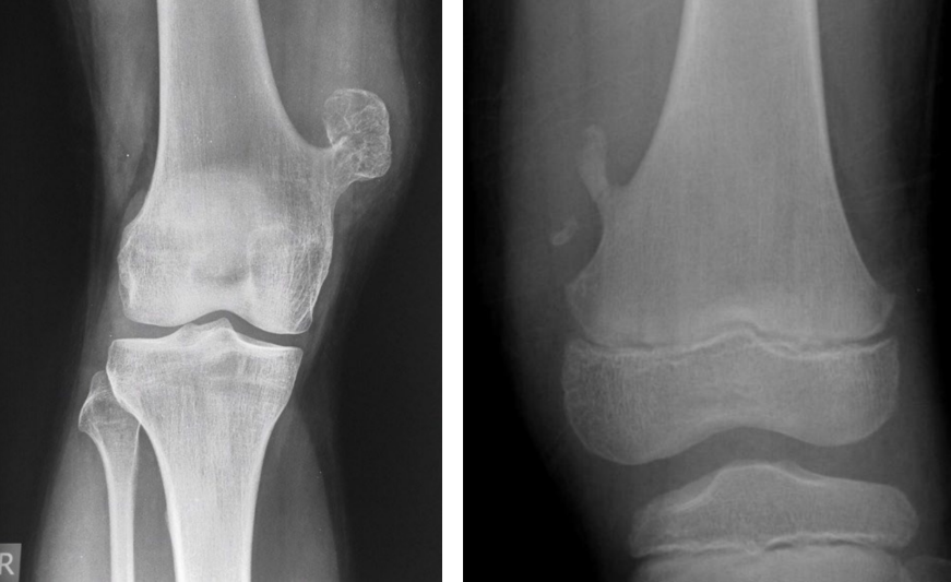
<p>What is this</p>

What is this

Osseous tumor → projected away from epiphysis = osteochondroma

29
New cards

What is osteochondroma

Most common BENIGN tumor in childhood → ASYMPTOMATIC
30
New cards
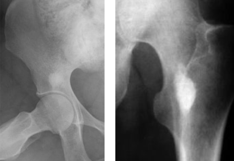
<p>What is this</p>

What is this

Bone island → enostosis

31
New cards

What is the difference between chondroid and osteoid matrix formations

Chondroid = form cartilage

Osteoid = form bone
32
New cards
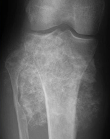
<p>What is this</p>

What is this

Fluffy, cloud like → chondroid matrix

33
New cards
<p>What is this</p>

What is this

Ivory like → osteoid matrix formation

34
New cards
<p>What is this</p>

What is this

Moth eaten destruction + sclerosis + sunburst/Codman triangle + osteoid matrix formation = osteosarcoma

35
New cards

What is osteosarcoma

Primary bone cancer in 10-20 y/o; secondary from Paget

36
New cards
<p>What is this</p>

What is this

Moth eaten + endosteal scalloping (affect 2/3 of cortical thickness) + fluffy = chondrosarcoma

37
New cards

What is chondrosarcoma

MALIGNANT cartilage tumor; most common primary tumor in >75

38
New cards

What is an enchondroma?

Usually benign cartilaginous tumor; malignant transformation is rare; expansile and lytic in phalanges

39
New cards
<p>What is this</p>

What is this

See sharp scalloped margin + chondroid matrix formation = enchondroma

40
New cards
What age clue helps distinguish Ewing sarcoma?
It occurs in very young patients.
41
New cards

Which periosteal reaction is commonly associated with Ewing sarcoma

Laminated (onion-skin); sunburst may also occur; varies a lot

42
New cards
What is the characteristic distribution of multiple myeloma lesions?
Numerous, uniform, well-circumscribed lytic lesions.
43
New cards
What is the key age distinction between myeloma and eosinophilic granuloma?
Myeloma occurs in older adults; eosinophilic granuloma in young adults.
44
New cards
What is eosinophilic granuloma?
A form of Langerhans cell histiocytosis (LCH) affecting bone.
45
New cards
What distinctive jaw finding can occur in LCH?
Floating teeth due to mandibular bone destruction.
46
New cards
What are the three subtypes of geographic bone destruction?
Type 1A: thin sclerotic margin; type 1B: well-defined border without sclerosis; type 1C: indistinct border.
47
New cards
<p>What is this</p>

What is this

Floating tooth → EG

48
New cards
<p>what is this</p>

what is this

Vertebral plana → EG

49
New cards
<p>What is this</p>

What is this

Many lytic bone lesion + osteopenia → Multiple myeloma

50
New cards
<p>What is this</p>

What is this

See punched out lesion in skull without sclerotic rim (geographic skull) → EG

51
New cards

What is Brodie abscess

Lytic lesion in oval shape along long axis of bone + surround by sclerotic rim

52
New cards
<p>What is this</p>

What is this

Oval shaped, on bone long axis + lytic → Brodie abscess

53
New cards
<p>What is this</p>

What is this

Widening of diploid space → Thalassemia

54
New cards
<p>Describe this</p>

Describe this

Lt = rib within rib esp in middle and anterior rib

Rt = wide MCP shaft

Both are thalassemia signs



