bio 225 exam 2
 Call Kai
 Learn
 Practice Test
 Spaced Repetition
 Match
 Flashcards
 Knowt Play
1/235

There's no tags or description
Looks like no tags are added yet.
236 Terms
the vertebrate central nervous system consists of:
the brain and spinal cord

brain
integrating center
spinal cord
mediates information flow between brain and body (and vice versa)
nuclei
groups of neuronal cell bodies within the CNS
tracts
bundles of many axons within the CNS
ascending vs descending tracts
ascending: carries info up to brain
descending: carries info from brain down along spinal cord
vertebrate peripheral nervous system consists of what?
all the neurons outside of the CNS
nerves and ganglia
nerves
bundles of many axons outside of the CNS (leaving CNS)
consists of myelinated and unmyelinated axons enclosed in connective tissue
may contain sensory or motor or both
cranial vs spinal - classified based on what CNS they’re coming off of
ganglia
groups of neuronal cell bodies outside of the CNS
white matter
shiny, bright appearance
primarily composed of myelinated axons
don’t typically see cell bodies + dendrites
grey matter
dulled appearance
contains dendrites, cell bodies, and unmyelinated axons
short axons
organization of white and grey matter in brain vs spinal cord
brain: grey matter surrounds white
spinal cord: white matter surrounds grey in a butterfly design

structure and function of spinal cord
mediates spinal reflexes
pathway for impulses to and from the brain
receives afferent/sensory signals via dorsal root
somatosensory neuron cell bodies in the dorsal root ganglion
sends out efferent/motor signals via ventral root
motor neuron cell bodies in the ventral horn
pic: blue is afferent/sensory going in, red is efferent/motor going out

how does the withdrawal reflex achieve a coordinated movement?
It simultaneously activates required motor neurons and inhibits opposing ones to ensure smooth movement.
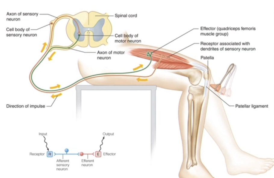
what are reflex arcs and how does divergence work in them?
reflex arc: control many involuntary behaviors, most of which are mediated by multiple neurons and employ divergence
divergence: 1 receptor spreads its signal to many effectors
What is the pathway of a reflex arc, and what is the role of interneurons?
pathway: sensory neuron → interneuron → motor neuron
interneurons: have short, branching axons that synapse onto multiple targets (motor neurons) to spread signal via divergence
simple reflex arcs
rare
contains only a single synapase/location of synapses
requires only 2 neurons (cluster of neurons)
1 sensory signal in, 1 motor signal out
ex: patellar reflex
contralateral - def and ascending vs. descending tracts
opposite side
most signals between the body and the brain (and vice versa) are contralateral
ascending tracts carry afferent/sensory signals within the CNS
in from lefthand side, crossing over to righthand side of spinal cord, up to the right somatosensory cortex
descending tracts carry efferent/motor signals within the CNS
right hand side of brain, carried down via descending tract, then some of the signals cross over to left side
left side of the brain moves right side of the body

ipsilateral
same side
signals stay/exit on the same side of the CNS
what does the spinal cord directly connect to?
the hindbrain
hindbrain main components
medulla oblongata
pons
cerebellum

medulla oblongata
regulates breathing, HR, blood pressure
pons
communicates between medulla, cerebellum, and forebrain
cerebellum
coordinates motor behaviors
forebrain/cerebrum
controls “higher” processes
cerebrum structure
frontal lobe
temporal loba
parietal lobe
occipital lob

corpus callosum
white matter tract
connects the left and right cerebral hemispheres
cerebral cortex
surface gray matter
can be divided into functional areas
motor areas in the frontal lobe
premotor cortex
primary motor cortex
premotor cortex
coordinates movements of groups of muscles
primary motor cortex
controls discrete skeletal muscles
stimulation results in specific movements on opposite side of body
what happens when the premotor cortex is damaged?
loss of skill that can often be relearned
what happens when the primary motor cortex is damaged?
paralysis of voluntary movements that are rarely regained
reflexes remain intact
somatosensory areas in the parietal lobe
primary somatosensory cortex
somatosensory association area
primary somatosensory cortex
primary input from touch, temperature, and pain receptors in skin & muscle on opposite side of body
what happens when the primary somatosensory cortex is damaged?
loss sensation that rarely returns
somatosensory association area
interpretation of sensation
integration with memory
ex: reach into pocket
what happens when the somatosensory association area is damaged?
loss of identification that can be relearned
ex of damaged somatosensory cortex vs somatosensory association area
normal: reach into pocket w/ key → recognize the ridges in denim material and coldness from key; recognize that it is a key
damaged: reach into pocket w/ key → cannot feel the coldness from key and cannot recognize that it is a key BUT can learn to recognize that it’s a key
visual areas of the CNS
primary visual cortex
visual association areas
far more ipsilateral signals than in most other systems
primary visual cortex
primary input from optic tracts
activated by visual stimuli
visual association areas
integration and perception
limbic system and 5 functions (actually 7)
many functionally important structures are situated below the cerebral cortex
functions: smell, homeostasis, emotions, fear & aggression, learning & memory, relay station
diencephalon key structures
the thalamus and hypothalamus
thalamus
filters and sorts sensory inputs
routes information
hypothalamus
diverse set of homeostatic functions
regulates pituitary gland
plays a key role in homeostatic regulation
hypothalamus: homeostatic regulation
invovled in the stress response
HPA (hypothalamus, pituitary, adrenal) axis
regulates food intake
regulates body temperature
helps maintain ion and water balance
regulates circadian rhythms
homeostasis requires what?
the balancing of sympathetic and parasympathetic nervous systems
sympathetic nervous system
most active during stress or physical activity
“fight or flight” response
increases heart rate & breathing, directs blood to working muscles during exerise
parasympathetic nervous system
most active during periods of rest
“resting and digesting”
redirects energy toward maintenance activities, such as digestion
sympathetic and parasympathetic neurons differ structurally and functionally
sympathetic:
preganglionic neurons have cell bodies in the CNS and axons that extend to their ganglion
postganglionic neurons release entirely different NT (neuroepinepherine)
ganglion near the CNS and long postganglionic axon
parasympathetic:
postganglionic neurons have cell bodies in the ganglion and axons that extend from the ganglion to their target
long preganglionic axon and ganglion near target organ
receptors that respond to ACh are different for each
acetycholine (ACh)
first NT to be discovered
synthesized at axon terminals of “cholinergic” neurons
making and releasing primarily ACh
NT released at neuromuscular junction and parts of central and peripheral nervous systems
ACh has 2 types of receptors (AChR): nicotinic and muscarinic
acetylcholine receptors
nicotinic AChR and muscarinic AChR
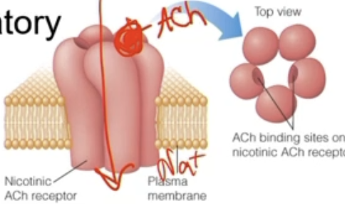
nicotinic AChR
ionotropic
when ACh binds, the channel will directly open and allow Na+ to enter
excitatory
produce EPSP in postsynaptic neuron
found in autonomic and motor neurons
non-specific cation channel (most often conduct Na+)
muscarinic AChR
metabotropic
G-protein coupled receptors (GPCRs)
linked to “second” messenger systems
found in smooth and cardiac muscle, glands, and CNS
excitatory or inhibitory (depending on receptor type and G-protein pair)

norepinephrine/noradrenaline
part of catecholamine: family of signaling chemicals made in your body that includes dopamine, norepinephrine, and epinephrine
like epinephrine/adrenaline, but functions mostly as a sympathetic neurotransmitter
triggers immediate, targeted responses vs epinephrine is released by adrenal glands directly into bloodstream to circulate everywhere and produce broad, body-wide emergency effects
changes in action potential frequency of sympathetic neurons alter what?
the target response
increase in sympathetic neurons → increase in AP firing rate → blood vessel constricts (in given example)
decrease sympathetic neurons → decreased AP firing rate → blood vesssel dilates

adrenergic receptors
receptor that norepinephrine/noradrenaline binds to
also respond to epinephrine from the adrenal gland
metabotropic
GPCRs linked to ion channel function via G-proteins
found throughout the body
diverse effects based on receptor subtype
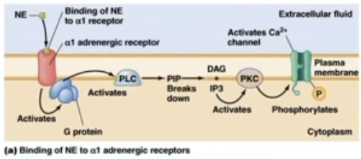
adrenergic receptors subtype alpha1
location: smooth muscle of blood vessels to skin and gut
pathway: Gq → PLC (phospho-lipase C)
breaking down of PIP2 into IP3 and DAG
further consequences related to Ca2+ (activation of Ca2+ channels)
effect: vasoconstriction
smooth muscles will contract and make diameter of blood vessels smaller
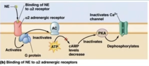
adrenergic receptors subtype alpha2
location: adrenergic axon terminals
pathway: Gi (inhibitory G protein) → ↓cAMP
when noroepinephrine binds, the G protein is Gi
inactive adenocyclase which is normally converting ATP to cAMP
decreases cAMP
inactives Ca2+, which is needed for noroepinephrine release
effect: inhibit NE release
adrenergic receptors subtype beta1&2
location:
beta1 - heart
beta2 - smooth muscle of blood vessels to heart & skeletal muscle
pathway: Gs (stimulatory G protein) → ↑cAMP’
Gs active adenocyclcase which will convert more ATP into cAMP
activates Ca2+ channels, increasing Ca2+, increasing rate and strength of contraction
effect:
increase heart rate and contraction strength
vasodilation

the adrenal medulla is innverated by what?
the sympathetic nervous system only
preganglionic sympathetic neuron release ACh → binds to receptor in chromaffin cells → release epinephrine → released into blood stream as a hormone
adrenal medulla innveration
the chromaffin cells release epinephrine into the blood stream in response to acetycholine from preganglionic sympathetic neurons
the developmental and evolutionary origins suggest chromaffin cells are modified post-ganglionic neurons
differences in receptor subtypes among effector organs
effector organs express different metabotropic adrenergic receptors
differences explain the diverse effects of epinephrine/norepinephrine in various tissues
what systems regulate homeostatic processes?
the endocrine, parasympathetic, and sympathetic nervous systems
sensory systems
detect stimuli and send info to an integrating center
receives a stimulus → convert into neuronal signal → carried via afferent neurons into CNS
sensory stimuli
detected in neurons or accessory cells
generator and receptor potential
generator potential
a graded potential that occurs in a sensory neuron as a direct result of the stimulus
dendrite of sensory neuron has receptor for stimulus embedded in plasma membrane→ direct depolarziation of primary afferent neuron, creating a generator potential
if this depolarization reaches the neuron’s trigger zone with enough strength, it initiates an AP that travels along axon to CNS
**the cell detecting the stimulus and the neuron sending the signal to the brain are the same single cell - direct pathway
receptor potential
a graded potential that occurs in a specialized receptor cell (accessory cell) due to a sensory stimulus and then must be transmitted to the neuron
stimulus triggers a receptor protein that is localized in an accessory cell, causing depolarization called a receptor potential
this depolarization triggers an influx of Ca2+ → vesicles release NT via exocytosis
the NT bind to the receptors on the dendrite of an adjacent afferent neuron, inducing a secondary graded potential in that neuron
does not actually fire AP
the detector cell is a separate accessory cell that must communicate via chemical neurotransmitters to activate the sensory neuron - indirect pathway
sensory receptors can be classified by their stimulus modality (6)
chemoreceptors: detect the presence of chemicals in the environment
mechanoreceptors: detect pressure and movement, including proprioception (detection of body and space)
photoreceptors: detect light
thermoreceptors: detect temperature
electroreceptors: detect electric fields
magnetoreceptors: detect magnetic fields
stimulus encoding and 4 impt features
sensory receptors convert info about the stimulus into changes in membrane potential and eventually action potentials
encode 4 important features:
stimulus modality - signal pathways
stimulus location - receptive field
stimulus intensity - frequency/strength
stimulus duration - tonic & phasic receptors
sensory pathways can encode stimulus modality
a particular afferent neuron is associated with one modality of receptor and stimulus (adequate stimulus)
each afferent neuron stimulates activity within a particular neural pathway
perception based on pathway activated, not the stimulus identity
ex: close eyes, rub, open - what do u see? speckled light pattern
this is a response to pressure stimulus - perceieve light even tho stimulus was pressure
receptive field
region of the sensory surface that causes a response when stimulated
varies in size and therefore acuity
smaller receptive fields allow more precise localization of the stimulus (i.e. greater spatial acuity)
receptive field and localization of stimulus
receptive fields encode stimulus location based on the signal pathway
acuity
ability to resolve fine detail of a stimulus
lateral inhibition
signals from neurons at the center of the receptive field inhibit surrounding neurons
improves acuity
enhances contrast: magnifies the difference in firing rates between the central point of stimulus and surrounding area
decreases “noise”: mutes weak, redundant background signals from surrounding neurons
improves edge detection/boundaries: allows the CNS to clearly identify where a stimulus begins and ends
action potential frequency encodes signal intensity
sensory neurons code stimulus intensity by changes in action potential frequency
ex: increased stimulus → increased frequency of action potentials
sensory receptors
encode a limited range of stimulus intensities
sensory neurons code stimulus intensity by changes in action potential frequency
ex: strong stimuli → high frequency
dynamic range
increase in magnitude of graded potential or frequency of AP

dynamic range
range of stimulus intensities over which a receptor exhibits changing responses
threshold: weakest stimulus that produces a response in a receptor
saturation: top of the dynamic range (maximal response - won’t change even if stimulus intensity increases)
dynamic range and discrimination
large dynamic range = poor sensory discrimination
large change in stimulus intensity causes a small change in AP frequency
narrow dynamic range = good sensory discrimination
small changes in stimulus intensity causes a large change in AP frequency
sensory discrimination
the ability to detect differences between stimuli
improves by distributing responses amongst different receptors
range fractionation: different receptors cover different parts of the stimulus intensity spectrum
expands the range of intensities the sensory intensities the sensory surface or system responds to

range fractionoism
different receptors cover different parts of the stimulus intensity spectrum
logarithmic encoding
some receptors use logarithmic encoding
logarithmic encoding of intensity allows for increased dynamic range without completely sacrificing discrimination
better discrimination in lower range

tonic and phasic receptors
encode stimulus duration
tonic receptors respond for the entire duration of a stimulus
phasic receptors respond to changes in a stimulus
phasic receptors
respond to changes in a stimulus
can encode stimulus changes in several ways
pic: on set (first column), combo, off
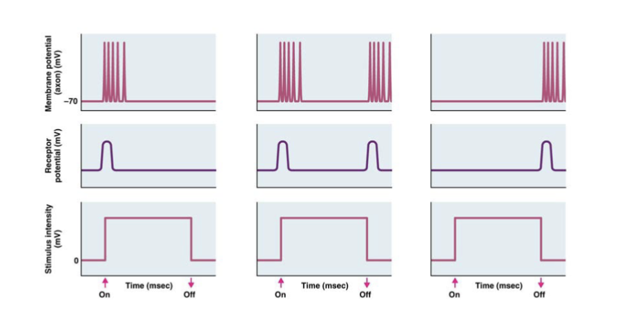
sensory adaptation
decreased response to a stimulus as duration increases
tonic receptors are typically slow adapting
phasic receptors adapt so quickly that they produce APs only when the stimulus changes
for prolonged stimuli, sensory adaptation can continue until the stimulus is tuned out
AP frequency and prolonged stimulus
AP frequency often decreases
photoreceptors
convert light energy into changes in membrane potential
photoreceptive opsins
seven-membrane-spanning GPCRs
opsins localize to membranes in the outer segments of vertebrate photoreceptors
phototransduction
occurs thorugh chromophore isomerization (chemical goes from cis to trans conformation)
summary of light-induced events
opsins covalently bind vitamin-A derived choromophores
photons cause isomerization of the choromophore
isomerization of chromophore leads to changes in the opsin
conformational change in opsin
dissociation of choromophore from obsin (“bleaching”)
G-protein singnaling events cause changes in membrane potential

2 classes of photoreceptor are found throughout the animal kingdom
rhabdomeric receptors
rhabdomeric PRs were recently discovered in vertebrates too → these PRs help entrain our internal clocks to environmental light/dark cycles
cilary receptors
rods and cone are ciliary receptors (vision)
both have evolved multiple times (not clustered when looking at evolutionary tree)
rhabdomeric photoreceptors signal thorugh Gq
11-cis 3-hydroxy retinal absorbs light and isomerizes into all trans 3-hydroxy retinal
all trans 3-hydroxy retinal dissociates from opsin
activated opsin activates a Gq protein
activated Gq activates PLC (phospholipase C), converting PIP2 to DAG and IP3
DAG activates a TRP cation channel (non-selective)
gated by IP3 and DAG
Ca2+ and Na+ enter the cell, depolarizing it
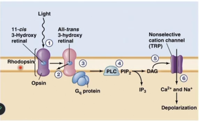
ciliary photoreceptors signal through G¡/transducin
11-cis Retinal absorbs light and isomerizes into all-trans retinal
all-trans retinal dissociates from opsin
activated opsin activates the G¡ protein (inhibitory G protein) transducin
transducin actives PDE (phosphodiesterose), which converts cGMP to GMP
signaling molecule that can open CNG (cyclic nucleotide-gated) channel
the decreased cGMP closes a Na+ channel
Na+ entry decreases, hyperpolarizing the cell

mammalian eye
allows formation of a bright, focused image
light enters through front of eye and hits cornea → travels through pupil (hole in iris) → hits the lens, which is held in place by ciliary body → lens does fine tuning so light gets targeted to fovea (specialized part in retina where rods/cones located) → activation of photoreceptors → light passes through and hits choroid (absorbs any remaining light energy) → exit eye via optic disk through optic nerve
muscle fibers of the iris change the pupil diameter and control the amount of light entering the eye
the cornea refracts light and focuses it onto the lens, which is responsible for fine-tuning the focus onto the retina
the lens changes shape depending on the distance of the light source in a process called accommodation
light passes through the retina and excites the photoreceptors
the choroid is a pigmented layer that absorbs the light
muscle fibers of the iris
change the pupil diameter in response to light
controled by SNS and PNS
radial muscle layer (like the rays of sun, spreads out)
dilates pupil via SNS
circular layer of muscles
contracts via PNS
choroids of nocturnal animals
contain a reflective layer
the tapetum lucidum reflects and amplifies dim light in nocturnal animals and make their eyes “glow”
cornea and lens
convex lenses that direct light onto the retina
when parallel light waves hit the lens, it will cause refraction that converges the waves onto a single point
most refraction occurs at the cornea
the lens is responsible for fine-tuning
most of the focusing is done by the cornea

accommodation
referes to the ability of the eye to focus light from different distances
during accommodation, the cillary muscle contracts, the ligaments slcak, and the lens bulges (to change focal length)
distant object
enters eye as parallel rays
short focal length
nearby object
enters eye as nonparallel rays
increased focal length (point of convergence is beyond the retina → dispersed light hitting retina → not focused)
nearby object w/ accommodation
focal length restored
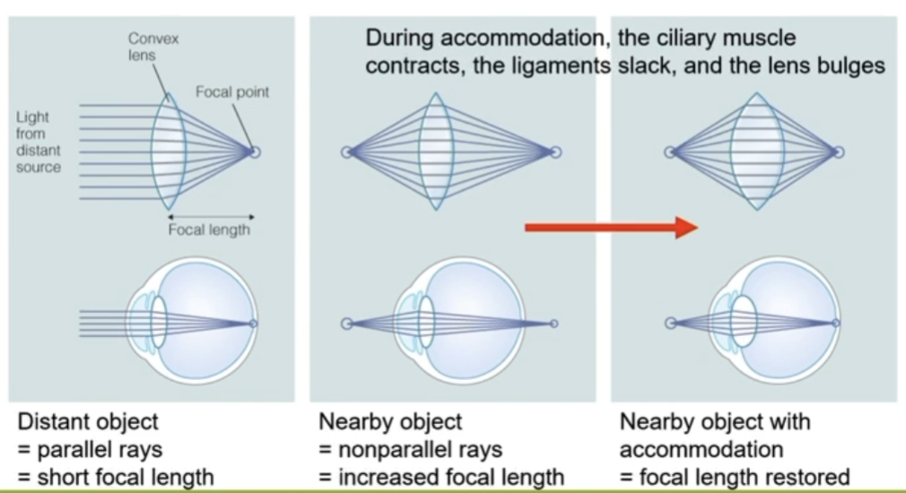
rods and cones
located at the back of the retina
few layers of cells light must pass through in retina to hit the photoreceptors
activate G protein coupled receptor → cause activation of PDE → cause hyperpolarization
decrease inhibitory NT (glutamate) release
activation of bipolar cells → activation of retinal ganglion cells → carry signal out via optic disk and optic nerve
