Lab Exam II
 Call Kai
 Learn
 Practice Test
 Spaced Repetition
 Match
 Flashcards
 Knowt Play
1/88

Earn XP
Description and Tags
89 Terms
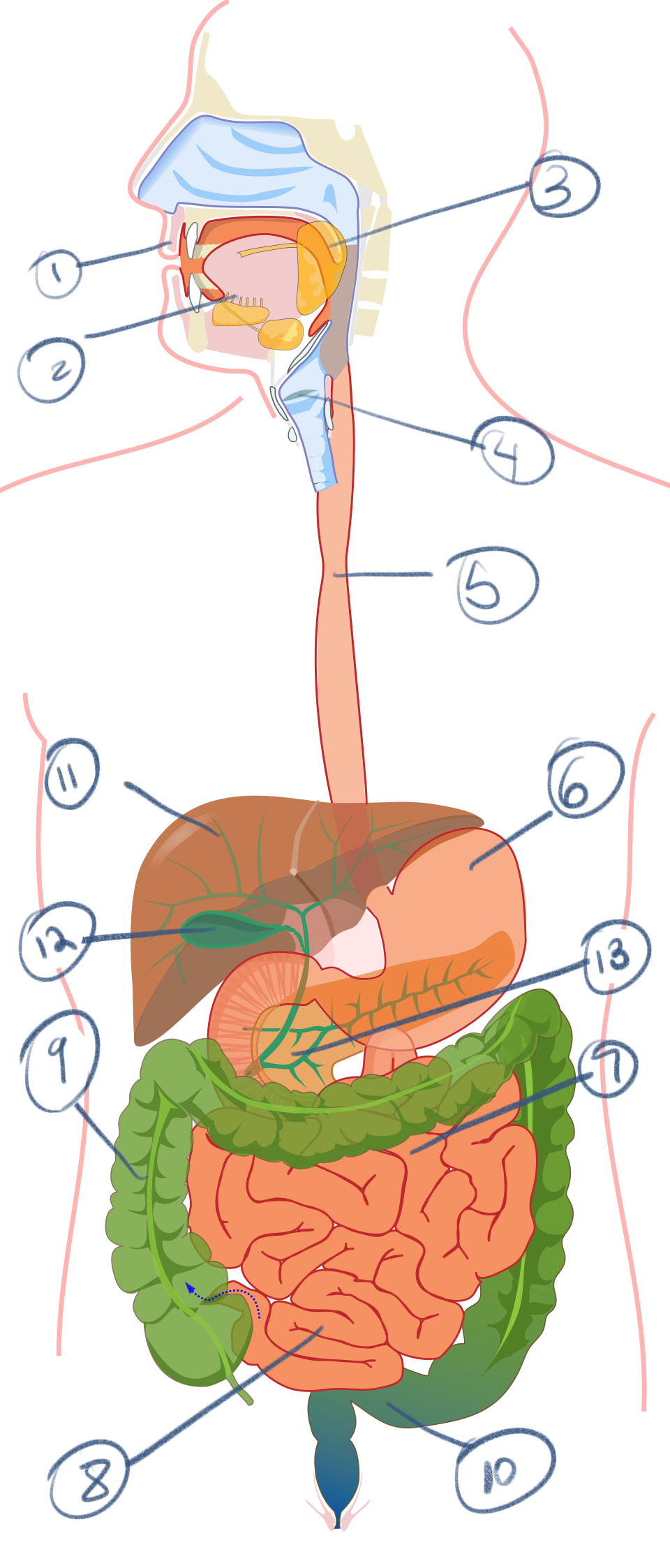
Identify and define digestive system
1) Mouth and teeth - ingestion of food and mechanical digestion. Saliva begins chemical digestion
2.) Tongue - muscle that manipulates food and contains taste buds
3.) salivary glands - secrete salvia for moistening food and amylase to digest carbohydrates
4.) pharynx - area in throat where food and air pathways cross
5.) esophagus - muscular tube that moves food via peristalsis from pharynx to stomach
6.) stomach - store food and contributes HCl and pepsin for digestion
7.) Small intestine (duodenum) - first section of small intestine, most chemical digestion with help from secretions of pancreas, liver, and gall bladder
8.) small intestine (jejuno-ileum) - jejunum and ileum - absorption of nutrients from food into blood
9.) large intestine (colon) - absorption of water
10.) rectum - feral matter is stored just prior to elimination via anus
11.) liver - produces bile, stores glycogen, detoxifying harmful molecules in blood
12.) gallbladder - concentrates bile and secretes as needed into duodenum
13.) pancreas - both exocrine and endocrine tissues. Exocrine portion secretes digestive enzymes and alkaline fluid into duodenum.
Respiratory vs Circulatory system
Respiratory - gas exchange with environment
Circulatory - gas movement within body
Cursorial Definition and modifications
Running - limbs directly under body, clavicle reduced or absent.
2 subtypes of cursorial definition and modifications
Digitigrade - walks on toes, lengthens limbs and reduces size of foot
Unguligrade - “tip toe” fusion of meta carpals and meta tarsals
Layers of artery and vein and what they are made of
Tunica intima - epithelium, dark purple stained internal elastic membrane
Tunica media - smooth muscle, more prominent in arteries than veins
Tunica adentitia - scattered mix of collagenous and elastic fibers
Saltatorial definition and modifications
Jumping - hind legs significantly longer than forelimbs. Hindlimbs used for jumping, forelimbs used for balancing/landing
Axial vs Appendicular skeleton
Axial skeleton - midline of body, skull, vertebrae, and ribs
Appendicular - limbs and supporting bones
Volant Definition and modifications
Flying - metacarpals and phalanges elongates to support skin that make up wing
Fossorial Definition and modifications
Digging/burrowing - thick and strong forelimbs for burrowing, wide bones, long claws, thicker forelimbs than hindlimbs
Plantigrade Definition and Modifications
Walking - walk on entire foot including phalanges, metatarsals, some tarsals. Large area of stability
Arboreal definition and modifications
Climbing - elongated forelimbs for large arc of rotation, large and prominent clavicle, long digits.
Homodont vs heterodont
Heterodont - different types of teeth
Homodont - all teeth have same shape
3 diets through teeth identification
Herbivores - flat premolars and molars, little or no canines. Prominent incisors
Carnivores - small incisors, premolars into carnassial shape. Molars either reduced or absent or in carnassial shape
Omnivore - both features of herbivores and carnivores: flat molars, carnassial premolars.
Three layers of tooth
Enamel - outer layer of tooth, mineralized but brittle
Dentin - less mineralized not as heart as enamels and also less brittle. Support underneath enamel
Dental pulp - tissue, blood vessels, nerves
Mammal spine sections
Cervical: skull to rib
Thoracic: ribs/chest
Lumbar: lower back, no ribs
Sacral: pelvis area
Caudal: tail
Human caudal: coccyx

Identify these bones (appendicular and axial)
Atlas
Suprascapula
Coracoid
Trunk vertebrae
Sacral vertebrae
Urostyle
Clavicle
Humerus
Radioulna
10. ilium
11. ischium
12. pubis
13. femur
14. tibiofibula
15. astragalus
16. calcaneum
Bones of pelvic girdle
Illium
Ischium
Pubis
Bird clavicle
Furcula
Premaxilla vs maxilla
Premaxilla - front teeth
Maxilla - back teeth
What is the jaw called and what bones make it up?
Mandible and made of two dentaries (singular: dentary)
“Great hole” in back of skull term and definition
Foramen magnum - opening in skull so spinal cord may connect to brain
Fused structure composed of carpals and meta carpals
Carpometacarpus
First two bones of cervical vertebrae and function
Atlas (first) - up and down movement
Axis (second) - left and right movement
2 different types of ribs term and definition
Sternal ribs - attached to sternum
Pleural ribs - attached to thoracic vertebrae
General mammalian forelimbs bones
Humerus, radius, ulna, carpals, metacarpals, phalanges
General mammalian hindlimb bones
Femur, tibia, fibula, patella(kneecap), tarsals(ankles), metatarsals, phalanges
What allows the skull to articulate with first vertebrae
Occipital condyles
Skeletal system definition
Organ system composed of bone and cartilage that supports vertebrate body
Neural vs haemal
Neural - notochord (nervous system)
Haemal - blood
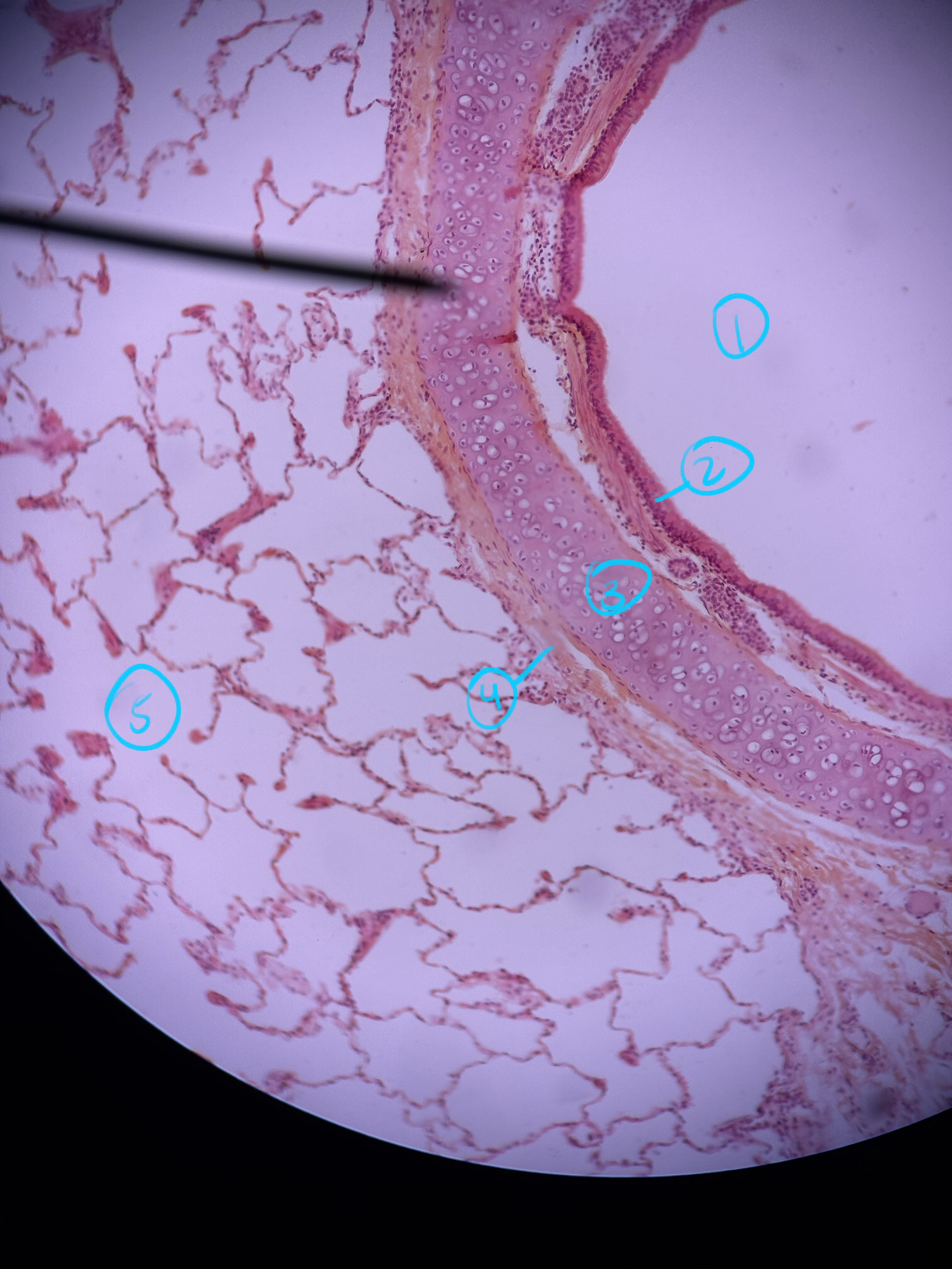
Identify what this entire structure is and what the numbers are
Lung and Bronchiole
1.) lumen of bronchiole
2.) epithelial tissue
3.) hyaline cartilage
4.) smooth muscle
5.) alveoli

Identify this image and what the numbers are
Artery and Vein
1.) lumen of artery
2.) internal elastic membrane
3.) tunica intima - epithelial layer
4.) tunica media - smooth muscle
5.) tunica adventitia - mix of collagenous and elastic fibers
Artery vs Vein
Artery - away From heart, more smooth muscle and more circular
Vein - towards heart, less smooth muscle, more flattened/collapsed
Enzyme that breaks down carbohydrates
Amylase
Stomach major layers and what they are made of
1.) Mucosa - epithelial and gastric glands
2.) submucosa - fibrous connective tissue
3.) muscularis externa - smooth muscle
4.) serosa - adipose and loose connective tissue

Identify this image and the numbers
Stomach layers
1.) lumen of stomach
2.) gastric pits
4.) mucosa
5.)muscularis mucosae
6.)submucosa
7.) muscularis externa
8.) serosa
9.) gastric glands
Cells in gastric glands, what they produce and what part of the gastric gland they in
Parietal cells - produce HCl, found all over gastric glands
Chief cell - produce pepsinogen, found in base of gastric glands
Mucous cells - produce mucus, found in neck of gastric glands
What can denature amylase?
Acid.
Organs of mammalian circulatory system and what they do
1.) spleen - destroys old erythrocytes and produces lymphocytes, in infants it also produces erythrocytes
2.) heart - muscular organ that elevates blood pressure in blood vessels to pump blood throughout body
3 vessels on aorta and where they lead to
(Left) brachiochepalic trunk - right shoulder, upper arm, head
(Middle) left common carotid artery - left side of neck and head
(Right) subclavian artery - left shoulder and upper arm
Reproductive system
Producing offspring
Excretory system
Osmoregulation (maintaining homeostasis in solute concentration of body fluids) and excreting nitrogenous waste
Endocrine system
Produces hormones, molecules that regulate activity of other tissues and organs in body
Three hormones produced by thyroid and functions
Thyroxin (T4)
Triiodothyroxin (T3) - both regulate metabolism
Calcitonin - stimulates storage of calcium in bones
Integumentary system
Outer covering of animal body (skin)
Nervous system
Transmitting and processing information via electrical signals
Three major layers of skin and what type of tissue are they made of
Epidermis - epithelium
Dermis - dense irregular connective tissue
Hypodermis - loose connective tissue and adipose tissue
Epidermis Five sublayers and what they are made of
Stratum corneum - top layer of dead cells
Stratum lucidum - borrow layer of dead cells, semi transparent
Stratum granulosum - top layer of living squamous epithelium, starting to flatten
Stratum spinosum - majority of living squamous epithelium, irregular shape
Stratum basale (or germinativum) - bottom layer of living epithelium cells, currently undergoing mitosis, may appear brown due to melanin
Sublayers of dermis and what they are made of
Papillary layer - upper layer, dermal papillae where dermis pushes up towards epidermis
Reticular - lower layer of dense irregular connective tissue
Sebaceous gland
Secretes oil onto hair
Arrector pili muscle
Muscle attached to hair follicle at angle and erects hair if cold or agitated
Chondrichthyes skin
Placoid scales (dermal denticles)
Lissamphibia skin characteristics
Smooth and moist skin, no scales. Mucous glands in dermis. Poison glands may be found in dermis.
Melanophores
Irregularly shapes cells on dermis that contain Pigment melanin in amphibians
Reptile skin characteristics
Epidermal scales - thick layer of protein keratin

Label the spine sections
1.) cervical
2.) thoracic
3.) lumbar
4.) sacral
5.) caudal

Identify the numbers (axial and appendicular skeleton)
Note: #5 is the hole in the skull
1.) Premaxilla | 2.) maxilla
3.) Mandible/Two dentaries | 4.) occipital condyles
5.) Foramen magnum | 6.) atlas
7.) axis | 8.) scapula
9.) humerus | 10.) ulna
11.) radius | 13.) carpals
14.) metacarpals | 15.) phalanges
16.) sternum | 17.) sternal ribs
18.) pleural ribs | 19.) pubis
20.) ilium | 21.)ischium
22.) femur | 23.) patella
24.) fibula | 25.) tibia
26.) tarsal | 27.) metatarsals
28.) phalanges

Label the numbers ( axial, appendicular, and spine sections)
Note: #2 is the hole in the skull, #3 is adjacent to #2; #11 is specifically the hook on the rib; #14 is the collarbone; #13 is the tip of the structure
1.) mandible | 2.) foramen magnum
3.) occipital condyles | 4.) atlas
5.) axis | 6.) cervical vertebrae
7.) thoracic vertebrae | 8.) synasacrum
9.) caudal vertebrae | 10.) pygostyle
11.) uncinate process | 12.) sternum
13.) keel | 14.) furcula
15,) scapula | 16.) humerus
17.) radius | 18.) ulna
19.) Carpometacarpus | 20.) phalanx
21.) ischium | 22.) ilium
23.) Pubis | 24.) femur
25.) tibia | 26,) tibiotarsus
27.) tarsometatarsus | 28.) phalanges

Label the numbers (axial and appendicular skeleton)
Extra: what is the skull called?
Notes: #1 is upper jaw #2 is lower jaw
1.) palatoquadrate cartilage
2.) meckel’s cartilage
3.) gill arches
5.) vertebrae
6.)pectoral girdle
7.)pectoral fins
8.) dorsal fin
9.) caudal fin
10.) pelvic fins
11.) pelvic girdle

Label the skeleton (axial and appendicular skeleton)
Note: #6 is the vertebrae behind the pelvis
1.) mandible | 2.) foramen magnum
3.) atlas | 4.) axis
5.) dorsal vertebrae | 6.) sacral vertebrae
7.) pleural ribs | 8.) caudal vertebrae
9.) scapula | 10.) coracoid
11.) humerus | 12.) radius
13.) ulna | 14.) carpals
15.) metacarpals | 16.) phalanges
17,) pubua | 18.) ilium
19.) ischium | 20.) fermur
21.) tibia | 22.) fibula
23.) tarsals | 24.) metatarsals
25,) phalanges

Identify this picture and label the numbers
Pacinian corpuscle (whole mount)
1.) lamellated connective tissue
2.) single nerve fiber
3.) fiberblast nuclei
Where would you find a pecinian corpuscle and what are their functions?
They are touch receptors deep within the skin (within the reticular layer of dermis or hypodermis)

What is this image and label the numbers
Taste buds (tongue)
1.) fungiform papillae
2,) taste buds
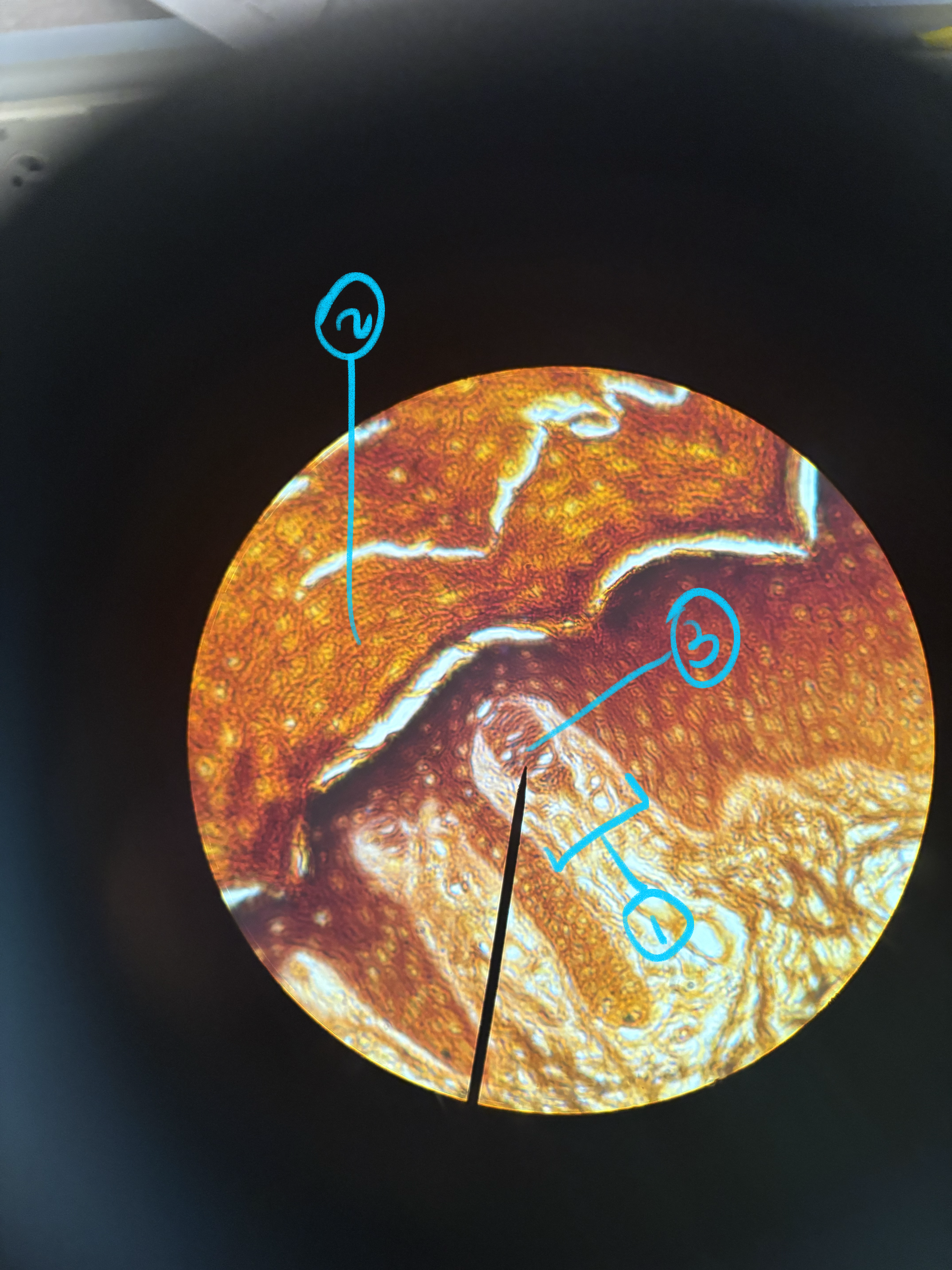
What is this image of and label the numbers
Meissner’s corpouscle
1.) dermal papillae
2.) epidermis
3.)vertical lamellar of Schwann cells
What is the function of meissner’s corpuscle and where is it found
It is a touch receptor that senses light touch, meaning it’s right under the epidermis, within some dermal papillae
meissner vs pacinian corpuscle
Meissner: in dermal papillae right under epidermis; for light touch
Pacinian: deep within skin reticular layer of dermis or hypodermis; senses pressure or vibrations

What is this image of and label the numbers
1.) hair
2.) hair follicle
3.) sebaceous gland
4.) sweat gland
5.) arrector pili muscle
Organs of mammalian nervous system
Brain - organ of central nervous system located in head; responsible for processing and integrating information
Spinal cord - located along dorsal midline of body, responsible for transmitting reflex responses and relaying info between peripheral nervous systems and brain
Dorsal root ganglion - cluster of nervous tissue just outside spinal cord containing perikarya of sensory neurons; transmits sensory info to central nervous system
Spinal nerve- one of a series of nerves branching off from spinal cord; carry both incoming sensory info and and outgoing motor signals

What is this image of and label the numbers
Neuromuscular junction
1.) motor end plate
2.) axon
3.) muscle fibers
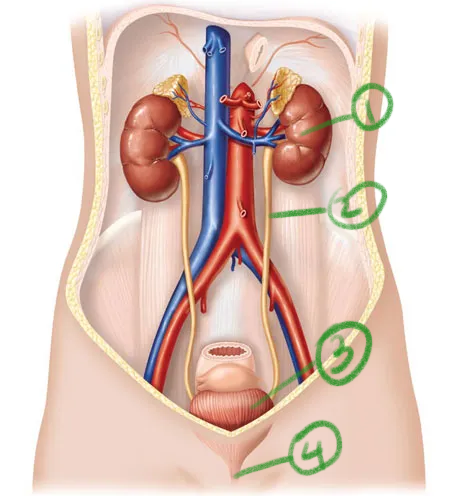
What organ system is this and label the numbers and state their functions
Excretory system
1.) kidneys - filter the blood, maintain osmotic balance in body fluids, and produce urine
2.) ureters - ducts that carry urine to bladder
3.) urinary bladder - expandable pouch that stores urine prior to elimination
4,) urethra - tube which urine is eliminated; in males, tube where semen leaves the penis

What organ system is this and label the numbers and its functions
Note: ignore #3,8,6
1.) vagina - female organ of sexual intercourse and birth canal
2.) cervix - narrow opening of uterus
4.) uterus - where embryo implants and develops
5.) oviduct (fallopian tube) - transports eggs from ovary to uterus. Fertilization usually happens here in humans
7.) ovary - female gonad; produces eggs and sex hormones

What organ system is this and label the numbers and its functions
Male reproductive system
1.) scrotum - sac of skin enclosing testis
2,) testis - male gonad; produces sperm and sex hormones
3.) epididymis - where sperm mature
4.) vas deferens - tube that carries sperm from epididymis toward urethra
5.) seminal vesicles - secrete sticky fluid that contains nutrients for sperm
6.) prostate gland - secrets alkaline fluid that protects sperm in acidic environment of vagina; force with which this fluid is expelled provides some propulsion of ejaculation
7.) bulbourethral glands - secrete mucoid substance (pre-ejaculatory fluid) into urethra to neutralize pH of urine residue and lubricate tip of penis
8.) penis - intromittent organ; contains spongy erectile tissue that fills with blood during arousal to cause erelection.

What is this image of and label the numbers
Thyroid and parathyroid
1.) parathyroid
2.) thyroid
3.) follicles
4.) colloid

What is this image, where can it be found and label the numbers as well as its functions
Pituitary gland (located just below hypothalamus of brain)
1.) anterior pituitary (adenohypophysis) - produces and secretes growth hormone, prolactin, thyroid stimulating hormone, follicle stimulating hormone, etc. it produces its own hormones
2.) posterior pituitary (neurohypophysis) - secretes hormones that are produced with it in specialized neurons (neurosecretory cells)
3.) infundibular stalk - nerve fibers that connect pituitary to hypothalamus

What is this image and what is its function. Label the numbers
Ovary - female gonad; produces gametes and sex hormones.
1.) follicle
2.) follicular cells
3.) oocyte

What structure image is this, what is its functions, and label the numbers
Testis - male gonad, responsible for producing gametes and sex hormones
1.) seminiferous tubules
2.) epididymis

What image is this of, label the numbers and its functions
Penis (cross section)
1.) urethra - tube that carries both semen and urine in males
2.) corpus spongiosum - surrounds urethra and prevents its from being compressed during ejaculation
3.) corpora cavernous - spongy structures that fill with blood during arousal to cause erection

What image is this of, identify the numbers and its functions
Note: #2 is the empty space around #1
Kidneys
1.) glomerulus - ball of capillaries containing blood to be filtered
2.) bowman’s capsule - surrounds glomerulus and where filtrate begins journey through nephron
3.) convoluted tubules - part of system of tubes that conduct secretion and reabsorption

What is this image and its function, identify the numbers and describe the functions of them
Mammary gland - location where milk is produced
Lobules of glandular tissue - where milk is produced
Interlobular connective tissue - fibrous connective tissue and adipose tissue

Be able to trace the direction of blood flow through the heart


Be able to identify sections of the heart, veins and arteries.

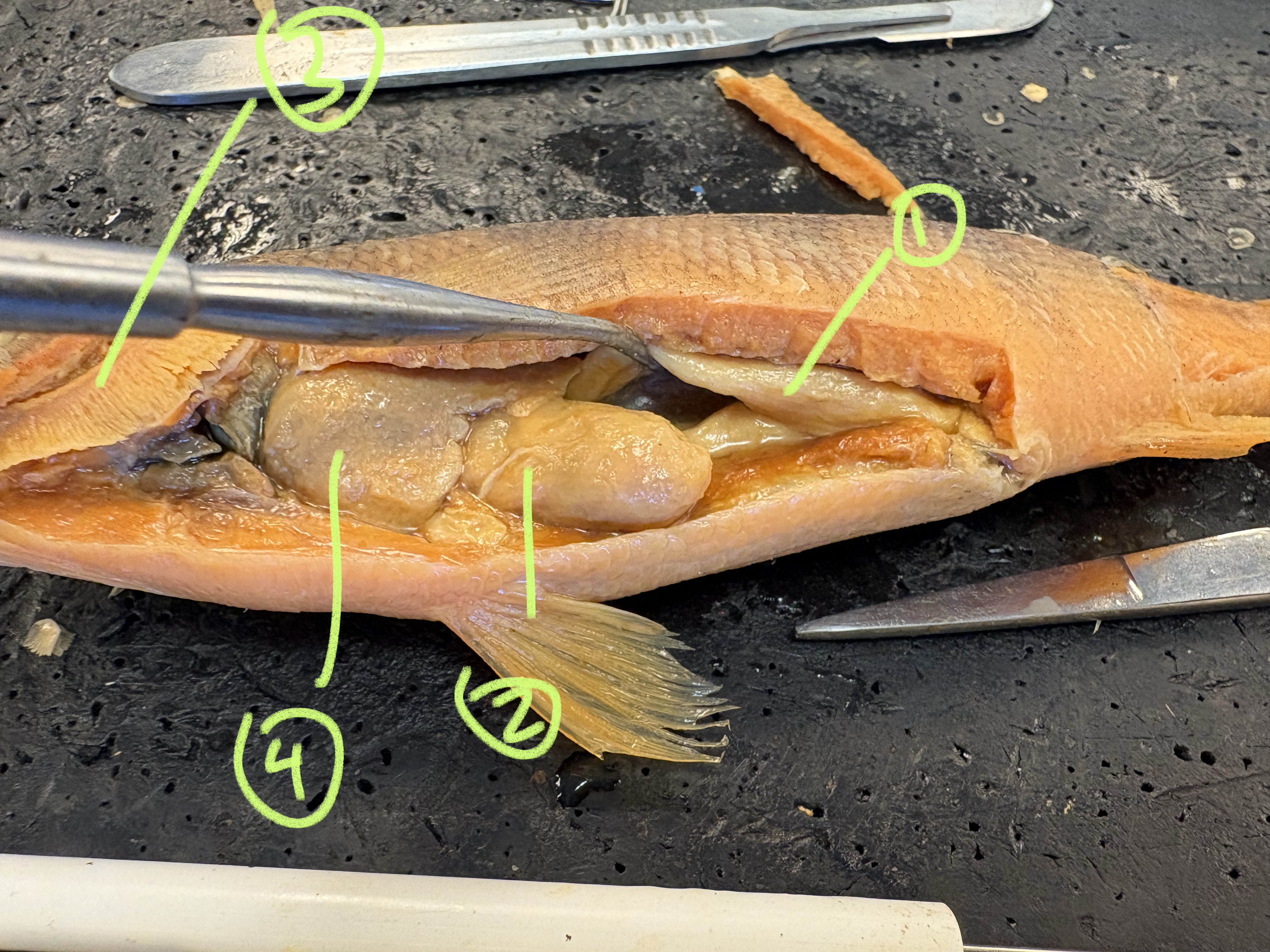
Identify the numbers
1.) testis
2.) stomach
3.)gills
4.) liver

Identify the numbers
1.) operculum
2.) first dorsal fin
3.) second dorsal fin
4.) pectoral Fins
5.) pelvic fins
6.) anal fins
7.)caudaL fin
8.) lateral line
How to identity if a fish is a male or female
Ovary vs. testis
Ovary is more circular and orange (may have presence of eggs)
Testis is kind of split in two, paler, skinnier

What type of scale is this
Ctenoid
Ctenoid vs Ganoid vs Cycloid vs placoid
Placoid - same layers as teeth, smooth and rough texture
Ganoid - bone, rhomboid or diamond shaped, interlocked, heavy, inflexible
Cycloid - round flat and smooth, layered, flexible
Ctenoid - rough or oval with come-like “teeth“, flexible
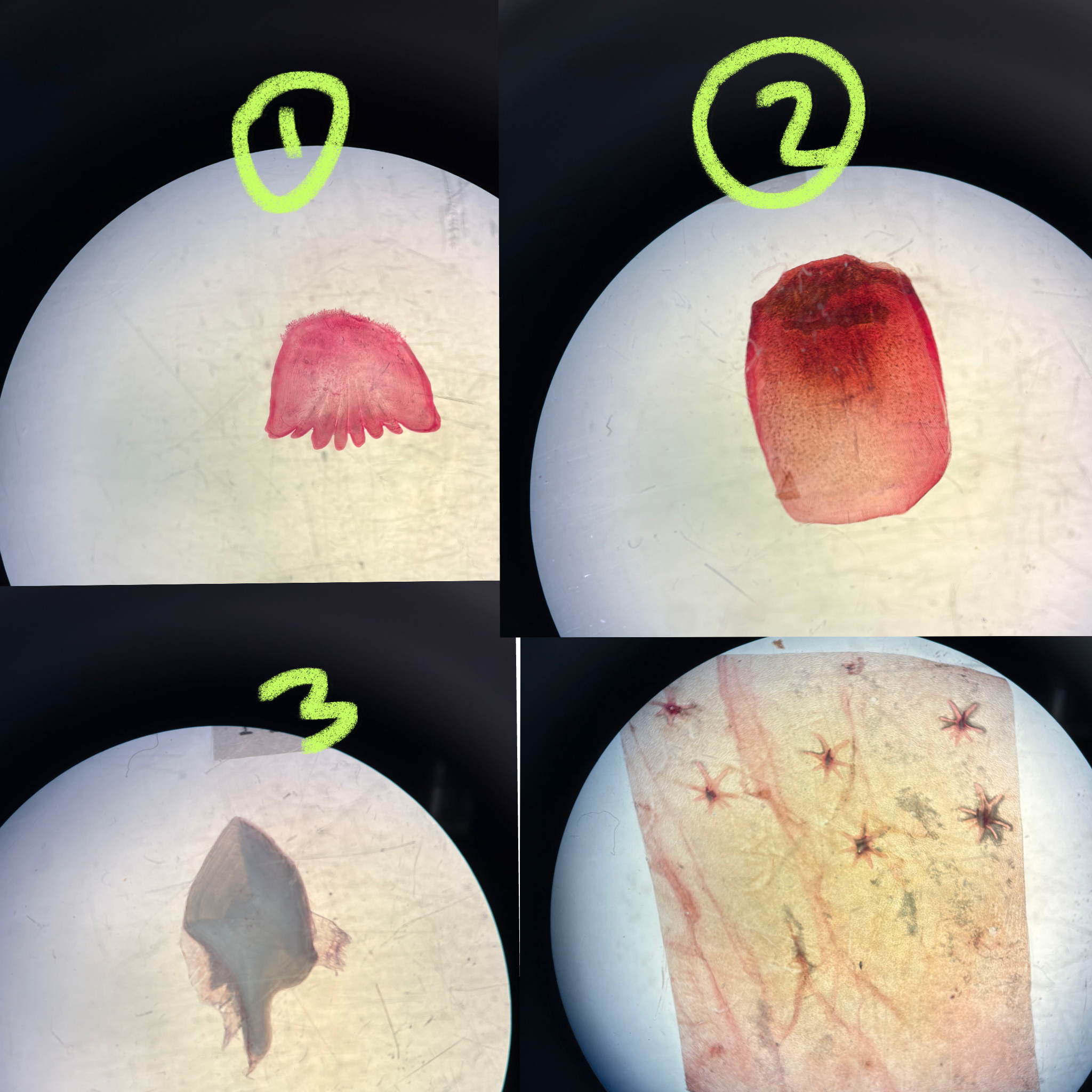
Identify these scales
Ctenoid
Cycloid
Ganoid
Placoid

Identify these frog ventral side muscles
Mylohyoid
Deltoid
Pectoralis major
Sartorius
Gracilis major
Gracilis minor
Gastrocnemius
Adductor magnus
Rectus abdominis

Identify frog dorsal side muscles
Dorsalis scapulae
Latissimus dorsi
Illiolumbar
Longissimus dorsi
Coccygeo-illacus
External oblique
Triceps femoris
Semimembranosus
Gracilis minor
10.)Gastrocnemius

Identify this slide and label the numbers
Note #2 are the black dots in between epidermis and dermis
Epidermis
Melanospheres
Mucous glands
Dermis
Poison glands