Nervous System (Anatomical Divisions of the CNS)
 Call Kai
 Learn
 Practice Test
 Spaced Repetition
 Match
 Flashcards
 Knowt Play
1/59

There's no tags or description
Looks like no tags are added yet.
60 Terms
What are the 2 main organs of the CNS
brain and spinal cord
what’s the main function and specific parts of the brain are in the CNS
brain: processing of conscious experiences, regulation of homeostasis
cerebrum
diencephalon
brain stem
cerebellum
what’s the function of the spinal cord in the CNS
coordination of reflexes; integration of motor and sensory pathways

function of the cerebrum & main parts
makes up most of the mass of the brain
cerebral cortex - wrinkled portion of gray matter; responsible for higher functions
longitudinal fissure - separates the cerebrum into left and right cerebral hemispheres
corpus callosum - white matter deep within the cerebrum providing communication between the 2 hemispheres
enumerate the 3 deep nuclei of the cerebrum
basal nuclei - cognitive processing and planning movements
basal forebrain - learning and memory
limbic cortex - part of the limbic system involved in emotion, memory, and behavior
what are the folds of the cerebral cortex
folding enables more gray matter to fit inside the cranial cavity of the skull
gyrus - ridge of one of the wrinkles
sulcus - groove between the 2 gyri
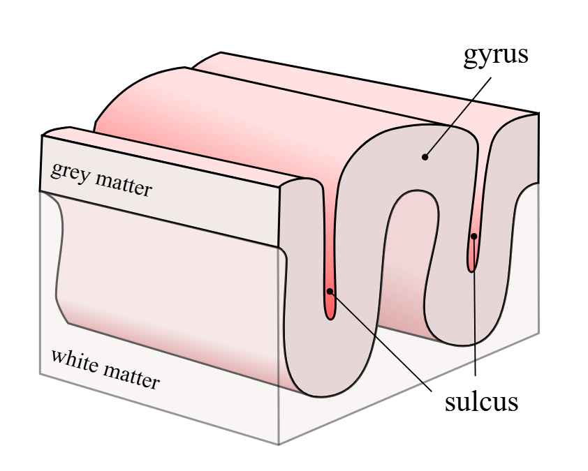
different types of sulcus
lateral sulcus - separates the temporal lobe from parietal lobe and frontal lobe and occipital lobe
central sulcus - separates frontal lobe and parietal lobe
parieto-occipital sulcus - separates occipital lobe from parietal and temporal lobes

different types of gyrus
postcentral gyrus - primary somatosensory cortex (BA 1,2,3); all tactile senses are processed in this area (touch, pain, pressure)
precentral gyrus - primary motor cortex (BA 4); contain upper motor neurons that instruct cells in the spinal cord to move skeletal muscles

explain broadmann’s areas
regions of the cerebral cortex associated with particular functions/localization of function
what are the BA numbers for primary visual perception and primary auditory sensation
Primary visual perception BA 17,18 (in occipital lobe)
Primary auditory sensation - BA 41,42 (in temporal lobe)
the lobe responsible for establishing long-term memory
temporal lobe
lobe associated with somatosensation
parietal lobe
BA responsible for thinking of a movement to be made
Premotor area (BA 6)
BA responsible for the production of language/controlling movements responsible for speech; usually on the left side
Broca’s area (BA 44, 45)
What’s the function of the prefrontal lobe
serves cognitive functions that can be the basis of personality, short-term memory, and consciousness
subcortical nuclei
clusters of grey matter located beneath the cerebral cortex
augment cortical processes (they actively filter, refine, amplify, power, and coordinate the high-level work done by the cerebral cortex)
list all subcortical nuclei structures
basal forebrain nuclei
hippocampus and amygdala
basal nuclei - caudate, putamen, globus pallidus
substantia nigra pars compacta
basal forebrain nuclei
primary location for acetylcholine production
hippocampus and amygdala
long-term memory formation and emotional responses
basal nuclei
reponsible for comparing cortical processing with the general state of activity in the nervous system to influence likelihood of movement taking place
what is the relationship of basal nuclei and brain stem nuclei
they form the motor pathway
Direct pathway (causes disinhibition of the thalamus) - Cortex >
striatum > globus pallidus internal segment (GPi) and substantia
nigra pars reticulata (SNr) -> thalamus -> excite the cortex
Indirect pathway (causes normal inhibition of the thalamus) -
Cortex > striatum > globus pallidus external segment (GPe) >
subthalamic nucleus (STN) > Gpi/SNr > thalamus > fail to excite
cortex
Function of the substantia nigra pars compacta
releases dopamine
direct pathway - activated by dopamine
indirect pathway - inactivated by dopamine
diencephalon
connection between cerebrum and the rest of the nervous system EXCEPT the system with olfaction
what are structures enclosed in the diencephalon
thalamus
hypothalamus
epithalamus - contains pineal gland
subthalamus - contains subthalamic nucleus, part of basal nuclei
thalamus
collection of nuclei relaying info between the cerebral cortex and periphery, spinal cord, or brain stem
all sensory info (except olfaction) pass thru before cortical processing
axons of peripheral sensory organs synapse in the thalamus
cerebrum send info down to communicate motor commands

hypothalamus
collection of nuclei largely involved in regulating homeostasis
in charge of the ANS and endocrine system thru regulation of pituitary gland
other parts involved in memory and emotion

what are the parts of the brain stem
midbrain
pons
medulla oblongata
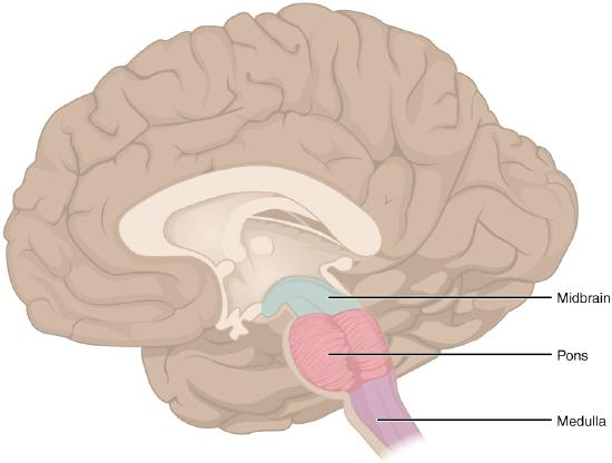
What major neural pathways and nerves connect through the brain stem?
Major ascending (sensory) and descending (motor) pathways running between the brain and spinal cord pass through the brain stem.
Cranial Nerves
What are the key functions of the hindbrain and the pons?
Hindbrain: Regulates vital life-support processes, including the cardiovascular and respiratory systems and their rates.
Pons: Acts as the primary bridge/connection between the brainstem and the cerebellum.
Where is the midbrain located, and what is its overall function and architecture?
Location: Positioned directly between the thalamus and the pons.
Overall Function: Coordinates sensory representations across visual, auditory, and touch/body (somatosensory) perceptual spaces.
Structural Boundaries: Divided into the tectum (roof) and tegmentum (floor), which border the cerebral aqueduct.

What is the tectum, and how do the inferior and superior colliculi differ in function?
Tectum: The roof of the cerebral aqueduct, composed of two pairs of sensory bumps called colliculi.
Inferior Colliculus: Relays hearing signals along the auditory brainstem pathway to the thalamus, which forwards them to the cerebrum.
Superior Colliculus: Combines visual, auditory, and somatosensory spatial information to coordinate reflexive movements (such as turning your eyes toward a sudden sight, sound, or touch).

What is the tegmentum, and what key neural structures does it contain?
Structure: Forms the floor of the cerebral aqueduct and is continuous with the brainstem's gray matter.
Cranial Nerve Nuclei: Contains nuclei extending across the midbrain, pons, and medulla that exchange signals via cranial nerves.
Autonomic Control: Houses neural regions responsible for regulating critical cardiovascular and respiratory functions.
what is the Pons
also known as the bridge
Thick bundle of white matter (anterior portion) attached to the cerebellum
Gray matter beneath is a continuation of the tegmentum of the midbrain
Input from the forebrain is sent to the cerebellum

what is the medulla
White matter found on the exterior, continuous with that from the spinal cord
Gray matter at the tegmentum
Reticular formation - associated with sleep and wakefulness (general brain activity and attention)
what is the function of the cerebellum and pathway
"little brain" also covered in gyri and sulci "
Responsible for comparing information from the cerebrum with sensory feedback from the periphery through the SC
Descending fibers from cerebrum > neurons in the pons > cerebellum (copy of motor commands sent to spinal cord) > sensory input from the periphery (spinal and cranial nerves) > inferior olive (comparison of commands from cerebrum) > midbrain (corrective command) > spinal cord
where do nerves pass at the spinal cord
intervertebral foramina
what’s the cauda equida
bundle of nerves developed as the vertebral column grows
what is the level of the upper lumbar vertebrae
sacral spinal cord

part of the brain shown in the picture
gray horns
Posterior horn – sensory processing
Anterior horn (multipolar motor neurons) – sending out of motor signals to the skeletal muscles
Lateral horn (only on thoracic, upper lumbar, and sacral regions) – central component of the sympathetic division of the ANS
What are the common carotid arteries, and what role do they play in the orthostatic reflex?
common carotid arteries: Arise from the aorta and contain stretch receptors that sense drops in blood pressure.
Orthostatic Reflex: Triggers a sympathetic nervous system response that increases heart rate when you stand up to maintain blood pressure.
How do the internal and external carotid arteries differ in their path and destination?
Internal Carotid Arteries: Branch off the common carotids and enter the braincase through the carotid canal of the temporal bone.
External Carotid Arteries: Supply blood to the superficial structures and surface of the cranium.
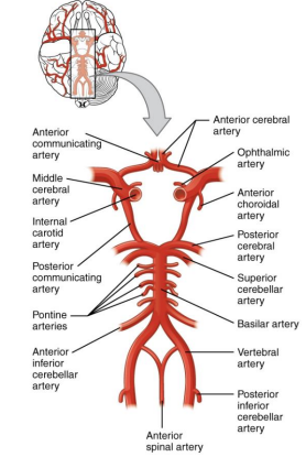
How do the vertebral arteries enter the skull, and what does the anterior spinal artery supply?
Vertebral Arteries: Pass upward through the transverse foramina of the cervical vertebrae and enter the cranial cavity via the foramen magnum.
Anterior Spinal Artery: Travels along the anterior median fissure to deliver blood to the front (anterior) aspect of the spinal cord.

What are the roles of the basilar artery and the Circle of Willis in CNS circulation?
Basilar Artery: Formed by the merging of the two vertebral arteries; supplies blood to the brain stem and cerebellum.
Circle of Willis: A junction (confluence) of interconnected arteries that maintains consistent blood flow (perfusion) to the brain even if one pathway becomes blocked or narrowed.

How does venous blood and cerebrospinal fluid (CSF) drain from the brain back into general circulation?
Superior Sagittal Sinus: Runs along the longitudinal fissure groove and absorbs CSF from the meninges.
Confluence of Sinuses: The shared meeting point where the superior sagittal, occipital, and straight sinuses drain together.
Drainage Pathway: From the confluence of sinuses, blood flows into the transverse sinuses → sigmoid sinuses → jugular vein.
What are the meninges, and what are the main features of the dura mater?
Meninges: The outer connective tissue membranes protecting the central nervous system.
Dura Mater: A thick, fibrous, and tough outer protective layer.
Anchoring & Coverage: Anchored to the inner surface of the cranium and vertebral cavity, forming a sheath over the brain, spinal cord, and major entering blood vessels.
What is the arachnoid mater, and what are the roles of the subarachnoid space and granulations?
Arachnoid Mater: A thin fibrous tissue forming a loose sac around the CNS, characterized by a spiderweb-like mesh of arachnoid trabeculae.
Subarachnoid Space: The compartment containing circulating cerebrospinal fluid (CSF), providing a protective liquid cushion for the brain and spinal cord.
Arachnoid Granulations: Specialized sites where drained CSF is filtered back into the venous bloodstream.
What is the pia mater, and how does it interact with brain surface contours and blood vessels?
Structure: Meaning "tender mother," it is a thin, continuous layer of cells forming a fluid-impermeable membrane.
Contour Following: Delicately clings to the brain surface, dipping directly into every fold, groove, and convolution (gyri and sulci).
Blood Supply Relation: The blood vessels that nourish CNS tissue sit directly between the pia mater and the underlying nervous tissue.
What is Cerebrospinal Fluid (CSF), how is it produced, and what are its main functions?
Production: Created within specialized membranes called choroid plexuses, where ependymal cells surround and filter capillary blood.
Circulation: Flows continuously both inside (through ventricles) and around the outside of the CNS.
Functions: Acts as a protective liquid cushion for the brain and spinal cord, perfuses neural tissue, and collects metabolic waste from interstitial fluid to return to the bloodstream.
What are brain ventricles, how do they form, and where does CSF go after leaving them?
Definition: Open fluid-filled spaces within the brain containing choroid plexuses that produce and circulate CSF.
Development: Originate from the hollow space within the embryonic neural tube (the central canal).
Ultimate Pathway: After traveling through all ventricles, CSF emerges into the subarachnoid space to be reabsorbed back into the blood.
How does CSF flow through the lateral ventricles, third ventricle, and cerebral aqueduct?
Lateral Ventricles (2): Located deep within the cerebrum; drain CSF into the third ventricle.
Third Ventricle: Lies in the space between the right and left diencephalon; receives CSF from the lateral ventricles through the interventricular foramina.
Cerebral Aqueduct: Runs through the midbrain, channeling CSF down from the third ventricle to the fourth ventricle.
What is the fourth ventricle, and how does CSF transition to the outside of the CNS?
Fourth Ventricle: Positioned in the space between the cerebellum, pons, and upper medulla; receives CSF from the cerebral aqueduct and narrows into the spinal cord's central canal.
Apertures: CSF exits the fourth ventricle via the median and lateral apertures.
Subarachnoid Space: Passing through these apertures allows CSF to flow into the subarachnoid space, enveloping the outer surface of the entire central nervous system.