Lab 2: Cardiovascular system
 Call Kai
 Learn
 Practice Test
 Spaced Repetition
 Match
 Flashcards
 Knowt Play
1/41

There's no tags or description
Looks like no tags are added yet.
42 Terms

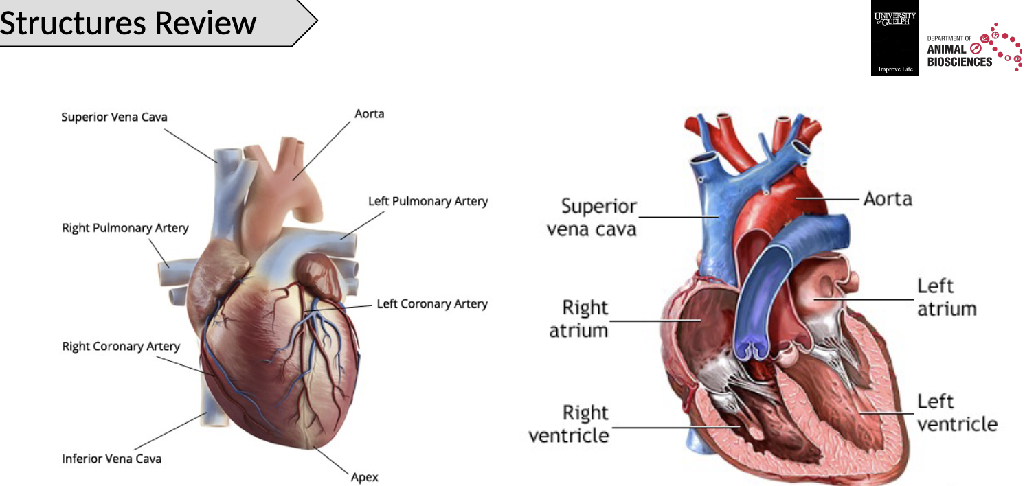

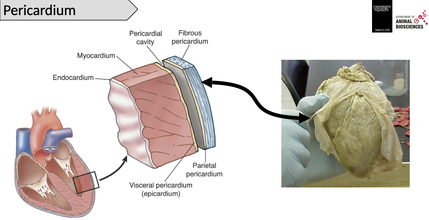


Valves in the heart include:
Atrioventricular: atrium to ventricle
tricuspid valve
bicuspid (mitral) valve
Semilunar: ventricle to artery
pulmonary valve
aortic valve


5- Left bundle branch
7- Atrioventricular node
1- Superior vena cava
3- Right bundle branch
6- Bundle of His
2- Sinoatrial node
4- Purkinje fibers
Self depolarization graph

Depolarization of the heart steps
Depolarization starts in SA node
Depolarization propagates through contractile cells with Gap Junctions
Continued depolarization of atria and AP passing through the Bundle of His and Purkinje fibre. *Note the electrical barrier
Starting at the apex, the ventricular heart muscles contract

ANS control of HR
SA node is under nervous regulation
Sympathetic activation will lead to increased heart rate; Increased Na+ leakage shortening the potential drift
Parasympathetic activation will lead to decreased heart rate; Increased K+ permeability
To maintain coordination between atrial and ventricular contraction, the sympathetic input proportionally increases the velocity of the AV node; This reduces the lag in conduction

Can the heart cramp?
The heart muscle itself can’t cramp, unlike skeletal muscles

Coupling of electrical activity, contraction, movement of blood, and valves diagram

Stage 1 of coupling of electrical activity, contraction, movement of blood, and valves
Refilling of the heart (diastole)
The heart is relaxed
Venous return fills the atria
Pressure pushes the AV valves open
Blood flows in the ventricles
Not enough pressure to open the semilunar valves

Status of valves in stage 1
AV Valves - OPEN
SL Valves - CLOSED
Stage 2 of coupling of electrical activity, contraction, movement of blood, and valves
Atrial Contraction (part of diastole)
SA node fires
Atrial contraction
Blood packed in ventricles through the AV valves which are pushed open
Pressure in ventricles not sufficient to force the semilunar valves open
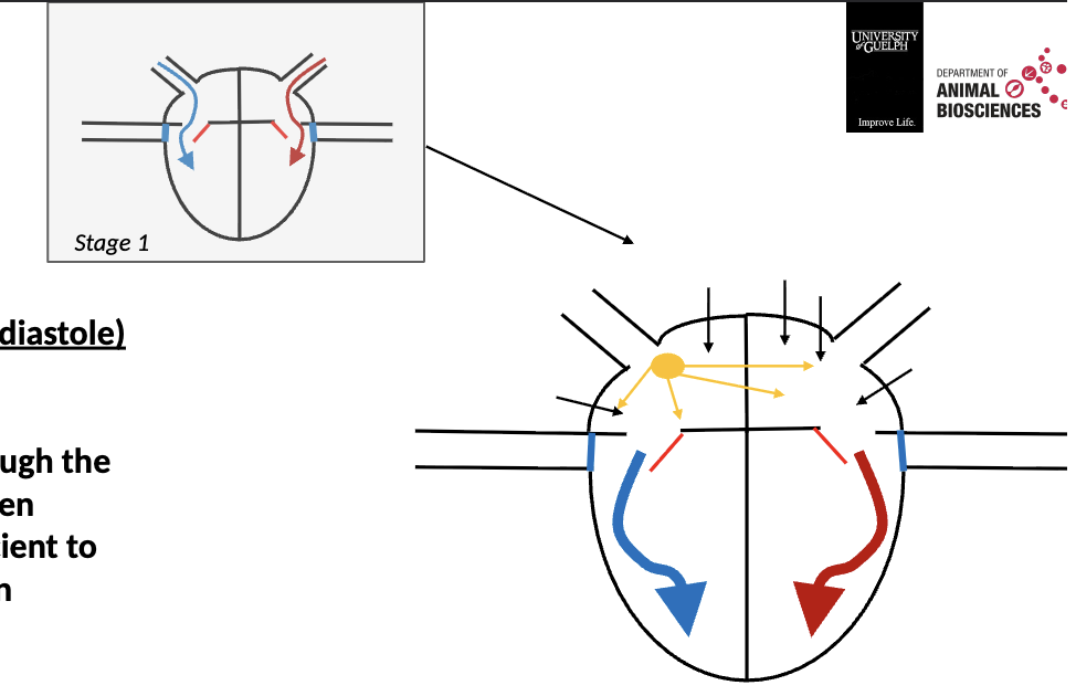
Status of valves in stage 2
AV Valves - OPEN
SL Valves - CLOSED
Stage 3 of coupling of electrical activity, contraction, movement of blood, and valves
Isovolumic contraction (part of systole)
AV node transmit the AP between atria and ventricles with a lag
Atria finished contracting, ventricles waiting for the AP wave
Blood packed in ventricles, all valves are closed
In between atrial and ventricular contraction

Status of valves in stage 3
AV Valves - CLOSED
SL Valves - CLOSED
Stage 4 of coupling of electrical activity, contraction, movement of blood, and valves
Ejection (systole per say)
AP passed on to the bundle of Hiss and Purkinje fibers
Ventricles depolarize and contract
Pressure in ventricles increased, sufficient to force open the semilunar valves
Blood rushes out of the ventricles into arteries

Status of valves in stage 4
AV Valves - CLOSED
SL Valves - OPEN
Stage 5 of coupling of electrical activity, contraction, movement of blood, and valves
Isovolumic relaxation (part of systole)
Contraction is over
The heart is empty
Low pressure throughout, all the valves are closed
Blood starts accumulating in the atria

Status of valves in stage 5
AV Valves - CLOSED
SL Valves - CLOSED
Full cycle diagram
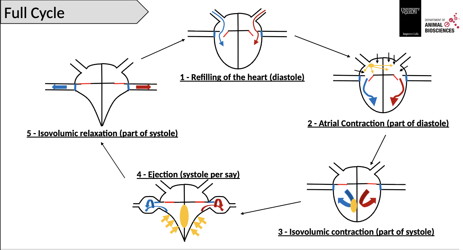
Electrocardiogram parts
P: Atrial depolarization
PQ interval: Time taken by the AP to pass the AV node
QRS: Ventricle depolarization
T: Ventricle repolarization

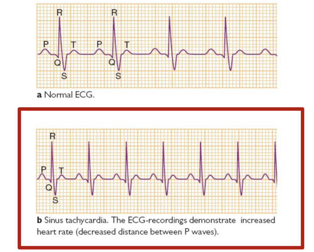
Normal ECG

Sinus tachycardia
Increased HR
decreases distance between P waves
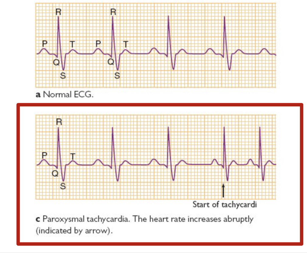
Paroxysmal supraventricular tachycardia
Paroxysmal tachycardia: HR increases abruptly
Atrial flutter
Frequent, but rhythmic atrial contractions (P waves). Ever second atrial depolarization is propagated to the ventricles (2:1 rhythm)

1rst degree AV block
The propagation of the atrial depolarizations to the ventricles is slow, and the PQ interval is abnormally long

2nd degree AV block
Some of the atrial depolarizations are not conducted to the ventricles

3rd degree AV block
There is a complete failure of impulse conduction from the atria to the ventricles. QRS complexes originate in the tissues below the AV node. The ventricular rhythms are slow, but usually regular.

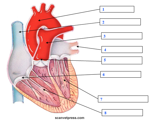
1 - aorta
2- superior vena cava
3 - pulmonary artery
4 - pulmonary vein
5 - pulmonary valve (semi-lunar)
6 - tricuspid valve
7 - left ventricle
8 - papillary muscle
Why is the aorta elastic
To absorb pressure coming from left ventricle after ejection
To keep pressure in the arterial side higher than in the veins
To maintain the flow of blood in the body

1 - lungs
2 - non-oxygenated
3 - oxygenated
Steps on how electrical activity in the heart propagates
SA node is self excited (fires an action potential)
Depolarization travels through the atria to the AV node with little delay
AP propagates to bundle of His and purkinje fibers quickly
The contractile cells depolarize from the apex through the ventricular wall (ventricular depolarization)

diastole
atrial contraction
isovolumic contraction
ejection phase (systole)
isovolumic relaxation
Describe the status of each of the valves listed below (open/closed) during phase 1 of the cardiac cycle
Open
tricuspid
bicuspid
Closed
pulmonary
aortic
Describe the status of each of the valves listed below (open/closed) during phase 2 of the cardiac cycle
Open
bicuspid
tricuspid
Closed
aortic
pulmonary
Describe the status of each of the valves listed below (open/closed) during phase 4 of the cardiac cycle
Open
pulmonary
aortic
Closed
bicuspid
tricuspid
Describe the status of each of the valves listed below (open/closed) during phase 5 of the cardiac cycle
Closed
bicuspid
tricuspid
pulmonary
aortic

Match the electrical waves (episodes) during a normal electrocardiogram (ECG) with the electrical event they represent
P - atrial depolarization
QRS - ventricle depolarization
T - ventricle repolarization
PQ - time taken by the action potential to pass to the AV node (from the atria to the ventricles)