Abdomen Protocol: Sections 10&11
 Call Kai
 Learn
 Practice Test
 Spaced Repetition
 Match
 Flashcards
 Knowt Play
1/18

There's no tags or description
Looks like no tags are added yet.
19 Terms
What structures are inculded in the Transverse and Sagittal gallbladder ?
1. Transverse GB Neck (TRV GB Neck)
2. Transverse GB Body (TRV GB Body)
3. Transverse GB Fundus (TRV GB fundus)
4. Sagittal GB Neck to Fundus
5. Sagittal GB Neck to Fundus
6. LDEC Transverse GB
7. LDEC Sag GB
Annotation for Sagittal
SAG GB (Don’t need to write neck, body, fundus in sag)
Transverse & Sagittal Gallbladder Depth, focal zone, section width
Depth: decreases, remember GB is relatively anterior
Focal Zone: center of GB and/or neck
Section width: Full or slightly decreased
Transverse & Sagittal Gallbladder General Tips
Superior is neck inferior is fundus
liver at MEDIUM Gray
Transverse & Sagittal Gallbladder Scanning Tips
Come subcostal, go superior and come down into the neck.
For alternate neck window: Go intercostal where you get the MPV and angle inferior. Neck – obstruction! Fundus – wall!
Document Main Lobar Fissure from GB to RPV.
Transverse & Sagittal Gallbladder note
Looking for pathology that may not have been seen supine and for stones to move). Upright / erect scanning if needed for stone/density evaluation, shadowing, movement etc.
LDEC Porta Hepatis, Portal Triad, CHD and Distal CBD
LDEC, subcoastal, go to the porta hepatis and identify the portal triad.
8. Bile duct measurement at HA
Common bile duct at head of pancreas
GB (looking for pathology that may not have been seen supine and for stones to move).
LDEC Porta Hepatis, Portal Triad, CHD and Distal CBD annotate, position, depth, focal zone, section width
Annotate: LDEC SAG
Position: Stay in decubitus position (right after you finished GB)
Depth: Include the porta hepatis & midline structures. IVC is a landmark for depth setting.
Focal zone: At the bile duct
Section width: Full or slightly decreased
LDEC Porta Hepatis, Portal Triad, CHD and Distal CBD Note
Transverse slightly oblique for CHD at Porta Hepatis. Angle more sagittal to follow the CBD to the head of the pancreas as distal as possible as it heads to the ampulla of Vater.
LDEC Porta Hepatis, Portal Triad, CHD and Distal CBD Note Anatomy
CBD courses through head of the pancreas and meets up with pancreatic duct. They both go into the ampulla of vater.
CHD is usually small in diameter due to the weight of the liver around it.
CBD will dilate a little (this is normal behavior of the bile duct) as it exits the liver because it no longer has the weight of the liver on it.
If you see the CHD dilated, despite the weight of the liver, that is true intrahepatic biliary dilatation.

What are the yellow and red veins pointing at ?
Yellow: CHD
Red: CBD
Risk factors for DVT

Signs & Symptoms of Deep Veins Thrombosis
1. Extremity pain and tenderness2. Swelling
3. Venous distention
4. Discoloration
5. Palpable cord
6. Pitting Edema
Symptoms related to PE include. What does PE mean??
Pulmonary Embolism:
1. Tachypnea
2. Chest pain
3. Tachycardia

Phlebitis (thrombus in superficial veins)
Most likely with co-existing palpable cord

pitting edema
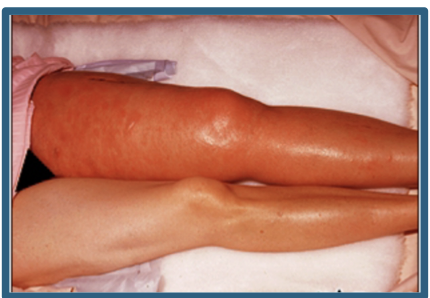
red, hot, swollen leg
focal zone
refers to the depth at which the ultrasound beam is narrowest and most intense.
• the area where the image quality is optimal due to the improved resolution and signal-to-noise ratio.
The focal zone is achieved by …
focusing the ultraound beam at a specific depth
focal zone is located between the point of divergence and the point where the beam becomes too wide to provide good resolution