Anatomy BIO 1030- Exam 2- PLNU
 Call Kai
 Learn
Learn Practice Test
 Spaced Repetition
 Match
Match Flashcards
 Knowt Play
1/76

There's no tags or description
Looks like no tags are added yet.
Name | Mastery | Learn | Test | Matching | Spaced | Call with Kai | Chat |
|---|
No analytics yet
Send a link to your students to track their progress
77 Terms
Extracellular matrix: composition
-proteins
-polysaccharides
Extracellular matrix: purpose
-hold cells together
-influence activity of cells
Epithelial tissue:
General features-uses
1. Covers body surfaces
----A. outside of the body
----B. lining of digestive and respiratory tracts
----C. body cavities
----D. blood vessels
2. Glands
----A. derived developmentally from body surfaces
----B. releases proteins or solutions into blood, cavity, or body surface
Epithelial tissue:
General features-basic functions
PAFESSr
-protection
-absorption
-filtration
-excretion
-secretion
-sensory reception
Epithelial tissue:
Special Characteristics
1. tightly packed cells
2. continuous sheets
3. polar
4. supported by connective tissue
5. avascular but innervated
6. high proliferative/regenerative capacity
Epithelial tissue:
Special Characteristic-t
tightly packed cells
Epithelial tissue:
Special Characteristic-c
continuous sheets
Epithelial tissue:
Special Characteristic-p
polar
Epithelial tissue:
Special Characteristic-Polar: Apical Surface
-Free side
-not in contact with other cells
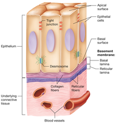
Epithelial tissue:
Special Characteristic-Polar: Basal Surface
-bottom layer
-attached to other cells via basement membrane
-basement membrane (basal and reticular lamina)
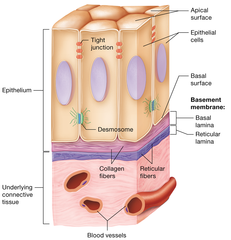
Basement membrane
-composed of two lamina "sheets"
-basal lamina
-on top: closest to epithelial cells
-selective filter that only allows certain substances to pass epithelial cells
-reticular lamina: deeper than basal, closer to connective tissue

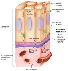
basal lamina
-on top: closest to epithelial cells
-selective filter that only allows certain substances to pass
-thin sheet of glycoproteins
-produced by epithelial cells
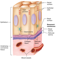
-reticular lamina
-deeper than basal, closer to connective tissue
-extracellular material from underlying connective tissue
-gives more resilience to the basement membrane and epithelium
Epithelial tissue:
Special Characteristic-s
supported by connective tissue
Epithelial tissue:
Special Characteristic-a
Avascular but not innervated
-blood vessels do not penetrate epithelial membranes (epi cells are next to blood vessels)
Epithelial tissue:
Special Characteristic-h
high proliferative/regenerative capacity
Why is epithelial tissue high proliferative/regenerative capactiy?
The skin gets constant damage (sun, abrasions, chemicals, weather, etc.) so it needs constant repair
What happens with chemotherapy treatment?
Chemo keeps your cells from dividing as fast
-makes it slow down to a point where the skin can't heal itself
number of cell layers
-simple: single (one) layer
-statified: layered
-pseudostatified: looks statified but is not

cell shapes
1. Squamous
2. Cuboidal
3. Columnar
4. Transitional

Simple Squamous Epithelium
A single layer of squamous epithelial cells and the underlying basal lamina
Funtion: rapid diffusion and filtration
Loction:
alveoli of the lungs,
kidneys, and lining the inner surface of blood vessels

Simple Squamous Epithelium
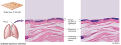
Simple Squamous Epithelium

Simple cuboidal epithelium
single layer cuboidal epithelial cells
Function: secretion and absorption
Location: Kidney tubules, secretion portions of small glands

simple cuboidal epithelium

simple columnar epithelium
Made up of a single layer of tall cells that fit closely together, contain goblet cells
Function:
secretion and absorption
location: lining of stomach and intestines, and glands (digestive tract)

simple columnar epithelium

Pseudostatified Columnar Epithelium
Is a simple columnar epithelium that appears stratified with cillia. Nuclei of cells are uneven.
Function:
trap and remove particles
associated with goblet cells (secrete)
Location:
Lining of respiratory tract

Pseudostatified Columnar Epithelium

stratified squamous epithelium
Several layers of squamous epithelial cells
Funtion:
-resist wear and tear
-protect from infection
Location:
Moist:
Oral cavity and esophagus
Anus and Vagina
Keratinized:
Skin (where friction usually is)

stratified squamous epithelium

statified cuboidal epithelium
Several layers of cuboidal cells; rare
Location
-Sweat and mammary glands (usually a bilayer)

statified columnar epithelium
Several layers of columnar cells; rare
Location:
Pharnyx

transitonal epithelium
Several layers of epithelial cells; cells can change shape from dome shape (ice cream cone) to squamous when stretched
Fucntion:
Stretch
Location:
Urinary system

transitional epithelium

summary of epithelial tissues

Endocrine glands
-release chemicals INTO body for long distance communication
-ductless
-release by exocytosis into the blood to move throughout the body
Structure:
-multicellular
-diffuse endocrine system (single cells scattered through an organ/organ system)
Exocrine glands
-release chemicals ONTO body surface
-multicellular: release into ducts
-unicellular: goblet cells
-merocrine: pack products into secreting vesicles for exocytosis
-holocrine: product accumulates in the cell until the cell bursts or ruptures
Connective tissue
**most abundant tissue in the body!!**
Connective tissue: General features
-Strength and Support
-Protection
-Insulation
-Transport substances
-degree in vascularity
-rich blood supply: adipose, bone
-poor blood supply: cartilage
Collagen
Protein fibers in tendons and ligaments
-most common protein in the body
-"rope" of three fibers wound
-flexible and strong, not elastic (no recoil)
-WHITE fibers
Elastin (Elastic Fibers)
-long thin proteins with branching networks
-elastic
-YELLOW fibers
Reticular fibers
-thin, short, collagen fibers
-continuous with collagen

Blast cells
produce/maker cells
epithelium vs. connective tissue

Serous Membrane
-made of thin layers of epithelial and connective tissues
-fist pushed into balloon (completely surrounds)
-in LUNGS, HEART, INTESTINES
-contatins serous fluid between two membranes
-visceral serous membrane
-partietal serous membrane

visceral serous membrane
Membrane
-covers organs
-touching organ

Parietal Serous Membrane
Membrane:
-Covers wall of cavity
-away from organ

Serous membrane: Pleura
Serous membrane around lungs
Serous membrane: Pericardium
Serous membrane around heart
Serous membrane: Perioneum
Serous membrane around intestines
Mesenchyme
-embryonic connective tissue
-all connective tissue comes from this tissue (develops into connective and skeletal tissue)
Structure
-loosly organized
-star shaped
-fluid ground substance
mesenchyme

Loose connective tissue, Areolar
Loosely arranged, gel-like matrix with all three fiber types
-irregular, loose, wool-like/fuzzy
Contains Cells:
-fibroblasts
-macrophages
-mast cells
-fat cells
Function:
-universal packing material
-allows diffusion
-reservoir for water and salts
Location:
-surrounds organs and capillaries
-attaches skin or underlying tissues

Loose connective tissue, areolar

Adipose Tissue
-fat tissue
-closely packed adipocytes
-nucleus pushed to side
Function:
-Energy storage
-insulation
-support and protect organs (padding)
Location:
-subcutaneous (skin): under basement membral
-around kidneys
-mammary glands

Adipose tissue

Reticular connective tissue
Structure:
-Reticular fibers and cells
Function
-support soft tissue or organs and blood vessels
Location
-lymph nodes
-spleen
-bone marrow

dense regular connective tissue
Structure
-matrix of parallel fibers
-fibroblasts
Function:
-Tensile strength and stretch resistance in one
Location:
-tendons
-ligaments

Dense Irregular Connective tissue
Strucuture
-collagen fibers in irregular bundles
-fibroblasts
-tye-die llings
Function
-tensile strength and stretch resistance in MULTIPLE directions
Location:
Dermis of skin
fibrous joint capsue
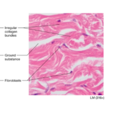
Elastic connective tissue
Structure
-wrinkly parallel fibers to recoil
Function
-elastic/recoil after stretching
Location:
-wall of arteries and bronchioles

Cartilage characteristics
-Avascular
-condrycytes in lacunae
-matrix
Hyaline Cartilage
Structure
-transparent matrix
-chondrocytes in lacunae
Function
-forms embryonic skeleton
-growth of long bones
-rigid and smooth, yet flexible
Location
-nose
-trachea
-articular cartilage
-epiphyseal plate
-costal cartilage of ribs

Fibrocartilage
Structure
-bundles of collagen fibers (semi-parallel)
-lacuna
Function
absorbs compression
-tensile strength
location
intervertebral disks
articualar disks
pubic symphysis

Elastic Cartilage
Structure
-Elastic fibers
lacuna
Function
-flexibility
-shape retention
Location
external ear
epiglottis

Bone Tissue
-most of skeleton
-solid matrix
-excellent blood suppy
-osteocytes in lacunae
-tree

Blood tissue
structure
-45% cells
-primary fluid: plasma
Function:
transport gases, nutrients, wastes, hormones
-fibrous proteins in the plasma help with clotting
Location
cardiovascular system

Summary of Connective tissues
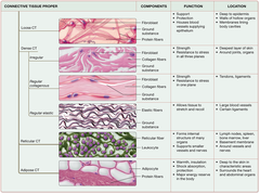
Summary of specialized connective tissue

integumantary system: Functions of Skin
-physical barrier
-chemical barrier
-biological barrier
-body temp regulation
-cutaneous sensation (touch, temp, pressure, pain)
-vitamin D synthesis
-blood reservoir
-excretion
Skin Strucure
Epidermis
Dermis
Hypodermis

Epidermis
-thick stratified squamous epithelium
-kertainocytes
-desmosomes
-melanocytes
-langheran cells
-phagocytes
-immune protection
-merkel cells (touch)
-stata: layers of dead cells
Dermis
-connective tissue
-strutural support of skin
-rich supply of blood and nerves
-relationship with accessory structures
-papillary layer
-reticular layer
Papillary layer of dermis
-upper layer of the dermis that forms dermal papillae
-areolar connective tissue
-papillae project into epidermal layer (hills)
-dermal ridges (assist with grip/friction)

Reticular layer of dermis
-dense irregualr connective tissue
-thick bundles of collage
hypodermis
-loose connective tissue
-adipose tissue
Thick vs. thin skin (picture)
