Anatomy of ovaries and fallopian tubes
 Call Kai
 Learn
 Practice Test
 Spaced Repetition
 Match
 Flashcards
 Knowt Play
1/31

There's no tags or description
Looks like no tags are added yet.
32 Terms
True/False: The only structure in the abdominopelvic cavity that is not covered by peritoneum is the ovary
True
The only true intraperitoneal structure is the
Ovary
Ovaries are connected to the uterine cornua medially by
Ovarian ligament
Anterior ovarian surface connected to the posterior surface of the broad ligament by the
Mesovarium
Infundibulopelvic ligament contains vessels and extends from the lateral pelvic wall to the …
Lateral ovary
Posterior to ovary
Ureter, internal iliac artery, and vein
Landmark for lateral aspect of adnexa
Iliopsoas muscle
Hilum of ovary is located on the _______ surface
Superoanterior
Premenopausal ovary size
3 × 2 × 1cm
Normal volume of ovary
6-13cc
Normal follicle size
Immature: 10mm or less
Dominant: 15-25mm
Another name for dominant follicle
Graafian follicle
Postmenopausal ovaries normally do not exceed __cm in any dimension or 8cc volume
2
Fossa of waldeyer (ovarian fossa)
Shallow peritoneal fossa on lateral pelvic wall
Bordered by ureter and iliac vessels
Cortex
Functional tissue of ovary
Composed of 1000s of oocytes embedded in connective tissue
Follicles form in cortex layer
Varies in thickness according to the age of individual
Several follicles can mature, but one egg is typically released from dominant follicle during each menstrual cycle
Medulla
Connective tissue and vessels that supply and drain cortex
No sonographic distinction between cortex and medulla
Blood supply to ovary comes from …
Uterine artery and ovarian arteries
Uterine arteries branch from …
Internal iliac arteries
Ovarian arteries branch from …
Anterior aorta below the renal arteries
Small branches of the uterine artery merge with branches of the ovarian artery near the uterine _______
Cornua
Right ovary vein empties into ____
IVC
Left ovarian vein empties into
Left renal vein
Fallopian tube is also known as
Oviducts, salpinges, or uterine tubes
Fallopian tubes
Trumpet-shaped muscular canals
8-14cm length
Tortuous
Extend from the cornua to medial ovary
Supplied with blood from vascular arch
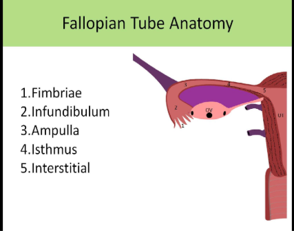
Tube segments: Interstitial
AKA intramural
Short section that passes through the uterine wall
Most dangerous place for ectopic
Tube segments: Isthmus
Middle portion of fallopian tube
Tube segments: Ampulla
Widest and longest portion
Egg fertilization normally occurs here
Most ectopic pregnancies occur in this segment
Tube segments: Infundibulum
Contains fimbriae
Trumpet - shaped ends opens into pelvic peritoneal cavity
Wall layers or fallopian tube
Mucosal lining - contains cilia to help propel egg through tube
Muscle
Serosal coating
The _________ ligament connects the ovary to the uterine cornua ________connects the ovary to the lateral pelvic wall
Ovarian, infundibulopelvic
What sonographic landmark is preferred for location the ovaries
Iliac vessels
Most dangerous location for an ectopic pregnancy
Intersitial portion