Ophthalmology Photos
 Call Kai
 Learn
 Practice Test
 Spaced Repetition
 Match
 Flashcards
 Knowt Play
1/42

There's no tags or description
Looks like no tags are added yet.
43 Terms

Anatomy of the Eye

Fundoscopic Exam

Abnormal Fundoscopic Exam

Emmetropia

Hyperopia

Myopia
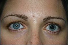
Do these eyes maintain PERRLA?
no, pupils are not the same size

Glaucoma

Look at one from class
Normal flow of fluid through the eye

Look at one from class
Open angle glaucoma flow

Look at one from class
Closed angle glaucoma flow

what exam is this
Tonometry
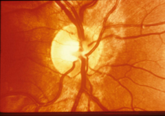
is this normal
Normal cup and disc
Distinct disc margins, well marked cup, healthy pink color of neuroretinal rim

Normal cup to disc ratio
2:1

is this normal
Abnormal cup to disc ratio

Notice vessels pushed nasally and abnormal size of the physiological cup
Cupping

what test
Perimetry test (visual field test)
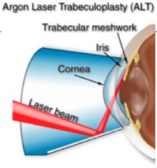
what is this
Trabeculoplasty

what disease with what presentations
closed angle glaucoma with corneal clouding and conjunctival injection

what is this
Laser peripheral iridotomy

Conjunctival injection

Ciliary Injection

Conjunctivitis

Bacterial non-Neisseria conjunctivitis

Gonococcal conjunctivitis

Allergic conjunctivitis

what is this
Subconjunctival hemorrhage
Note that bleeding remains outside of the limbus

what anatomical structure
Cornea
Note that it is 5 layers and is bad if all 5 are penetrated due to aqueous humor leakage

Classic linear abrasion
Found in corneal abrasion; Note that it should NOT be dendritic in nature; Indicates something scratching on eye repeatedly

what is this and what does it indicate
Dendritic lesion with terminal buds
Indicates Herpes in the eyew
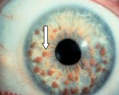
Dendritic lesion that is easy to miss

Herpetic lesions around the eye

Cataract

Red reflex in right eye, opacity in left eye indicating cataract
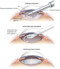
Surgical removal of a cataract

Vision with cataracts

As Pt rotates head, blood will move
Hyphema

Fundoscopic exam of Pt with Central Retinal Artery Occlusion

Note diminished blood flow to eye results in a pale fundus
CRAO

Retinal detachment on fundoscopic exam

Occurs with retinal detachment
Posterior vitreous separation

Rhegmatogenous retinal detachment

Retinal hemorrhage