506: The Eye & Orbit
 Call Kai
 Learn
Learn Practice Test
 Spaced Repetition
 Match
Match Flashcards
 Knowt Play
1/219

There's no tags or description
Looks like no tags are added yet.
Name | Mastery | Learn | Test | Matching | Spaced | Call with Kai | Chat |
|---|
No analytics yet
Send a link to your students to track their progress
220 Terms
Lateral canthus
Identify the structure at #1

Palpebrae (eyelid)
Identify the structure at #2
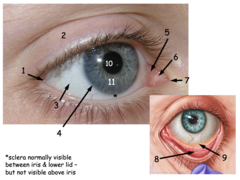
Bulbar conjunctiva
Identify the structure at #3
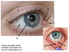
Border between sclera & cornea
Identify the structure at #4

Lacrimal lake
Identify the structure at #5

Caruncle
Identify the structure at #6
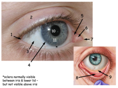
Medial canthus
Identify the structure at #7

Palpebral conjunctiva
Identify the structure at #8

Fornix
Identify the structure at #9

Pupil
Identify the structure at #10

Iris
Identify the structure at #11

Ophthalmic (V1)
- Supraorbital n.
- Supratrochlear n.
Identify the dermatome in yellow. What nerve(s) participate in this sensation?

Maxillary (V2)
- Infraorbital n.
Identify the dermatome in red (middle). What nerve(s) participate in this sensation?

Mandibular (V3)
Identify the dermatome in peach (bottom)

lacrimal n.
frontal n.
nasociliary n.
what are the 3 main branches of the opthalmic branch (V1) of CN V?
frontal n.
the supraorbital n. is a branch of the:
frontal n.
the supratrochlear n. is a branch of the:
Frontal n.
What ophthalmic division provides sensory to the forehead and scalp?
Lacrimal n.
What ophthalmic division provides sensory to the lacrimal gland and upper eyelid?
Nasociliary n.
What ophthalmic division provides sensory to the medial orbit and upper nose?
Supratrochlear n.
Identify the structure at #1
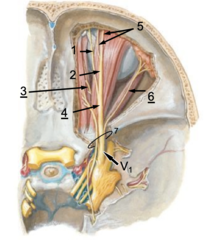
Supraorbital n.
Identify the structure at #2

Nasociliary n.
Identify the structure at #3

Frontal n.
Identify the structure at #4

Medial & lateral branches of supraorbital n.
Identify the structure at #5

Lacrimal n.
Identify the structure at #6

Superior orbital fissure
Identify the structure at #7
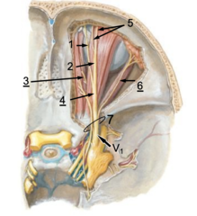
Lacrimal n.
Identify the structure at #1

Nasociliary n.
Identify the structure at #2
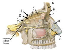
Frontal n.
Identify the structure at #3

Anterior & posterior ethmoidal n.s
Identify the structure at #4

Supraorbital n.
Identify the structure at #5

Supratrochlear n.
Identify the structure at #6

Infratrochlear n.
Identify the structure at #7

External nasal branch (from anterior ethmoidal)
Identify the structure at #8

Infraorbital n. (from V2)
Identify the structure at #9

anterior ethmoidal n.
posterior ethmoidal n.
what are the two branches of the nasociliary n.?
nasociliary n.
the anterior ethmoidal n. is a branch of the:
nasociliary n.
the posterior ethmoidal n. is a branch of the:
anterior ethmoidal n.
the external nasal n. branch is a branch of the:
V2
the inferior eyelid's sensory innervation comes from:
V1
the superior eyelid's sensory innervation comes from:
nasociliary n.
which branch of V1 is the only nerve that supplies the sensory info to the eyeball?
obicularis oculi m. (orbital & palpebral parts)
identify the structure:

medial palpebral ligament
identify the structure (red arrow):

lateral palpebral ligament
identify the structure:

- Support for eyelids (eyelid skeleton)
- Anchors tarsal muscles
- Contains tarsal glands
Function of the tarsal plates:
Tarsal plates
Identify the structure:

CN VII to the orbicularis oculi (palpebral part)
motor control for the blink reflex is done by:
V1, nasociliary n.
sensory innervation to activate the blink reflex triggered by touch is done by:
CN II
sensory innervation to activate the blink reflex triggered by visual threat or bright light is done by:
CN VIII
sensory innervation to activate the blink reflex triggered by sudden sounds (>50-60 dB) is done by:
frontal
maxilla
zygomatic
lacrimal
ethmoid
sphenoid (greater and lesser)
name the 6 bones that make up the orbit:
Optic canal
Identify the structure at #1

Supraorbital foramen
Identify the structure at #2

Posterior ethmoidal foramina
Identify the structure at #3

Anterior ethmoidal foramina
Identify the structure at #4

Superior orbital fissure
Identify the structure at #5

Inferior orbital fissure
Identify the structure at #6
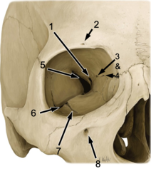
Infraorbital groove
Identify the structure at #7

Infraorbital foramen
Identify the structure at #8

Frontal
Identify the structure at #1

Ethmoid
Identify the structure at #2

Lesser wing of sphenoid
Identify the structure at #3

Greater wing of sphenoid
Identify the structure at #4
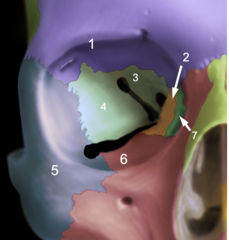
Zygomatic
Identify the structure at #5

Maxilla
Identify the structure at #6
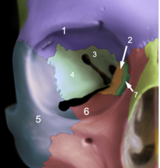
Lacrimal
Identify the structure at #7

Orbital floor & infraorbital canal (maxilla)
Identify the structure at #1

Orbital plate of ethmoid bone (lamina papyracea)
Identify the structure at #2

superior orbital fissure
V1 enters the orbit via the:
frontal bone
the supraorbital foramen is located on the:
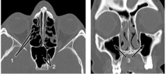
Ethmoid air cells
Identify the structure at #1
maxilla bone
the infraorbital foramen is located on the:
Sphenoid sinus
Identify the structure at #2

Maxillary sinuses
Identify the structure at #3

orbital blowout fracture
what is the diagnosis?

- diplopia (occurs if both eyes don't track equally)
- infraorbital nerve damage (facial numbness)
- infections
What are the complications of an orbital blowout fracture?
The inferior rectus disrupts the infraorbital groove (usually into the maxillary sinus but could go into ethmoid sinus)
What muscle is involved with an orbital blowout fracture?
Special sensory only
What type of nerve is the optic nerve (CN II)?
Dura mater
Arachnoid mater
Pia mater
What meninges surround the optic "nerve"?
Forebrain
Optic tracts are an extension of the:
Hydrocephalus
High CSF pressure:
Vision problems due to swelling of optic disc
Symptoms of hydrocephalus:
Subarachnoid space
Identify the structure at #1

Optic disc
Identify the structure at #2

CSF
what causes the pressure exerted on the optic nerve in papilledema?
papilledema (usually bilateral - if unilateral, likely indicative of ocular pathology, e.g. optic nerve glioma)
patient presents with bilateral blurred vision. An eye exam was done and displays bulging of the optic disc. What is the diagnosis?
anterior (to equator)
the rectus muscles of the eye insert onto the eyeball on the _______ aspect
posterior (to equator)
the oblique muscles of the eye insert onto the eyeball on the _______ aspect
superior rectus m.
identify the structure:

medial rectus m.
identify the structure:

inferior rectus m.
identify the structure:

lateral rectus m.
identify the structure:
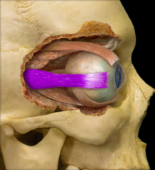
superior oblique m.
identify the structure:

inferior oblique m.
identify the structure:

levator palpebrae superioris m.
identify the structure:

Levator palpebrae superioris
What muscle elevates the upper eyelid?
Superior oblique
Identify the structure at #1

Superior rectus
Identify the structure at #2
