Anatomy I: Bone Development
 Call Kai
 Learn
 Practice Test
 Spaced Repetition
 Match
 Flashcards
 Knowt Play
1/81

There's no tags or description
Looks like no tags are added yet.
82 Terms
osteocalcium
What requires vitamin K to function?
- vascular
- innervated
- with lymph vessels
- surrounded by the periosteum (irregular dense CT)
What are the characteristics of bone tissue?
Internal surface
Where is the endosteum found?
External surface
Where is the periosteum found?
- osteocytes
- matrix: 50% mineral salts; 25% proteins (collagen type I); 25% water
What is the composition of bone tissue?
Bone
What is the blue sticker pointing to?

Periosteum
What is the pink sticker pointing to?

Bone marrow
What is the yellow sticker pointing to?

A mesenchymal stem cell line
What cell line do osteoprogenitor cells come from?
We know it is a trabeculae (spongy) because it lacks a Haversian canal
Is this a section of a spongy or compact bone?

Osteoid and calcium hydroxyapatite
What is bone matrix composed of?
Unmineralized matrix composed of type I collagen and glycosaminoglycans (GAGs)
What is osteoid?
calcium hydroxyapatite
What part of the bone gives bone its strength and rigidity?
Collagenous mesenchymal
Bone development begins with the replacement of ____________ ____________ tissue by bone
Woven
The first type of bone formed is called _______ bone and is characterized by having the collagen fibers randomly organized
Lamellar
Woven bone is later replaced by ___________ bone, which has a parallel ring of collagen
Woven
*irregular mesh of collagen
What type of bone is this?

Lamellar
*highly organized collagen
What type of bone is this?

Collagen fibers are organized like a "mesh"; irregular organized pattern. Less mineral than mature bone.
How are collagen fibers organized in immature, or woven bone?
Osteons
Woven bone does not have _________.
T
T or F woven bone is restricted in size
intramembranous ossification
What type of bone formation goes directly from embryonic connective tissue or mesenchyme into bone?
endochondral ossification
What type of bone formation goes from mesenchyme into cartilage, then into bone?
endochondral ossificiation
What type of bone development do long bones, such as the femur, go through?
1. Mesenchyme becomes highly vascularized; mesenchymal cells aggregate.
2. Mesenchymal cells differentiate into osteoblasts that begin to produce osteoid, this is called a bone blastema. Osteocytes become interconnected, forming a functional syncytium. Osteoblasts line the surface of the bone blastema.
3. Calcium is transported by blood vessels, which initiates the mineralization process. (Using collagen for guidance)
What are the steps of intramembranous ossification?
Osteoblasts
_________ line the surface of the bone blastema in the second step of intramembranous ossification
first step of intramembranous ossification
If we see a group of mesenchymal cells means what?
Mineralization process
In the third step of intramembranous ossification, calcium is transported by blood vessels, which initiates what?
Committed cartilage (mesenchymal cells)
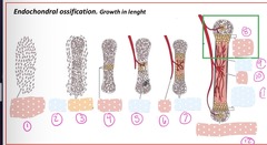
Endochondral ossification; growth in length: what is 1

Compact nodules
Endochondral ossification; growth in length: what is 2

Proliferating chondrocytes
Endochondral ossification; growth in length: what is 3

hypertrophic chondrocytes
Endochondral ossification; growth in length: what is 4

Osteoblasts (bone)
Endochondral ossification; growth in length: what is 5

Blood vessel
Endochondral ossification; growth in length: what is 6

Proliferating chondrocytes
Endochondral ossification; growth in length: what is 7

Epiphyseal growth plate
Endochondral ossification; growth in length: what is 8 and 11

Bone marrow
Endochondral ossification; growth in length: what is 9

Bone
Endochondral ossification; growth in length: what is 10

secondary ossification center
Endochondral ossification; growth in length: what is 12
Normal cartilage (individual cells)
What is 1

Cell proliferation (growth here)
What is 2
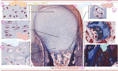
Hypertrophic individual cells
What is 3

Areas of bone marrow
What is 4

Compact bone area
What is 5

hypertrophic cells
What is 6

Cartilage
What is 7

Type II collagen
What type of collagen is in the matrix of cartilage?
Type I collagen
What type of collagen is in bone?
(1) WNT pathway to repress chondrocytic differentiation instead of becoming cartilage
(2) the transcription factor Runx2, and differentiate into osteoblasts
- in a later stage, these Runx2 - positive cells express osterix and then become osteocytes
What are the molecular features of membranous ossification?
Osterix
Runx2-positive cells express _______ and then become osteocytes
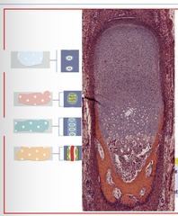
epiphyseal cartilage
Which zone of endochondral ossification is 1?

Reserve zone
Which zone of endochondral ossification is 2?

Proliferative zone
Which zone of endochondral ossification is 3?

Hypertrophic zone
Which zone of endochondral ossification is 4?

vascular invasion zone
Which zone of endochondral ossification is 5?

Reserve zone
What zone is the blue sticker?

Proliferative zone
What zone is the pink sticker?

Hypertrophic zone
What zone is the green sticker?

Vascular invasion zone
What zone is the yellow sticker?
Blood vessels
What is required to form bone?
Bone grows from hypertrophic zone towards proliferative zone
"Running away"
What is the direction of bone growth in regard to the proliferation zone?
Ossification-osteoclast front
The____________ invades and replaces chondrocytes as it moves past the site previously occupied by hypertrophic chondrocytes
Epiphyseal cartilage
What is 2

Secondary growth plate
What is 3

Perichondrium
What is 4

Primary growth plate
What is 5

zone of provisional calcification (ZPC)
What is 6

Primary metaphyseal spongiosa
What is 7

Metaphysis
What is 8

Reserve zone
Which zone is the light blue sticker

Proliferative zone
Which zone is the pink sticker

Hypertrophic zone
Which zone is the green sticker

Vascular invasion zone
Which zone is the yellow sticker

Diameter
In appositional growth, bone increases in _________.
Endosteal, Periosteal
In appositional bone growth, the bone is absorbed on the _________ surface and added to the _________ surface
A mutation in the FGFR3 (fibroblasts growth factor receptor 3) gene, which plays a role in the formation of collagen and contributes to the ossification of bone. This mutation prevents adequate bone formation in utero, resulting in a shortened stature and a large head
Clinical correlation: What is achondroplasia due to?
Cartilage
Identify the tissues and growth zones: yellow sticker

Beginning of bone formation
Identify the tissues and growth zones: blue sticker

Mature bone
Identify the tissues and growth zones: pink sticker

This is in the hypertrophic zone; collagen type 10 is only made in this zone (collagen type X)
The unique type of collagen found in the yellow circled area is.....

1. Mesoderm-derived mesenchymal cells differentiate into chondrocytes.
2. Chondrocytes proliferate rapidly and secrete extracellular matrix to form a hyaline cartilage model
3. Chondrocytes near the center of the model begin to undergo hypertrophy and alter the matrix they produce, which allows calcification
4. Calcification prevents nutrients from reaching the chondrocytes, so they die
5. Blood vessels invade the space left by dead chondrocytes, and the blood vessels bring osteogenic cells and trigger the transformation of the perichondrium into periosteum
6. Osteoblasts of the periosteum form a collar of compact bone on the surface, forming the ossification center
What are the steps of endochondral ossification?
1. Mesoderm-derived mesenchymal cells express the transcription factor Sox9, which leads to proliferation and differentiation into chondrocytes
2. An increase is reactive oxygen species (ROS) leads to chondrocyte hypertrophy, the cells start secreting collagen type 10, and increase the secretion of fibronectin. They also secrete VEGF and attract macrophages, which will degrade the matrix once the chondrocyte die
3. Osteoprogenitor cells carried by blood vessels express Runx2 and differentiate into osteoblasts
4. Runx2-positive cells express osterix and become osteocytes
What are the molecular features of endocondral ossification?