Quiz 5 Developmental Biology
 Call Kai
 Learn
 Practice Test
 Spaced Repetition
 Match
 Flashcards
 Knowt Play
1/45

There's no tags or description
Looks like no tags are added yet.
46 Terms
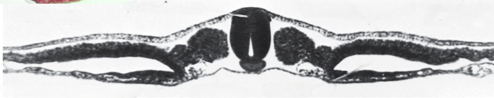
Be able to recognize and name on a cross section the paraxial mesoderm and that this is region from which somites develop.
The paraxial mesoderm is a segmental tissue located adjacent to the notochord & neural tube, giving rise to somites which differentiate into skeletal muscle, vertebrae, and dermis.

What molecular signals are important for differentiating paraxial mesoderm from other?
Paraxial mesoderm develops where there is low BMP signaling from endoderm
Because repressed by noggin and chordin in notochord; repress BMP in endodermt o allow differentiation into somites.

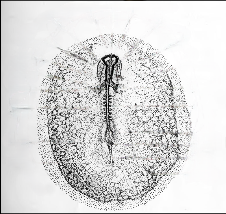
Be able to recognize and label somites on a developing embryo
Somites are block-like structures that form along the sides of the developing neural tube, arising from the paraxial mesoderm and differentiating into skeletal muscle, vertebrae, and dermis.


Explain how somites develop
As node regresses, somites come from cells in the stem zone, but can’t see them as somites until later.

Explain or recognize the "stem zone". Explain why it is important for somite development.
A posterior population of multipotent stem cells (Neuromesodermal Progenitors or NMPs) that continously proliferates to fuel the elongating body axis. As the embryo moves forward, cells exiting the stem zone are deposited into the presomitic mesoderm (PSM)

What genes are expressed in the stem zone? What is their function? What do you expect to happen if you overexpressed or knocked down these genes?
Driven by high Fgf8 and Wnt3a
Fgf8 and Wnt3a work synergistically to keep axial stem cells in an undifferentiated, proliferative state

What do you expect to happen if you overexpressed or knocked down these genes in the stem zone?
Continual high Fgf8/Wnt3a prevents cells from maturing or segmenting. The presomitic mesoderm remains unsegmented and overly elongated, causing a failure in somite formation
Knocking down Fgf8/Wnt3a causes premature differentiation of progenitors. Axis elongation terminatess abruptly, resuliting in a serverely truncated embryo with fewer total somites

Explain and/or draw and/or describe the "clock and wavefront" model of somite development
The wavefront is established by a mutual antagonism between an anterior Retinoic Acid (RA) gradient and a posterior Fgf8/Wnt3a gradient
The clock relies on cyclic autonomous negative feedback loops driven by Notch signaling and downstream targets like Hairy, L-fng, and Axin

What molecular signals are important for the "clock" and in the "wavefront" for actually specifying somites?
Every time a wave of Hairy expression sweeps from the posterior to the anterior PSM, one cycle is completed
When a wave hits the determination front, Mesp2 is upregulated to set the boundary, while Ephrin and Eph receptors control cell sorting and physical separation of the somite block

What is the determination front?
Exact threshold location where Fgf8 levels drop low enough to allow cells to respond to the clock signal and epithelialize into a new pair of somites

What do you expect to happen if you overexpressed or knocked down genes in the stem zone (Fgf8/Wnt3a)?
Overexpression pushes the determination front further posterior, delaying somite boundaires and creating fewer, abnormally large somites
Knockdown shifts the front anteriorly, causing premature epithelization and leading to smaller somites
What do you expect to happen if you overexpressed or knocked down genes in the stem zone (Notch/Hairy)?
Notch/Hairy knockdown abolishes the clock. Somites lose their periodicity, resulting in unsegmented, disorganized paraxial mesoderm blocks (similar to human congenital scoliosis)
How does this clock and wavefront model allow for different numbers of somites in different vertebrates?
Total number of somites is determined by the ratio between the speed of the clock (oscillation rate) and the rate of posterior axis elongation
Snakes have a clock that ticks much faster relative to axis elongation speed, generating many tiny somites

What four main tissue types do somites contribute to?
Sclerotome: Vertebrae, ribs, cartilage
Myotome: Skeletal muscles of the back, ribs, body wall, and limbs
Dermatome: Dermis of the dorsal skin
Syndetome: Tendons
What molecular signals pattern somites along the A-P axis of the organism
Somite identity is dictated strictly by the combinatorial expression of Hox genes, which are activated seqeuntially along the chromosome matching their physical expression layout along the body
Be able to name which germ layers (and subdivision of mesoderm) that contribute to limbs. Could you recognize and label these parts on an embryo cross section?
Ectoderm forms outer covering. At the very tip of limb bud, it thickens into a specialized structure called the Apical Ectodermal Ridge (AER)
Lateral Plate Mesoderm (Somatic Layer): forms body cavity, pelvis, limb bones
Paraxial Mesoderm (Somites): Specifically, myotome subdivision. Cells from myotome migrate out of the somites and into the limb bud to form all the skeletal muscles of limbs, as well as Dermatome to form dermis and more skeletal muscle.

Be able to name, recognize and/or label the three axes of a limb.
Axis | Anatomical Direction | Primary Organizing Center | Key Molecular Morphogen |
|---|---|---|---|
Proximal–Distal | Shoulder to Fingertip | AER (Apical Ectodermal Ridge) | FGF8 / FGF4 |
Anterior–Posterior | Thumb to Pinky | ZPA (Zone of Polarizing Activity) | Sonic Hedgehog (Shh) |
Dorsal–Ventral | Knuckles to Palm | Dorsal/Ventral Ectoderm | Wnt7a (Dorsal) / Engrailed (Ventral) |


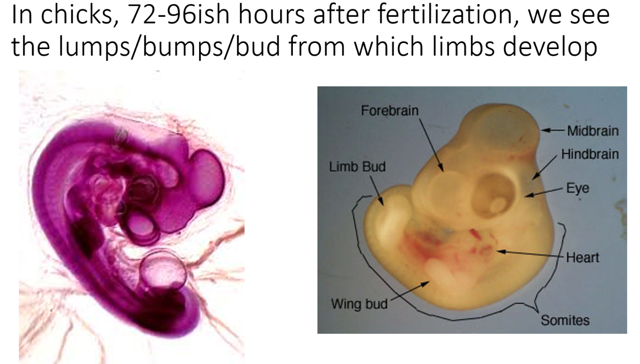
Be able to recognize and label a limb bud on a ~96hr chick or developmentally equivalent mouse. Describe what a limb bud is
A limb bud is a small, mound-shaped proliferation of embryonic tissue that serves as the precursor structure from which arms, legs, wings, or fins develop
Be able to describe important regions of the developing limb bud: Apical Ectodermal Ridge (AER)
The AER is a specialized thickened epithelium at the distal-most tip of the limb bud. It keeps the underlying mesoderm cells (the Progress Zone) in a highly proliferative, undifferentiated state
Predict what you'd expect if Apical Ectodermal Ridge (AER) was expanded or eliminated
Remove AER early: Limb development stops immediately; only the most proximal structures (humerus) form
Remove AER late: Forearm forms, but digits are missing
Add an extra AER: Duplicates the limb along the proximal-distal axis
Replace AER with an FGF bead: Normal limb development is completely rescued
Be able to describe important regions of the developing limb bud: Zone of Polarizing Activity (ZPA)
Located in the posterior mesoderm block of the limb bud, the ZPA secretes Sonic Hedgehog (Shh) to establish the identity of the digits. High concentrations specify posterior digits (pinky), while lower concentrations specify anterior digits (thumb)
Predict what you'd expect if Zone of Polarizing Activity (ZPA) was expanded or eliminated
Graft an extra ZPA to the anterior margin: Causes a mirror-image digit duplication
Shh knockout: Complete loss of posterior digits; only digit 1 forms
Be able to describe important regions of the developing limb bud: Progress Zone
The highly active mesoderm layer directly beneath the AER. Proximal-distal identity is determined by how long a cell spends proliferating within the PZ before exiting or by an early specification model balancing proximal Retinoic Acid against distal FGFs
Predict what you'd expect if Progress Zone was expanded or eliminated
If expanded we would see cells proliferating longer than normal so you could develop an extra radius or ulna or extra set of digits
If it was eliminated the embryo loses the pool of dividing cells required to build successive layers of the limb. Resulting in truncation or deletion of skeletal structures, phocomelia

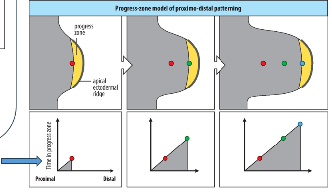
Progress-Zone Model
States that positional identity is determined strictly by a cellular biological clock based on the amount of time cells spend dividing inside the progress zone


Two-Signal Model / Early Allocation Model
More modern. States that proximal-distal identity is determined by a concentration gradient of opposing morphogens acting on a pre-allocated pool of cells. So Retinoic Acid diffuses from proximal flank toward limb bud and FGFs secreted by distal AER at the very tip of limb bud.
High RA / Low FGF: Specifies proximal cells (red dot)
Intermediate RA / Intermediate FGF: Specifies middle cells (green dot)
Low RA / High FGF: Specifies distal cells (blue dot)

Describe/explain how the limbs are positioned along the A-P axes (and why we only have 2 sets of limbs)
Limbs only form at precise coordinates along the A-P axis where Hox genes (Hox4, Hox5, Hox8, Hox9, Hox10) create a permissive molecular window. These Hox boundaries induce local expression of Retinoic Acid, which activates down-stream signaling to initiate budding
How are the location of forelimbs and hindlimbs differentiated from each other.
The identity of the limb is determined strictly by the expression of specific T-box transcription factors within the mesoderm:
Forelimbs (Wings/Arms): Express Tbx5
Hindlimbs (Legs): Express Tbx4 and Pitx1
Hox 4 & 5 Function, Overexpression, & Knockdown
Normal function: pattern the transition from the neck (cervical) to the chest (throacic) vertebrae and help position the forelimb field (arms/wings)
Overexpression: Forelimb tissue expands into the neck region
Knockdown: Forelimb missing or shifted toward head
Hox 8 & 9 Function, Overexpression, & Knockdown
Normal Function: Pattern the lower thoracic region and set the upper boundary for where the hindlimb field (legs) will initiate
Overexpression: Anterior vertebrae transform into lower thoracic/lumbar identity, shifting the hindlimb field forward
Knockdown: Thoracic/lumbar boundary is disrupted; hindlimb field fails to align properly with axial skeleton
Hox10 Function, Overexpression, & Knockdown
Normal Function: Specifically suppresses rib formation to create the lumbar (lower back) region. It is essential for specifiying the hindlimb territory
Overexpression: Thoracic vertebrae lose their ribs (entire lower body looks like ribless lumbar segments)
Knockdown: Lumbar vertebrae grow ribs; hindlimbs fail to form properly
Retinoic Acid (RA) Function, Overexpression, & Knockdown
Normal Function: Acts as the primary upstream master initiator. It acts within the permissive Hox windows to turn on the limb-specific transcription factors (Tbx5 for arms, Tbx4 for legs), which then activate Fgf10 to start the budding protrusion
Overexpression: Ectopic extra limbs form along the body flank
Knockdown: No limbs form at all (complete amelia)
Wnt3a Function, Overexpression, & Knockdown
Normal Function: Works downstream of RA as a mandatory co-factor. Wnt3a signaling interacts with Tbx factors to directly activate and maintain Fgf10 expression in the mesoderm, which is required to push the limb bud outward and signal the ectoderm to form the AER
Overexpression: Hyper-elongated limbs or duplicate outgrowths
Knockdown: Budding arrests immediately; limb elongation is deleted
Step-by-Step Sequence of Molecular Signaling: Step 1: Induction & Initiation
Retinoic Acid activates Tbx5 (forelimb) or Tbx4/Pitx1 (hindlimb)
Tbx transcription factors upregulate Fgf10 within the lateral plate mesoderm
Fgf10 protein signals direct to the overlying ectoderm to induce the formation of AER

Step-by-Step Sequence of Molecular Signaling: Step 2: Limb Elongation
The newly formed AER begins secreting Fgf8
Fgf8 signals back down to the mesoderm to sustain Fgf10 expression. This creates an unyielding positive feedback loop driving persistent distal outgrowth: Mesodermal Fgf10 → Ectodermal Fgf8 → Mesodermal Fgf10

Step-by-Step Sequence of Molecular Signaling: Step 3: Multi-Axis Coordination & Handshaking
The separate axes must coordinate so that fingers develop only at the distal tip of the hand.
The Loop: Distal FGF8 from the AER activates posterior Shh in the ZPA
The Hook: Shh stimulates the expression of Gremlin
The Break: Gremlin acts as an inhibitor against BMP4 (since BMP4 normally acts to shut down the AER and stop Fgf expression). By blocking the blocker, Gremlin keeps the AER alive and elongating
Eventually, as the limb grows too long, the distance dilutes this signaling network, BMP takes over, the AER regresses, and elongation stops so digits can differentiate
Step-by-Step Sequence of Molecular Signaling: Step 4: Dorsal-Ventral Patterning
Wnt7a is restricted to the dorsal ectoderm, inducing Lmx1b expression in the underlying mesoderm to specify dorsal traits (nails, knuckles)
Engrailed is expressed in the ventral ectoderm, blocking Wnt7a to specify ventral features (pads, palms)
Knockdown of Lmx1b / Wnt7a: Results in a limb that is ventralized on both sides (“two-palmed limb)
What is the specific embryonic tissue origin for all the bones, cartilage, and joints of the vertebrae limb?
Lateral plate mesoderm (specifically the local limb bud mesenchyme)
By what specific developmental process do limb bones form, and what are the two main cellular steps?
Endochondral ossification
Local mesenchymal cells condense and differentiate into a cartilage model (chondrocytes)
Blood vessels invade and bring in osteoblasts to replace cartilage matrix with hardened bone
Do limb muscles develop from the local lateral plate mesoderm of the limb bud? If not, where do they come from?
No. Limb muscles originate from the paraxial mesoderm. Precursor cells (myoblasts) must migrate a longdistance out of the ventrolaterla myotome of the somites into the growing limb bud
Once migratory muscle precursor cells enter the limb bud, into what two distinct anatomical masses do they split, and what do they become?
Dorsal muscle mass: Differentiates into extensor and abductor muscles
Ventral muscle mass: Differentiates into flexor and adductor muscles
What specific embryonic structure gives rise to limb tendons, and what is its physiological role during budding?
The syndetome (the topmost compartment of the sclerotome within the somites/paraxial mesoderm). They migrate into the bud to structurally connect migrating muscles to the developing cartilage bone models
Human and chick hands start as a solid, webbed paddle. What cellular mechanism is responsible for separating the individual digits?
Apoptosis (programmed cell death), specifically within the interdigital necrotic zone to clear the webbing between the finger rays
What clinical phenotype occurs if the interdigital necrotic zones fail to undergo sufficient apoptosis during development?
Syndactyly (webbed or fused fingers/toes)
Endoderm Major Derivatives
Forms the continous inner-epithelial linings of both the digestive gut tube and the respiratory tract (trachea, bronchi, lungs), along with glandular organs like the liver, gallbladder, and pancreas
Key Specifiying Morphogen Gradient for Endoderm
Patterned along its A-P axis by a posterior-high gradient of Wnt, Fgf, and BMPs, which induces localized expressions of regional master-genes:
Anterior Gut (pharynx/esophagus): Specified by Sox2
Midgut/Stomach: Transition managed by Pdx1 (critical for pancreas development)
Posterior Gut (intestines): Driven by CdxA / CdxB (Hox-related homeobox genes)