Ch4: Abdomen Flat/Upright
 Call Kai
 Learn
 Practice Test
 Spaced Repetition
 Match
 Flashcards
 Knowt Play
1/102

There's no tags or description
Looks like no tags are added yet.
103 Terms
what is the superior portion of the abdominopelvic cavity?
abdominal cavity
what the smaller inferior part of the abdominopelvic cavity?
pelvic cavity
the abdominal cavity extends from the _____ to the _______
diaphragm, superior aspect of the bony pelvis
the stomach can be found in which part of the abdominopelvic cavity?
abdominal cavity
the small and large intestines can be found in which part of the abdominopelvic cavity?
abdominal cavity
the liver can be found in which part of the abdominopelvic cavity?
abdominal cavity
the gallbladder can be found in which part of the abdominopelvic cavity?
abdominal cavity
the spleen can be found in which part of the abdominopelvic cavity?
abdominal cavity
the pancreas can be found in which part of the abdominopelvic cavity?
abdominal cavity
the kidneys can be found in which part of the abdominopelvic cavity?
abdominal cavity
the pelvic cavity lies within the margins of the _____
bony pelvis
the rectum can be found in which part of the abdominopelvic cavity?
pelvic cavity
the sigmoid of the large intestine can be found in which part of the abdominopelvic cavity?
pelvic cavity
the urinary bladder can be found in which part of the abdominopelvic cavity?
pelvic cavity
the reproductive organs can be found in which part of the abdominopelvic cavity?
pelvic cavity
the double-walled seromembranous sac that encloses the abdominalpelvic cavity is called the ____
peritoneum
the outer portion of the peritoneum that is in close contact with the abdominal wall is called the ____
parietal peritoneum
the part of the peritoneum that is positioned over and around the contained organs is called the ____
visceral peritoneum
the _____ and ______ are folds formed by the peritoneum that provide support for the viscera in position
mesentery, omenta
the space between the two layers of the peritoneum that contain serous fluid is called the _____
peritoneal cavity
the cavity behind the peritoneum is called the ____
retroperitoneum
the kidneys and pancreas are located in the retroperitoneum, meaning they ___
lie behind the peritoneum

what is A in this image?
pleural cavity

what is B in this image?
pericardial cavity

what is C in this image?
abdominal cavity

what is D in this image?
pelvic cavity

what is E in this image?
thoracic cavity

what is F in this image?
Abdominalpelvic cavity

what is A in this image?
parietal peritoneum
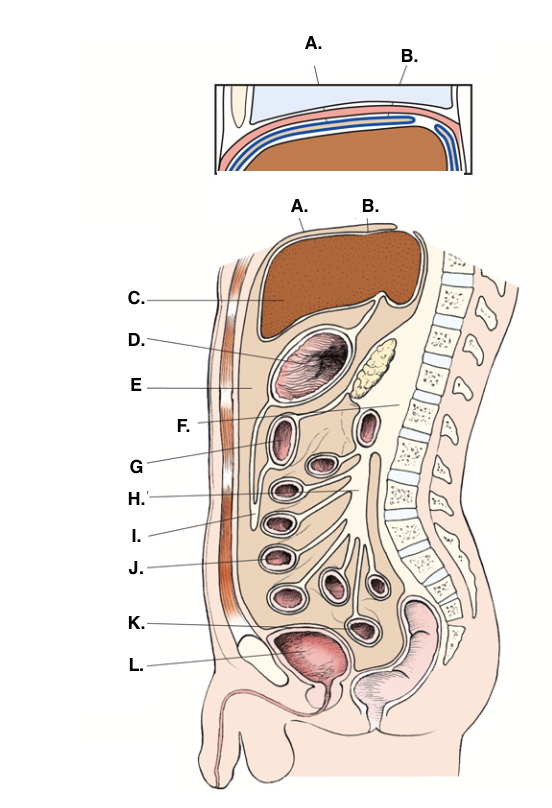
what is B in this image?
visceral peritoneum

what is C in this image?
liver
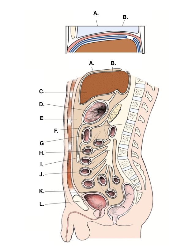
what is D in this image?
stomach

what is E in this image?
peritoneal cavity

what is F in this image?
retoperitoneum

what is G in this image?
transverse colon

what is H in this image?
mesentery

what is I in this image?
omentum

what is J in this image?
jejunum

what is K in this image?
sigmoid colon

what is L in this image?
urinary bladder

what is A in this image?
diaphragm

what is B in this image?
left lobe of liver
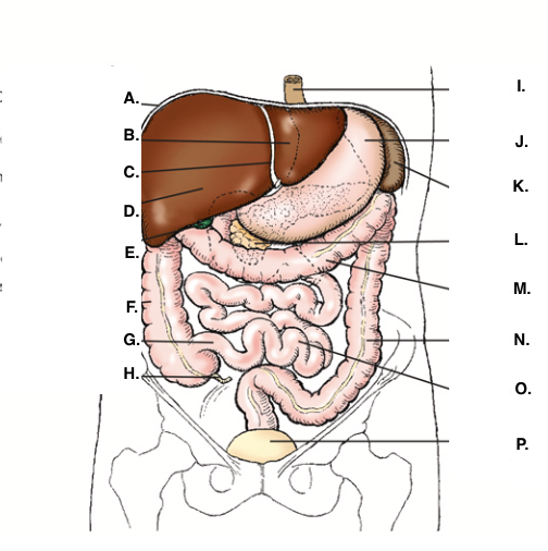
what is C in this image?
falciform ligament

what is D in this image?
right lobe of liver

what is E in this image?
gallbladder

what is F in this image?
ascending colon

what is G in this image?
ileum
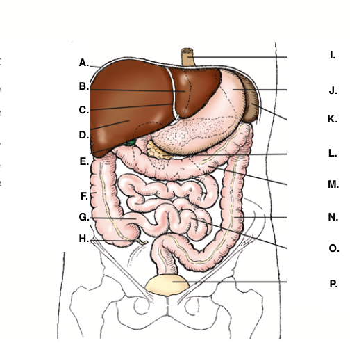
what is H in this image?
appendix

what is I in this image?
esophagus

what is J in this image?
stomach

what is K in this image?
spleen

what is L in this image?
pancreas

what is M in this image?
transverse colon

what is N in this image?
descending colon

what is O in this image?
small intestine

what is P in this image?
urinary bladder
abdominal aortic aneurysm (AAA)
localized dilation of abdominal aorta
ascites
fluid accumulation in the peritoneal cavity
bowel obstruction
blockage of bowel lumen
ileus
failure of bowel peristalsis
metastasis
transfer of cancerous lesion from one area to another
pneumoperitoneum
presence of air in the peritoneal cavity
tumor
new tissue growth where cell proliferation is uncontrolled
sharply defined outlines of the ____ is criteria for a good abdomen image
psoas muscles
for a good abdomen image, the lower boarder of the ____ must be visualized
liver
the ______ should be seen in a good abdomen image
kidneys
____ and ______ are also seen in a good abdoment image
ribs, transverse process of lumbar vertebrae
to allow the patient to come to rest and to let involuntary movement of the viscera to subside, you should ____
not start the exposure for 1 to 2 seconds after the patient stops breathing
voluntary motion during exposure may cause movement in structures that do not have involuntary movement like the ___, _____, and ____
liver, psoas muscles, and spine
why shouldn’t the patient breath during the exposure?
to not cause the blurring of bowel gas outlines in the upper abdomen as the diaphragm moves
what does the KUB for a supine AP projection stand for?
kidneys, ureters, and bladder
which projections are often paired with a supine AP projection of the abdomen?
upright AP abdomen or AP/PA projection in the lateral decubitus position
what does the AP upright and AP/PA later decubitus allow radiographers to see?
possible pneumoperitoneum and the presence/location of air-fluid levels
why would a PA chest be included with images of the abdomen?
to capture any air escaping form the gastrointestinal tract into the peritoneal space; the air usually rises and rest beneath the diaphragm
a supine and upright abdomen is often called a ___ and ____
flat, upright
recommended kVp for abdomen exposure technique
70-90 kvp
for a asthenic/pediatric(4-8) patient, kVp should be
between 60-70
for a hyposthenic(10-12in) patient, kVp should be
75
for a sthenic(12-14) patient, kvp should be
85
for a hypersthenic (15+) patient, kVp should be
90
for an asthenic/pediatric patient, mAs should be
6
for a hyposthenic patient, mAs should be
8
for a sthenic patient, mAs should be
10
for a hypersthenic patient, mAs should be
20
SID for abdomen projections
40 inches
for an abdomen supine (KUB) the IR should be centered to the ___
iliac crests
central ray (CR) should be ____ to the IR
perpendicular
for a abdominopelvic radiograph, shielding should
not be used
for an abdomen upright the IR needs to be centered ____
2 inches (5cm) above the iliac crests
an abdomen upright needs to be high enough to included the ___
diaphragm
for larger patients, the abdomen may be radiographed in ___ or ____ parts
two, four
a prone abdomen reduces ____ and ______, thus it reduces ___
superimposition, part thickness, dose
in this body habitus, the stomach sits low/medial; in the pelvis when standing
asthenic
in this body habitus, the colon sits low and folds on itself
asthenic
this body habitus, the gallbladders sit low and nearer the midline
asthenic
this body habitus has a short abdomen
asthenic
the stomach sits highest in this body habitus
hypersthenic
in this body habitus, the colons goes around periphery of abdomen
hypersthenic
in this body habitus, the gallbladder sits high and lies more parallel
hypersthenic
the longest abdomen is found in this body habitus
hypersthenic