Anatomy test 3
 Call Kai
 Learn
 Practice Test
 Spaced Repetition
 Match
 Flashcards
 Knowt Play
1/66

There's no tags or description
Looks like no tags are added yet.
67 Terms
Cranium
Fused bones that hold the brain
“Old People From Texas Eat Spaghetti”
Frontal
Temporal
Parietal
Occipital
Ethmoid
Sphenoid
Frontal bone
Forehead, front and top of cranium
Covers the frontal lobe
Squamous portion: forehead
Nasal portion: top of nose
Orbital: roof of eye sockets (eyebrows)

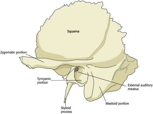
Temporal bone
Paired
Lower sides of the head
“Some Men Style Many Perilous Tiny Zebras”
Mastoid: bump behind the ear, site of muscle attachments
Squamous: thin fan forming temple
Petrous: dense, houses cochlea and semicircular canals
Tympanic: walls of the ear canal
Zygomatic: forms zygomatic arch with cheekbone
Styloid process
Mandibular: socket for the TMJ
Parietal bone
Paired
Top and sides of the head
Covers parietal lobes
Sagittal suture
Lambdoid suture
Squamosal suture

Occipital bone
Back and base of skull
Covers occipital lobes
Foramen magnum: hole that brainstem passes through
Condylar fossae: where atlas (C1) meets skull

Ethmoid
Seperates nasal cavity from cranium
Crista galli: points up into cranium
Cribiform plate: holes for olfactory nerve
Perpendicular plate: upper nasal septum
Labyrinths: superior and middle conchae

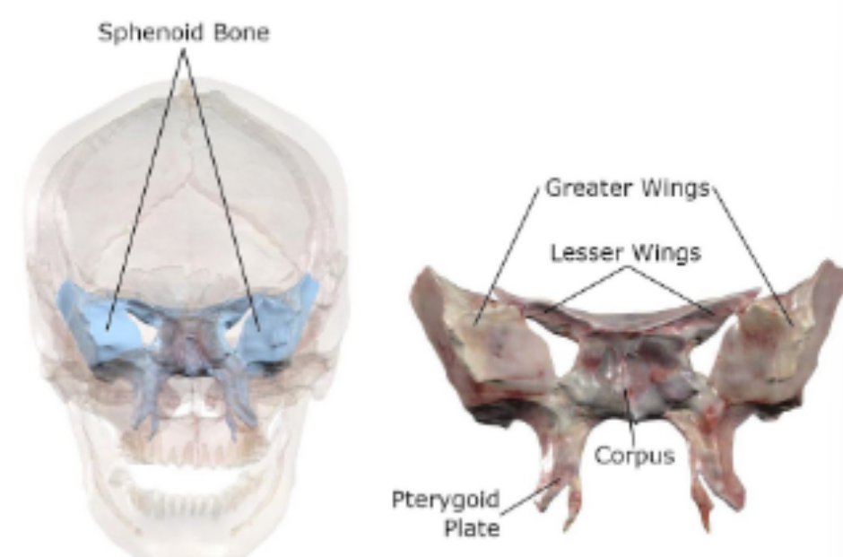
Sphenoid
Front base of the skull
Corpus: central body
Greater wings: cranial floor, sides of orbits
Lesser wings: support frontal lobe
Pterygoid plates: attach pterygoids, tensor veli palatini
Facial bones
"My Very Pretty Zebra Never Invites Lazy Men"
Maxillae
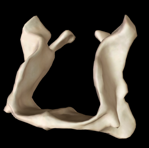
Mandible
Palantine
Zygomatic
Nasal
Inferior nasal conchae
Vomer
Lacrimal
Maxillae
Largest facial bone
Forms the upper dental arch
Roof of mouth
Floor and sides of nasal cavity
Parts of orbit
Alveolar: holds the upper teeth
Zygomatic: joins the cheekbone
Frontal: up to the fronal and nasal bones
Palatine: front 2/3 of the hard palate
Mandible
Halves fuse before we turn 1
Body: holds lower teeth
Ramus: vertical back portion
Angle: where body meets ramus
Coronoid process: temporalis attachment site
Condylar process: forms the TMJ
Mental symphysis: midline fusion (chin)

Palatine
Paired
Behind maxillae
Horizontal plates: forms back 1/3 of hard palate, anchors soft palate
Perpendicular plates: forms nasal walls, part of orbit
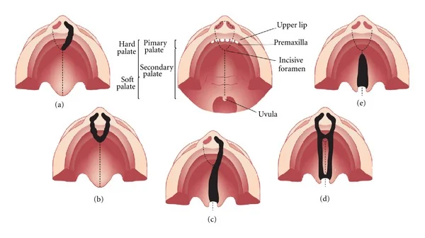
Hard palate
Seperates oral and nasal cavity
Front 2/3 = maxilla
Back 1/3 = palatine
Intermaxillary suture: midline
Incisive foramen: behind central incisors
Premaxillary sutures: outline the premaxilla
Rugae: ridges on the front

Micrognathia
Small, underdeveloped mandible
Tounge can block the airway
Affects breathing, feeding, and speech
Cleft lip/palate
Anterior cleft: along premaxillary suture
Posterior cleft: along intermaxillary suture
Unilateral, bilateral
Complete or incomplete
Affects feeding and speech
Zygomatic bone
Paired
“Cheekbones”
Frontosphenoidal: sides of the orbit
Maxillary: joins the maxilla
Temporal: joins temporal bone at zygomatic arch
Orbital: floor of the orbit

Nasal bone
Paired
Forms the bridge of the nose
Above is the frontal bone
Behind is the perpendicular plate of the ethmoid
Below is the maxilla

Inferior nasal conchae
Scroll like shape
Paired
Side walls of nasal cavity
Joins the maxilla in the front in the palatine behind

Nasal conchae
Turbinates:
Superior
Middle
Inferior
Lining: moist, blood-rich mucus
Function: warms and humidfies inhaled air, filters particles
Vomer
Divides the nasal cavity at midline
Forms lower posterior part of nasal septum

Lacrimal
Paired
Inner front wall of orbit near tear ducts
Smallest facial bone
Temporomandibular joint
Mandibular condyle attaches at mandibular fossa in temporal bone
Articular disk cushions the joint
Fibrous capsle surrounds joint with synovial fluid

Vocal tract
Cavities: oral, buccal, nasal, pharynx, larynx
Speech: filters voice, shapes resonance
Valves: velum and tongue
Life functions: breathing, swallowing, chewing
Nasal cavities
Nares: front openings
Choanae: back openings to pharynx
Septum: cartilage at midline
Side walls: conchae warm and humidify
Floor: hard palate
Nasal sounds: velum lowers to open
Oral cavity
Front: lips
Back: posterior faucial pillars
Sides: dental arches
Roof: hard and soft palate
Floor: mylohyoid
Vestibule: lips, cheeks, front teeth
Tongue
Tip: behind front teeth
Blade: below the alveolar ridge
Dorsum: below hard and soft palate
Root: faces pharynx and epiglottis
Body: central muscle mass
Muscular hydrostat: all muscle, no bone, extreme flexibility
Taste
Tounge (more)
Median sulcus: midline groove
Sulcus terminalis: v shaped groove that separates dorsum and base
Circumvallate papillae: taste buds infront of V
Lingual tonsils: immune tissue behind V
Lingual frenulum: tongue to floor of mouth
Ankyloglossia
Tongue-tie
Short frenulum near tip
Limits tip elevation and protrusion
Sometimes impacts speech or feeding
Salivary glands
Create saliva for swallowing to compact bolus of food, helps oral hygeine
Sublingual: under the tongue
Submandibular: most saliva
Partoid: around ramus, near CN VII
Dental arches and tooth surfaces
Upper arch: maxillae
Lower arch: mandible
Surfaces
Occlusal: biting surface
Lingual: towards tongue
Labial: towards lips
Buccal: towards cheeks
Deciduous teeth
Develop before birth
Erupt from 8-12 months until 2 ½
First lower teeth then upper centreal incisors
20 total, 10 per arch
Replacement begins around 6 and is done around 11-12
Permanent teeth
32 total, 16 per arch
More due to larger adult jaw
Replacing deciduous teeth
Successional: replace a baby tooth
All except molars
Bicuspids replace baby molars
Superadded: no baby predecessor
All permanent molars
Errupts around 6
Occlusion
Raising the mandible to teeth meet (biting)
Class I: normal
Class II: lower molar set back (overbite)
Class III: lower molar forward (overbite) - most likely to impact chewing and speech
Palates
Hard palate: chewing surface, alveolar and palatal sounds
Soft palate (velum): muscle behind hard palate, acts as a valve for swallowing and nasal sounds
Faucial pillars
Made of muscle
Anterior pillar: palatoglossus
Posterior pillar: palatopharyngeus (back of oral cavity)
Palatine tonsils inbetween
Tonsillitus
Infencted palatine tonsils
Severe cases can block breathing or swallowing
Buccal cavity
Between teeth and cheeks
Shapes oral resonance
Cheek tension for /p/ and /b/
Keeps food out of the cheeks
Nasopharynx
Behind nasal cavity and above velum
Adenoids: lymph tissue on back wall
Eustachian tubes open here
Faucial isthmus
the narrow opening and passageway that connects the oral cavity proper to the oropharynx
Superiorly: The soft palate and the uvula
Inferiorly: The base of the tongue
Laterally: The palatoglossal and palatopharyngeal arches (also known as the anterior and posterior pillars of the fauces)
Oropharynx
Behind oral cavity
Superiorly: velum
Inferiorly: hyoid bone
Front wall: base of the tongue
Valleculae: pocket between tongue base and epiglottis
Laryngopharynx (Hypopharynx)
Behind larynx
Superiorly: epiglottis
Inferiorly: esophagus
Tooth parts
Pulp: cavity that houses the neurovascular tissue supplying blood and nerve sensation to the tooth
Cementum: boneline tissue that covers the tooth root to attach it to the surrounding socket
Orbicularis oris
CN VII
Only intrinsic muscle of the lips
Ring surrounding lips
Closes and protrudes lips, presses lips against teeth
Labial sounds, whistling
Attaches to other facial muscles
Transverse muscles
Buccinator
Risorius
Elevator muscles
Levator labii superioris
Levator labii superioris alaeque nasi
Zygomatic minor
Zygomatic major
Levator anguli oris
Depressor muscles
Depressor labii inferioris
Depressor angulioris
Mentalis
Parallel muscles
Incisivus labii superioris
Incisivus labii inferioris
Supplementary muscles
Epicranius: raises eyebrows
Corrugator: wrinkles forehead
Orbicularis Oculi: closes eyelids
Procerus: wrinkles root of the nose
Platysma: see other flashcard

Buccinator
CN VII
Main cheek muscle
Back of jaws to mouth corner
Presses cheek against teeth
Keeps food out of the cheeks
Sucking during infant feeding
Pull lip corners sideways
Forceful air expulsion (blowing)
Risorius
CN VII
Cheek fascia to mouth corner
Pulls corner back out out
Smiling, laughter
Levator labii superior
CN VII
Elevates the upper lip

Levator labii superioris alaeque nasi
CN VII
Elevates upper lip
Flares the nostrils
Snarl, disgust, contempt (Elvis)

Zygomatic minor
CN VII
Cheekbone to upper lip
Elevates upper lips
Smiling

Zygomatic major
CN VII
Cheekbone to mouth corner
Pulls corners up and back
Smiling, laughter

Levator anguli oris
CN VII
Raises mouth corners up and in
Helps close mouth

Depressor labii inferioris
CN VII
Pulls lower lip down and out
Frowning

Depressor anguli oris
CN VII
Pulls mouth corners down
Frowning

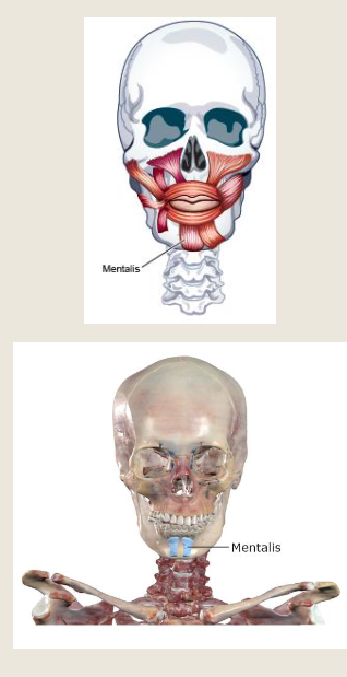
Mentalis
CN VII
Mandible to chin skin
Raises and pushes out lower lip
Pouting, wrinkles chin
Incisivus labii superioris
CN VII
Parallel to upper lip fibers
Rounds and protrudes upper lip

Invisivus labii inferioris
CN VII
Parallel to lower lip fibers
Rounds and protrudes lower lip

Platysma
CN VII
Thin sheet over the neck
Depresses mandible and lower lip
Suprise, fright

The tongue
Lingual septum: midline attachment
Submucous layer: surface attachment
Muscles of the tongue
Intrinsic: within the tongue → changes shape
Superior longitudinal
Inferior longitudinal
Transverse
Vertical
Extrinsic: from outside → changes position
Styloglossus
Palatoglossus
Hyoglossus
Genioglossus
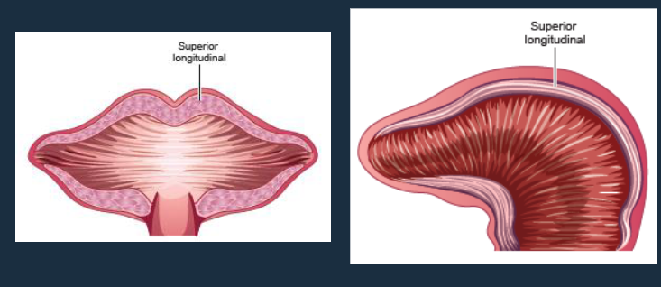
Superior longitudinal
CN XII
Along top of the tongue
Shortens tongue, tip up
Unilateral contraction → tip towards that side
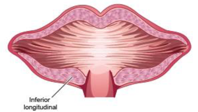
Inferior longitudinal
CN XII
Along underside
Shortens tongue, tip down
Unilateral contraction → tip towards that side
Vertical
CN XII
Top to bottom fibers
Top surface to underside
Flattens and widens

Transverse
CN XII
Side to side fibers
Midline septum to sides
Narrows and lengthens
