L24- Anaemia 2
 Call Kai
 Learn
Learn Practice Test
 Spaced Repetition
 Match
Match Flashcards
 Knowt Play
1/164

There's no tags or description
Looks like no tags are added yet.
Name | Mastery | Learn | Test | Matching | Spaced | Call with Kai | Chat |
|---|
No analytics yet
Send a link to your students to track their progress
165 Terms
What 8 factors should be considered when approaching and classifying anaemia?
RBC indices (MCV, MCH, MCHC, RDW);
acute vs chronic;
congenital vs acquired;
unwell vs well;
isolated vs associated cytopenias;
blood film findings;
reticulocytosis vs reticulocytopenia (production vs survival); and
clinical features including age, sex and underlying conditions.
How are causes of normocytic anaemia classified? (2)
Low/normal reticulocytes:
renal failure,
bone marrow failure (infiltration, Parvovirus B19 infection, aplastic anaemia) and
mixed deficiencies.
Increased reticulocytes:
acute blood loss and
haemolytic anaemias.
How are causes of macrocytic anaemia classified? (3)
Megaloblastic:
B12 deficiency,
folate deficiency and
medications.
Non-megaloblastic:
liver disease,
myelodysplastic syndrome and
alcoholism.
Reticulocytosis
Why is the reticulocyte count important in normocytic anaemia?
It is the best differentiator because it measures marrow response to anaemia (production vs survival).
What 5 further investigations may be required in normocytic anaemia?
blood film,
renal function,
haemolysis screen,
haematinics
bone marrow aspirate
some causes are clinically apparent.
What is haemolysis?
Haemolysis is red cell destruction causing shortened red cell survival.
Can haemolysis occur without anaemia and when does it result in haemolytic anaemia?
It can occur without anaemia if marrow compensation is adequate. Haemolytic anaemia develops when the marrow cannot compensate; reticulocytosis generally reflects this compensatory response.
What are the 3 intrinsic (inherited) and 4 extrinsic (acquired) causes of haemolysis?
Intrinsic:
membrane defects,
haemoglobinopathies (e.g. sickle cell disease) and
enzyme deficiencies.
Extrinsic:
immune haemolysis,
microangiopathic haemolysis,
infections such as malaria, and
medications.
How are intravascular and extravascular haemolysis distinguished by site?
Intravascular haemolysis occurs within the circulation
Extravascular haemolysis occurs in the spleen.
Give 2 examples of intravascular haemolysis
Mechanical haemolysis from heart valves
paroxysmal nocturnal haemoglobinuria.
How do we test for haemolysis? (5)
Marrow response: Reticulocytosis and polychromasia.
Red cell breakdown: ↑ Unconjugated bilirubin and ↑ LDH.
Free haemoglobin markers: Free haemoglobin in plasma/urine, ↓ haptoglobin, urinary haemosiderin/urobilinogen present or increased.
Blood film: Spherocytes, sickle cells and red cell fragments.
Specific testing: Direct Coombs Test (Direct Antiglobulin Test).
What is the typical laboratory pattern of extravascular haemolysis?
Indirect bilirubin?
LDH?
haptoglobin?
urine haemoglobin?
urine haemosiderin?
Indirect bilirubin ↑
LDH ↑
haptoglobin ↓ or unchanged
urine haemoglobin unchanged
urine haemosiderin unchanged
What is the typical laboratory pattern of intravascular haemolysis?
Indirect bilirubin?
LDH?
haptoglobin?
urine haemoglobin?
urine haemosiderin?
Indirect bilirubin ↑↑
LDH ↑↑
haptoglobin ↓↓↓
urine haemoglobin ↑↑
urine haemosiderin ↑↑
These laboratory distinctions are not always clear or useful in practice.
What do vertical and horizontal red cell membrane defects produce in inherited haemolytic anaemias?
Vertical defects: Destabilise the lipid bilayer, causing membrane loss and spherocyte formation → Hereditary spherocytosis (HS).
Horizontal defects: Destabilise the membrane skeleton, causing defective shape recovery or fragmentation → Hereditary elliptocytosis (HE) and Hypophosphatasia (HPP).

What are vertical and horizontal red cell membrane interactions, and which proteins are involved?
Vertical: Between the lipid bilayer and membrane skeleton — Band 3, ankyrin and spectrin.
Horizontal: Within the membrane skeleton — spectrin and actin.

What are the prevalence, inheritance and genetic defects of hereditary spherocytosis?
Prevalence:
Inheritance:
Genetic defects:
Prevalence: 1 in 1,000–3,000 Caucasians.
Inheritance: Usually autosomal dominant (AD).
Genetic defects: Mutations in Band 3, spectrin, actin or ankyrin.
Explain the pathophysiology of hereditary spherocytosis.
Mutations cause loss of membrane support and membrane material, producing spherocytes. These are less deformable and prematurely destroyed in the spleen.
What are the 3 clinical features of hereditary spherocytosis?
May be asymptomatic.
Anaemia.
Jaundice.
How is hereditary spherocytosis suspected- what are the three clues?
Haemolysis,
spherocytes on blood film
possible family history.
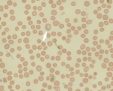
How is hereditary spherocytosis confirmed and what other test may be performed?
Confirm: Eosin-5-maleimide (EMA) binding assay.
Other test: Osmotic fragility testing (now rarely performed).
How is hereditary spherocytosis managed? (4)
Folic acid,
management of gallstones,
monitoring for aplastic crisis
splenectomy if severe.
What are the two main types of inherited red cell enzyme defects that cause haemolysis, and give an example of each?
Glycolytic enzyme defects: Pyruvate kinase (PK) deficiency.
Pentose shunt enzyme defects: Glucose-6-phosphate dehydrogenase (G6PD) deficiency.
How do inherited glycolytic enzyme defects cause haemolysis such as pyruvate kinase (PK) deficiency?
Usually glycolysis converts glucose through intermediates to pyruvate with ATP production, involving pyruvate kinase.
Impaired energy production in red blood cells.
Red cells cannot meet their energy requirements.
This shortens red cell lifespan, causing haemolysis.

How do inherited pentose shunt enzyme defects cause haemolysis such as glucose-6-phosphate dehydrogenase (G6PD) deficiency?
Usually the pentose shunt involves G6PD, NADP/NADPH and glutathione cycling in protection against oxidative stress.
Increased susceptibility of red blood cells to oxidative stress.
Leads to the formation of methaemoglobin and haemolysis.

What are the clinical features, blood film findings and management of pyruvate kinase deficiency?
Clinical features: Rare cause of chronic haemolysis.
Blood film: Polychromasia and spheroacanthocytes (prickle cells).
Management: Splenectomy may be required in severe cases.
What are the inheritance, prevalence and ethnic associations of G6PD deficiency?
Inheritance: X-linked recessive.
Prevalence: Relatively common.
Ethnic associations: Mediterranean and Afro-Caribbean populations.
What are the 2 clinical presentations of G6PD deficiency?
Neonatal jaundice
Acute episodic haemolysis
What 3 things trigger haemolysis in G6PD deficiency?
Infections
Broad beans
Medications (e.g. dapsone)
How is G6PD deficiency diagnosed and confirmed?
Initial diagnosis: Clinical findings and blood film during an acute haemolytic episode.
Confirmation: G6PD activity assay performed outside the acute episode.
What 3 blood film findings should be recognised in G6PD deficiency?
Heinz bodies
bite cells
blister cells, with spherocytes

Explain the sequence of oxidative red cell damage in G6PD deficiency.
Oxidative denaturation of haemoglobin → membrane-bound Heinz body formation → membrane damage → splenic macrophage attack → bite cells, blister cells or, with less severe damage, spherocytes

What is immune haemolysis, and what are its 2 main antibody categories?
Antibody-mediated red blood cell destruction.
Allo-antibodies: Can cause transfusion reactions.
Auto-antibodies: Cause autoimmune haemolytic anaemia.
**How is autoimmune haemolytic anaemia classified, and what secondary causes are listed?
Primary or secondary; warm or cold; and drug-induced. Secondary associations include SLE, rheumatoid arthritis and CLL. Drug examples include penicillin and quinidine.
How are red cells destroyed in autoimmune haemolytic anaemia?
Antibodies coat red cells, which are consumed in the spleen and lose membrane, producing spherocytes.
How is autoimmune haemolytic anaemia diagnosed and treated?
Diagnosis: evidence of haemolysis and positive Direct Antiglobulin (Coombs) Test. Treatment: immunosuppression with steroids or rituximab; splenectomy rarely.
What is the principle and positive result of the Direct Coombs Test?
Patient red cells with surface-bound human antibodies are washed and incubated with antihuman antibodies (Coombs reagent). A positive test produces red cell agglutination as the reagent links antibodies on the red cell surfaces.
What is microangiopathic haemolysis, and what is its characteristic blood film finding?
Red cell destruction due to abnormalities in small vessels; characterised by red cell fragmentation on blood film.
What are the principal causes of microangiopathic haemolysis?
Haemolytic uraemic syndrome (HUS), thrombotic thrombocytopenic purpura (TTP) and disseminated intravascular coagulation.
Why is malaria-associated anaemia multifactorial?
Haemolysis occurs particularly with falciparum malaria and high parasite loads during the erythrocytic phase. Raised hepcidin also contributes an anaemia of chronic disease element.
What are the principal low-reticulocyte causes of normocytic anaemia?
Renal failure, bone marrow failure (infiltration, Parvovirus B19 infection, aplastic anaemia) and mixed deficiencies.
What proportion of patients with GFR <25 are anaemic, and what diagnosis must be considered with new unexplained anaemia and renal failure?
90% are anaemic; always consider possible myeloma.
What mechanisms contribute to anaemia in renal failure?
Inadequate EPO, uraemia causing mild haemolysis, blood loss (e.g. haemodialysis) and secondary hyperparathyroidism.
How is renal failure-associated anaemia managed?
Treat the underlying cause, use recombinant EPO (rEPO) in some cases and ensure iron repletion; ferritin is unreliable.
What findings suggest bone marrow failure, and what investigation may be required?
Pancytopenia and an abnormal blood film; blast cells suggest malignancy and teardrop cells suggest fibrosis/infiltration. Bone marrow aspirate may be required.
How do healthy marrow and aplastic anaemia differ in the illustrated marrow specimens?
Healthy marrow contains substantial haematopoietic tissue; aplastic anaemia shows markedly reduced marrow cellularity.
What are the listed causes of bone marrow failure in normocytic anaemia?
Marrow infiltration, infection with Parvovirus B19 and aplastic anaemia.
What is Parvovirus B19 also called, and how common is previous infection in elderly people?
Erythrovirus B19; 85% of elderly persons have serological evidence of previous infection.
How does Parvovirus B19 typically present in children and adults?
Children: slapped cheek syndrome. Adults: transient arthritis or asymptomatic infection.
In which settings may Parvovirus B19 cause symptomatic anaemia?
Immunosuppression and chronic haemolysis.
What visual findings are illustrated in Parvovirus B19 infection?
A child with a characteristic red cheek rash (slapped cheek syndrome) and a marrow image showing abnormal enlarged erythroid precursor cells.
What are the causes of megaloblastic and non-megaloblastic macrocytic anaemia?
Megaloblastic: B12 deficiency, folate deficiency, medications including methotrexate and anti-cancer drugs. Non-megaloblastic: liver disease, MDS and alcoholism; hypothyroidism is also described as a rare cause.
What investigations are included in the macrocytosis work-up?
Reticulocyte count, blood film (megaloblastic or MDS changes), vitamin B12 and folate levels, TFTs, liver function tests, and ± haemolysis screen or bone marrow biopsy.
What is the fundamental defect in megaloblastic anaemia?
Impaired DNA production, producing a characteristic megaloblastic bone marrow appearance.
What blood film and marrow appearances are illustrated in megaloblastic anaemia?
Macrocytic red cells, including oval forms, and hypersegmented neutrophils on blood film, with abnormal megaloblastic erythroid maturation in marrow.
Which medications are specifically associated with megaloblastic anaemia?
Methotrexate and anti-cancer drugs.
What two major processes are illustrated in the B12 and folate metabolism diagram?
Nucleic acid synthesis and methylation reactions.
How are folate and vitamin B12 linked in the metabolic pathway?
Folate participates through THF, 5,10-methylene THF and 5-methyl THF; vitamin B12 participates in methionine synthase linking homocysteine, methionine and folate metabolism.
Which enzymes, cofactors and metabolites are labelled in the B12–folate diagram?
Methionine synthase, methylene THF reductase, riboflavin (FAD), NADPH/NADP, THF, 5,10-methylene THF, 5-methyl THF, methionine and homocysteine.
What is stated about vitamin B12 body stores, and what systems are needed for absorption?
The body stores B12 in excess. Absorption requires salivary proteins, stomach acid and intrinsic factor, pancreatic calcium supply and the small intestine.
What are the roles of haptocorrin and intrinsic factor in B12 absorption?
Haptocorrin is a salivary protein protecting B12 from stomach acid; intrinsic factor is produced by gastric parietal cells and forms complexes with B12 for intestinal absorption.
What is the role of the pancreas and small intestine in vitamin B12 absorption?
The pancreas provides calcium; the small intestine absorbs B12–intrinsic factor complexes.
What sequence is depicted in the vitamin B12 absorption diagram?
Dietary B12 enters the gastrointestinal tract, interacts with binding proteins and intrinsic factor, and the B12–intrinsic factor complex is absorbed in the intestine and transported into the circulation.
What are the main categories and causes of vitamin B12 deficiency?
Dietary: vegan diet. Gastric: pernicious anaemia, atrophic gastritis, gastrectomy. Medications/exposures: PPIs, antacids, metformin, nitrous oxide. Pancreatic: pancreatic insufficiency. Small bowel: coeliac disease, Crohn's disease.
What causes vitamin B12 deficiency in pernicious anaemia?
Autoimmune gastritis causing deficiency of stomach acid and intrinsic factor.
Which antibodies are detectable in pernicious anaemia, and which are most specific?
Antibodies to gastric parietal cells and intrinsic factor; intrinsic factor antibodies are most specific.
How do pernicious anaemia and atrophic gastritis differ in their effects on B12 absorption?
Pernicious anaemia causes deficiency of acid and intrinsic factor; atrophic gastritis causes loss of acid production only, so free B12 from supplements may still be absorbed.
What replacement approaches are mentioned for pernicious anaemia?
Intramuscular replacement or high-dose oral replacement; lifelong replacement is required.
What are the major clinical consequences of vitamin B12 deficiency?
Symptoms of anaemia, neurological symptoms, glossitis, macrocytosis/anaemia, pancytopenia if severe and raised LDH from red cell destruction.
What neurological manifestations occur in vitamin B12 deficiency?
Peripheral neuropathy, subacute combined degeneration of the cord and dementia.
Which spinal cord regions are labelled in the subacute combined degeneration diagram?
Posterior columns and lateral corticospinal tracts.
What oral and blood film findings are illustrated in B12 deficiency?
Glossitis (abnormal red tongue), macrocytic red cells including oval forms, and hypersegmented neutrophils.
What haematological abnormalities may occur in severe B12 deficiency?
Macrocytosis/anaemia, potentially pancytopenia, and raised LDH from red cell destruction.
What are the dietary causes of folic acid deficiency?
Alcoholism, elderly persons with a “tea and toast” diet, and total parenteral nutrition.
What malabsorptive conditions cause folic acid deficiency?
Coeliac disease and Crohn's disease.
What situations increase folic acid requirements?
Pregnancy, growth spurts, chronic haemolysis and haemodialysis.
What important fetal risk is associated with folate deficiency during pregnancy?
Neural tube defects.
How do the clinical features of folate deficiency compare with B12 deficiency?
Haematological features are similar, but folate deficiency causes no neurological disturbance.
Why may mild jaundice or haemolytic features occur in folate deficiency?
Mild jaundice results from ineffective red cell production and haemolysis; features of haemolytic anaemia may occur when deficiency is due to increased requirements.
How are B12 and folate results interpreted using the stated thresholds?
B12 >300 and folate >4: no further testing recommended. B12 <125 and/or folate <2: consistent with deficiency.
How are indeterminate B12 or folate results investigated?
Red cell folate (more specific), methylmalonic acid levels, clinical context, trial of treatment and consideration of other causes.
Which biochemical marker is raised only in B12 deficiency in the diagnostic approach?
Methylmalonic acid.
How is folate deficiency treated, and what precautions are required?
Oral folate 5 mg; consider need for B12 replacement and investigate the underlying cause.
How is vitamin B12 deficiency treated?
Usually parenteral B12 (intramuscular); consider the underlying cause. Neurological symptoms generally resolve; pernicious anaemia requires lifelong replacement.
What haematological response indicates effective B12 or folate treatment when anaemia is present?
Reticulocytosis, a marker of response.
What distinguishes non-megaloblastic macrocytosis from megaloblastic macrocytosis?
Cell maturation is maintained, but cells are proportionally larger.
What is the most common cause of macrocytosis, and what other causes are highlighted?
Alcohol excess is most common; liver disease and MDS are also causes, while hypothyroidism is rare.
What is myelodysplastic syndrome (MDS)?
A spectrum of malignant bone marrow disorders characterised by ineffective or abnormal blood cell production.
What symptoms may result from reduced blood counts in MDS?
Reduced RBCs: fatigue; reduced WBCs: infection; reduced platelets: bleeding.
What serious progression risk is associated with MDS?
Transformation or progression to acute myeloid leukaemia (AML).
What is the structure and oxygen-binding function of haemoglobin?
Four polypeptide globin chains forming two heterodimers, with iron-containing haem groups and reversible oxygen-binding capability.
What are the normal haemoglobin types, their globin chains and adult proportions?
HbF: α2γ2, <1%; HbA: α2β2, 95.5–97.5%; HbA2: α2δ2, 3–3.5%.
How does haemoglobin production change from fetal life to infancy?
HbF predominates during fetal life and decreases after birth; HbA rises and becomes predominant during infancy, with HbA2 present at a smaller proportion.
What time points and haemoglobin types are shown in the developmental haemoglobin graph?
Conception, birth and 9 months of age; embryonic haemoglobins, HbF, HbA and HbA2.
What does the haemoglobin oxygen dissociation diagram illustrate?
The relationship between oxygen partial pressure (PO2) and percentage oxygen saturation, with changes in oxygen affinity.
Which factors are labelled as increasing or reducing haemoglobin oxygen affinity?
Increased affinity: reduced temperature, alkalosis, reduced 2,3-DPG and HbF. Reduced affinity: increased temperature, acidosis and increased 2,3-DPG.
What is the fundamental difference between thalassaemia and haemoglobinopathy?
Thalassaemia is a quantitative disorder involving impaired globin chain production and imbalance; haemoglobinopathy is a qualitative disorder involving structurally abnormal variant haemoglobin.
What are the two principal types of thalassaemia, and how may they be classified in relation to haemoglobinopathies?
Alpha and beta thalassaemia; some sources consider thalassaemia a subtype of haemoglobinopathy.
Which geographical distributions are illustrated for thalassaemia and haemoglobin variants?
Thalassaemia across Mediterranean, Middle Eastern, South Asian and Southeast Asian regions; sickle cell anaemia prominently in Africa; HbC, HbD and HbE have distinct distributions, including HbE in Southeast Asia.
Which haemoglobin disorders are labelled on the geographical distribution map?
Thalassaemia, sickle cell anaemia, HbC, HbD and HbE.
How does impaired globin chain production cause microcytosis and haemolysis in thalassaemia?
Reduced haemoglobin production causes microcytosis; excess unmatched globin chains precipitate, damaging red cells and causing haemolysis.