Biology Topic 2 - Cells
 Call Kai
 Learn
 Practice Test
 Spaced Repetition
 Match
 Flashcards
 Knowt Play
1/39

There's no tags or description
Looks like no tags are added yet.
40 Terms
What are the distinguishing features of eukaryotic cells
Cytoplasm containing membrane-bound organelles
DNA Enclosed in a nucleus
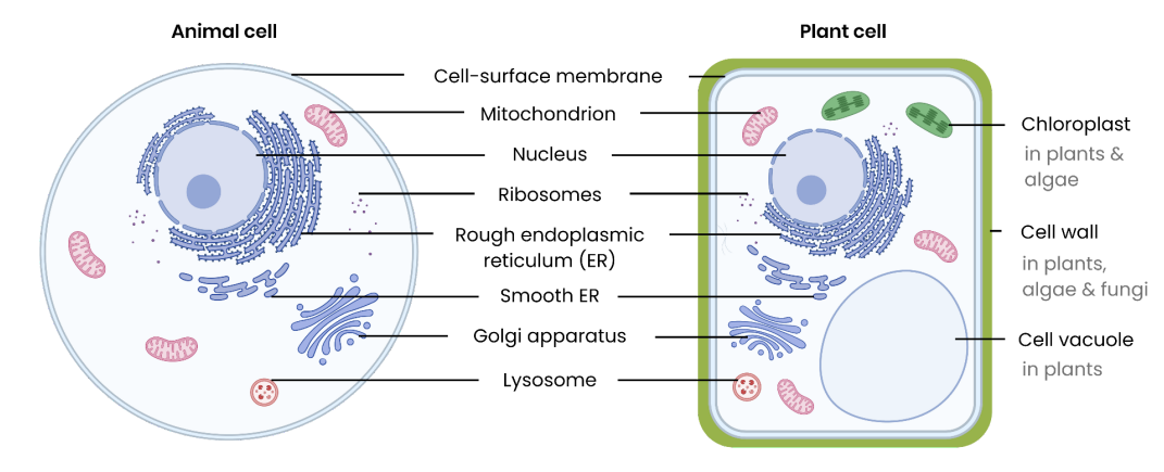
Describe the general structure of eukaryotic cells
Describe the structure of the cell-surface membrane
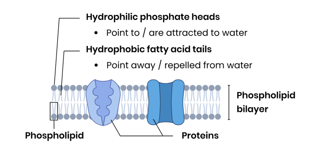
Describe the function of the cell-surface membrane
Selectively permeable → enables control of passage of substances in/out the cell
Receptors on surface → allows cell recognition
Describe the structure of the nucleus

Describe the function of the nucleus
Stores genetic information which codes for polypeptides
Site of DNA Replication
Site of transcription, producing mRNA
Nucleolus makes ribosomes / rRNA
Describe the structure of a ribosome
Made of ribosomal RNA and protein (2 subunits)
Not a membrane-bound organelle
Describe the function of a ribosome
Site of protein synthesis (translation)
Describe the structure of rough endoplasmic reticulum and smooth endoplasmic reticulum
*ribosomes on the surface of the RER

Describe the function of rough endoplasmic reticulum
Ribosomes on surface synthesise proteins
Proteins processed/folded/transported inside
Proteins packaged into vesicles for transport (e.g. to Golgi apparatus)
Describe the function of smooth endoplasmic reticulum
Synthesises and processes lipids (e.g. cholesterol and steroid hormones)
Describe the structure of the Golgi apparatus and Golgi vesicles

Describe the function of the Golgi apparatus
Modifies proteins and lipids (e.g. adds carbohydrates → glycoproteins or glycolipids)
Packages proteins/lipids into Golgi vesicles
Produces lysosomes (a type of Golgi vesicle)
Describe the function of Golgi vesicles
Transport proteins/lipids to their required destination (e.g. move to and fuse with the cell-surface membrane)
Describe the structure of lysosomes

Describe the function of lysosomes
Release hydrolytic enzymes (lysozymes)
To hydrolyse pathogens or worn-out cell components
Describe the structure of mitochondria

Describe the function of mitochondria
Site of aerobic respiration
To produce ATP for energy release (e.g. for protein synthesis / vesicle movement / active transport)
Describe the structure of chloroplasts in plants and algae
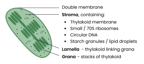
Describe the function of chloroplasts in plants and algae
Absorbs light energy for photosynthesis
To produce organic substances eg. carbohydrates / lipids
Describe the structure of the cell wall in plants, algae and fungi
Plants/algae: mainly cellulose (a polysaccharide)
Fungi: chitin (a nitrogen-containing polysaccharide)
Describe the function of the cell wall in plants, algae and fungi
Provides mechanical strength to cell
So prevents cell changing shape or bursting under pressure due to osmosis
Describe the structure of the cell vacuole in plants

Describe the function of the cell vacuole in plants
Maintains turgor pressure in cell → stopping plant wilting)
Contains cell sap → stores sugars, amino acids, pigments and any waste chemicals
How are eukaryotic cells organised in complex multicellular organisms
Cells become specialised for specific functions
Tissue → group of specialised cells with similar structure working together for a specific function
Organ → aggregation of tissues performing specific functions
Organ system → group of organs working together for specific functions
What are the distinguishing features of prokaryotic cells
Cytoplasm lacking membrane-bound organelles
So genetic material is not enclosed in a nucleus
e.g. bacteria and archaea (always unicellular)
Describe the general structure of prokaryotic cells

Compare and contrast the structure of eukaryotic and prokaryotic cells

Explain why viruses are described as acellular and non-living
Acellular → not made of cells (e.g. no cell membrane / cytoplasm / organelles)
Non-living → have no metabolism, cannot independently move / respire / replicate / excrete
Describe the general structure of a virus particle
Nucleic acids surrounded by a capsid (protein coat)
Attachment proteins to attach to specific host cells
No membrane bound organelle
Some also surrounded by a lipid envelope eg. HIV

Describe magnification
How many times larger the image is than the real object (I = A × M)
Describe resolution
minimum distance apart two objects can be to be seen as separate
Compare optical microscopes, transmission electron microscopes and scanning electron microscopes

How do you measure the size of an object under an optical microscope
Line up eyepiece graticule scale with stage micrometre scale
Calibrate: use stage micrometre to find size of one graticule division
Remove micrometre; count graticule divisions across object
Size = number of divisions × size of one division
Recalibrate at each magnification
Describe the basic steps in cell fractionation and ultracentrifugation
Homogenise tissue (blender) to break open cells and release organelles
Place in cold, isotonic, buffered solution
Filter to remove large debris (whole cells, connective tissue)
Ultracentrifuge at increasing speeds: remove pellet of heaviest organelles, respin supernatant at higher speed, repeat
Why is the homogenate kept cold, isotonic and buffered
Cold → reduces enzyme activity so organelles aren’t broken down
Isotonic → no osmosis, so organelles don’t burst
Buffered → constant pH so enzymes don’t denature
In what order are organelles separated by ultracentrifugation?
(*heaviest to lightest) Nuclei → chloroplasts → mitochondria → lysosomes → endoplasmic reticulum → ribosomes
What are the 3 general stages of the cell cycle in eukaryotic cells
Interphase
Mitosis
Cytokinesis
Describe the S phase in interphase
DNA replicates semi-conservatively → 2 chromatids (identical copies) join at a centromere
Describe the G1 and G2 phases in interphase
Number of organelles & volume of cytoplasm increases → high rates of protein synthesis