13. physiology of bone growth and development
 Call Kai
 Learn
 Practice Test
 Spaced Repetition
 Match
 Flashcards
 Knowt Play
1/24

There's no tags or description
Looks like no tags are added yet.
25 Terms
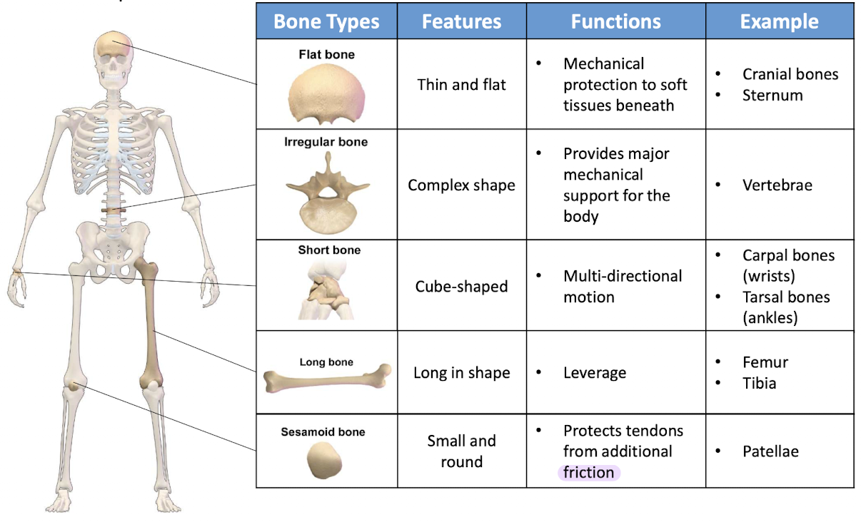
classification of bones
flat
irregular
short
long
sesamoid
flat bone

irregular bone

short bone

long bone

sesamoid bone
epiphysis
outside covered by articular cartilage (joints) or double layered fibrous membrane → periosteum (elsewhere)
inside contains compact bone (/cortical) + spongy bone (/cancellous/ trabecular)

diaphysis
outside covered by periosteum
secured to compact bone by Sharpey’s fibres
inside composed of compact bone that surrounds a central medullary cavity lined by delicate membranous lining → endosteum

composition of bone
mineral: hydroxyapatite (= crystalline calcium phosphate)
organic matrix
cells
water
4 types of cells within bone tissue
osteoblasts
osteocytes
osteogenic cells
osteoclasts*
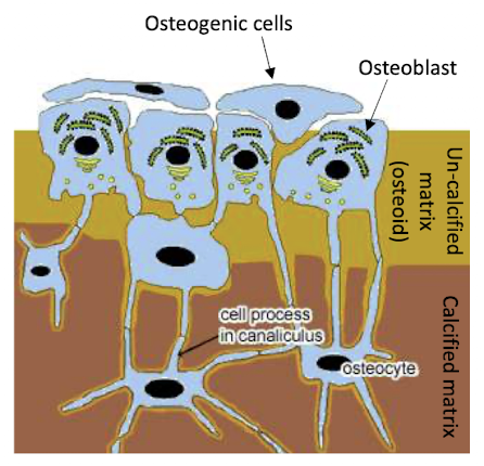
osteoblasts
responsible for bone formation by secreting collagen matrix and calcium salts
(first form uncalcified)
found in periosteum and endosteum
osteocytes
mature bone cells that form from osteoblast when entrapped by their secreted matrix after calcification
occupy spaces → lacunae (where cell body found) in bone
communicate with each other via long cytoplasmic processes that extend thru canaliculi
osteogenic cells
progenitor cell that can differentiate into osteoblast
(like stem cells)
osteoclasts
hematopoietic-origin (blood-forming cells)
responsible for bone resportion
found in periosteum and endosteum
compact bone
denser than spongy
microstructure consists of osteons (basic structural unit)
composed of concentric rings of calcified matrix → lamellae
only cells - > osteocytes
found on edges of each lamella
lamellae
consisted of mineralise collagen fibrils arranged in geometrical patterns
fibrils composed of tropocollagen (?) + nanocrystal of hydroxyapatite
arrangement
within each lamella
fibrils parallel
>force/ strength
from one lamella to next
direction rotated

spongy bone
> porous
lacunae and osteocytes → lattice-like network of matrix spikes called trabeculae
few cell layers thick as nourished by diffusion of nutrients that travel thru canaliculi from marrow cavity
spaces in between lattice filled with red bone marrow where hematopoiesis occurs
*open spaces of trabeculated network of spongy bone allow it to support shifts in weight distribution
function of spongy bone
decreased weight of bone

bone formation and development (ossification/ osteogenesis)
2 pathways
intramembranous ossification
endochondral ossification
intramembranous ossification
formation of bone directly from mesenchymal cells
differentiate into osteoblasts
flat bones e.g. skull, mandible, clavicles
mesenchymal cells cluster in vascular membrane and differentiate into osteoblasts
osteoblasts → osteocytes
spicule form and join into trabeculae
enclosing blood vessels as they grow
=> network of woven spongy bone
outer layers are remodelled
near surface, trabeculae thicken and fill in to form compact bone plates
middle stays spongy

endochondral ossification
development of bone from hyaline cartilage e.g. all long bones
condensation + differentiation of mesenchymal cells → chondrocytes
chondrocytes proliferate → hyaline cartilage
with formation of membrane → perichondrium that covers cartilage
cartilage matrix calcifies and chondrocytes degenerate
leaving cavity invaded by blood vessels
osteogenic cells migrating with blood vessels differentiate into osteoblasts
begin producing spongy bone at primary ossification centre
diaphysis → remodelling to create medullary cavity
end of bone → cartilage and chondrocytes continue to grow
capillaries and osteoblasts migrate into epiphyses
=> secondary ossification centres
hyaline cartilage remains only at joint surface (as articular cartilage) + at epiphyseal plates (growth plate) for prod and maturation of chondrocytes (bone elongation)

bone formation by osteoblasts
osteoblasts create nano-composite structure of bone by secreting collagenous extracellular matrix
control its mineralisation by releasing calcium phosphate-containing vesicles known as matrix vesicles (MV)
*process of matrix mineralisation depends on adequate supply of calcium and phosphate
bone respiration by osteoclasts
ability to polarise and reorganise its membrane into 4 distinct and unique domains
sealing zone (SZ)
ruffled border (RB)
basolateral domain (BL)
functional secretory domain (FSD)
sealing zone
allows establishment of isolated extracellular microenvironment between osteoclast and bone
ruffled border
where protons and proteases secreted to demineralise and degrade bone matrix
bone demineralisation - mediated by vacuolar H+-ATPare and Cl-channel
(→ pH 4.5)
degradation of org component (collagen fibre) of bone by lysosomal protease
functional secretory domain
express Cl-/ HCO3- exchanger
responsible for releasing bone degradation products
