Protozoa
 Call Kai
 Learn
Learn Practice Test
 Spaced Repetition
 Match
Match Flashcards
 Knowt Play
1/25

Name | Mastery | Learn | Test | Matching | Spaced | Call with Kai | Chat |
|---|
No analytics yet
Send a link to your students to track their progress
26 Terms

20 um
Entamoeba histolytica Trophozoite

12-15 um
Entamoeba histolytica Cyst wet mount saline

12-15 um
Entamoeba histolytica cyst Trichrome

20 um
Entamoeba histolytica Trophozoite wet mount Iodine
Entamoeba histolytica
Only pathogenic intestinal amoeba
Worldwide distribution
“Flask shaped” lesions in intestines - Dysentary
Forms sterile abscesses
Co-infections by bacteria causing GI disease may occur
serological test to differentiate from E. dispar
Entamoeba dispar
Amoeba
Identical to E. histolytica, need serological test
Does not ingest RBCs
Commensal
Entamoeba hartmanni
Amoeba
Identical to E. histolytica
Slightly smaller than E. histolytica
Cysts: 6-8 um
Troph: 8-10 um
Commensal
Entamoeba coli
Largest amoeba
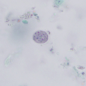
Trophs: up to 50 um, oval, single nucleus with irregular chromatin ring, eccentric karyosome
Cyst: up to 35 um, 5+ nuclei, coarse chromatin ring, splintered chromatoid bar
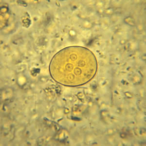
10-35 um
Entamoeba coli cyst iodine wet mount

15-50 um
Entamoeba coli troph Trichrome
Endolimax nana
Amoeba
Troph has dense karyosome with thin nuclear membrane - “ball and socket”, 6-12 um
Cyst looks like potato with eyes, 4 nuclei, 6-8 um
Reservoir hosts: humans, pigs, primates
6-12 um
Endolimax nana troph Trichrome
Iodamoeba butschlii
Amoeba
commensal
Troph: large dense karyosome, heavier chromatin, “dirty” cytoplasm
Cyst:single nucleus, crescent halo around karyosome, glycogen vacuole

8-20 um
Iodamoeba butschlii Troph Trichrome

5-20 um
Iodamoeba butschlii Cyst
Blastocystis
Pathogenic potential, opportunistic
Risk factors: immunocompromised, poor hygiene, tropical country
Vacuolated form most common: 5-40 um, 90% vacuole, nuclei pushed to periphery
Disease: associated with IBD, self limiting, non-specific

5-40 um
Blastocystis sp. iodine wet mount

5-40 um
Blastocystis sp Trichrome
Amoebae
Pathogenic: Entamoeba histolytica
Commensal: Entamoeba dispar, Entamoeba hartmanni, Entamoeba coli, Endolimax nana, Iodamoeba butschlii
Unknown: Blastocystis
Flagellates
Pathogenic:
Dientamoeba fragilis, Giardia lamblia/intestinalis, Trichomonas vaginalis
Commensal:
Trichomonas hominis, Trichomonas tenax, Chilomastix mesnili
Flagellate Life Cycle
Divide via binary fission
ingestion of cysts in contaminated food/water or from hands
Dientamoeba fraglilis
No external flagellum, looks similar to amoebas
worldwide
occasionally pathogenic, symptoms: colicky pains, fatigue, weight loss
NOT SEEN ON WET PREPS
Trophs: 4-15 um, 20-40% have one nucleus, fragmented karyosome (3-5 granules), no peripheral chromatin, dirty cytoplasm
likes to co-infect with pinworm

4-15 um
Dientamoeba fragilis Trichrome
Giardia lamblia/intestinalis/duodenalis
Most common parasite in USA
Pathogenic, infects upper small intestine, does not invade tissues
Beavers and others are reservoirs, Common in campers
Troph: 10-20 um, 2 nuclei, 2 paranasal bodies, 1 axostyle, 8 flagella, suck onto intestine
Cyst: features of troph inside cyst, may have 4 nuclei, 11-14 um
Giardia Symptoms
Acute:
similar to food poisoning, lasts a few days, gas, foul-smelling stool, watery diarrhea, gray-green color
Chronic:
recurrent brief episodes of loose, foul-smelling, gray, foamy stools