Renal Week 1
 Call Kai
 Learn
 Practice Test
 Spaced Repetition
 Match
 Flashcards
 Knowt Play
1/276

There's no tags or description
Looks like no tags are added yet.
277 Terms
Define renal blood flow (RBF) + equation
Renal blood flow is the total volume of blood delivered to both kidneys per minute (~1.2 L/min), it is ~25% of cardiac output; calculated as RPF / (1 – hematocrit).
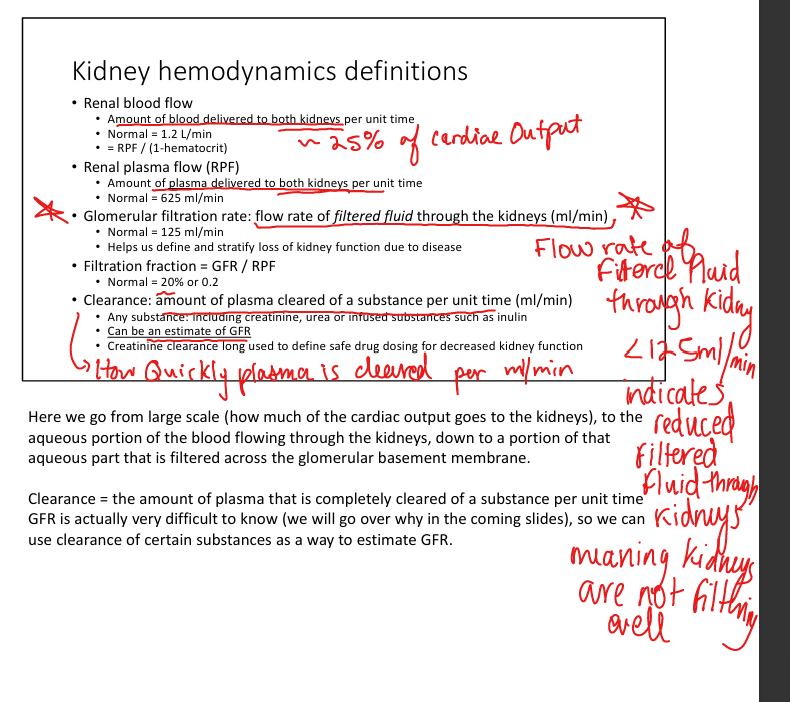

Define renal plasma flow (RPF)
Renal plasma flow is the plasma portion of blood delivered to both kidneys per minute (~625 mL/min)

Define glomerular filtration rate (GFR).
GFR is flow rate of filtered fluid through the kidneys (~125 mL/min), represents total filtration across all nephrons.

Define clearance + equation
Clearance is the amount of plasma completely cleared of a substance per minute, calculated as (Ux × V) / Px ;


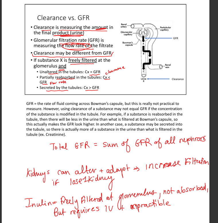
When does clearance= GFR
Only if the substance is freely filtered and neither reabsorbed nor secreted.
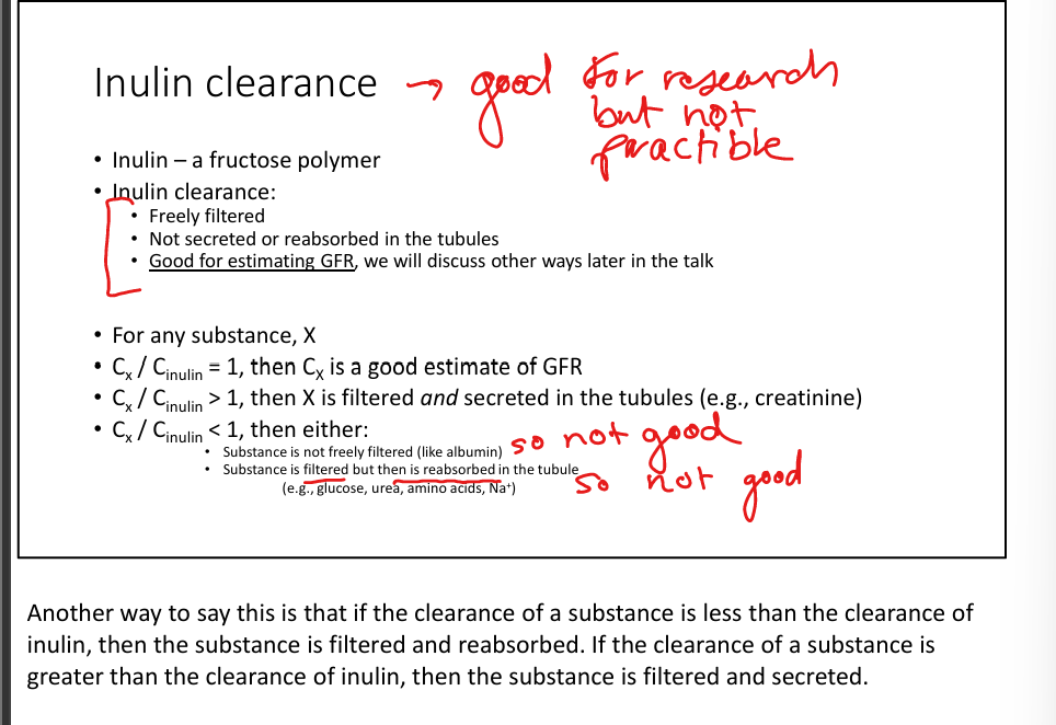
Explain why inulin is the gold standard for GFR.
Inulin is freely filtered, not reabsorbed, secreted, synthesized, or metabolized, making its clearance equal to true GFR; impractical clinically because it requires IV infusion.
Best substance for evaluating Renal Plasma Flow
PAH because it is freely filtered, completely secreted, and 90% is cleared on first pass


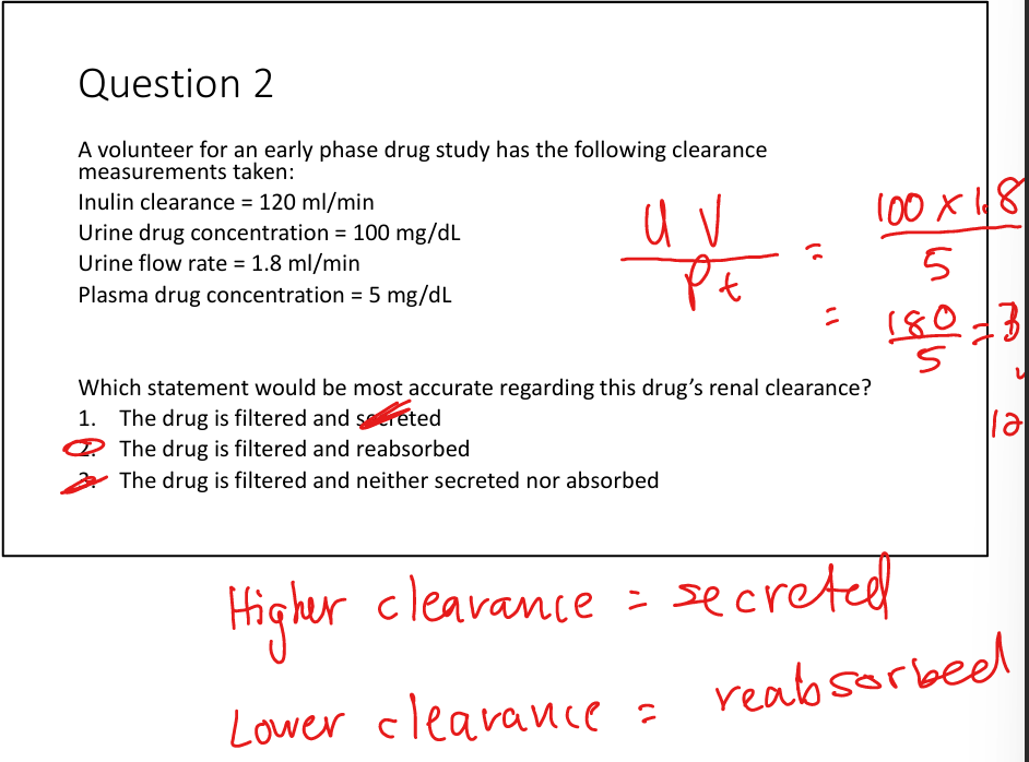
Higher clearance= ; Lower clearance=
Higher clearance= secreted; Lower clearance= reabsorbed
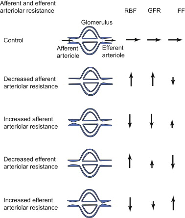
Explain how afferent arteriole tone affects GFR.
Afferent constriction decreases RPF(renal plasma flow) and Pgc(hydraulic pressure glomerular capillary) → ↓GFR;
Afferent dilation increases RPF(renal plasma flow) and Pgc(hydraulic pressure in glomerular capillary) → ↑GFR.

Explain how efferent arteriole tone affects GFR.
Efferent constriction increases Pgc(hydaulic pressure in glomerular capillary) → ↑GFR despite ↓RPF(renal plasma flow); excessive constriction eventually ↓GFR
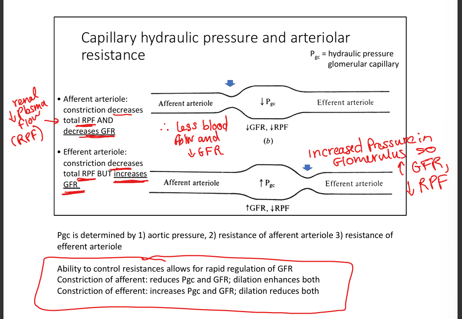
What three things govern arteriolar resistance?
Renin-Angiotensin-Aldosterone System (RAAS), Autoregulation and Tubuloglomerular feedback

Define tubuloglomerular feedback (TGF).
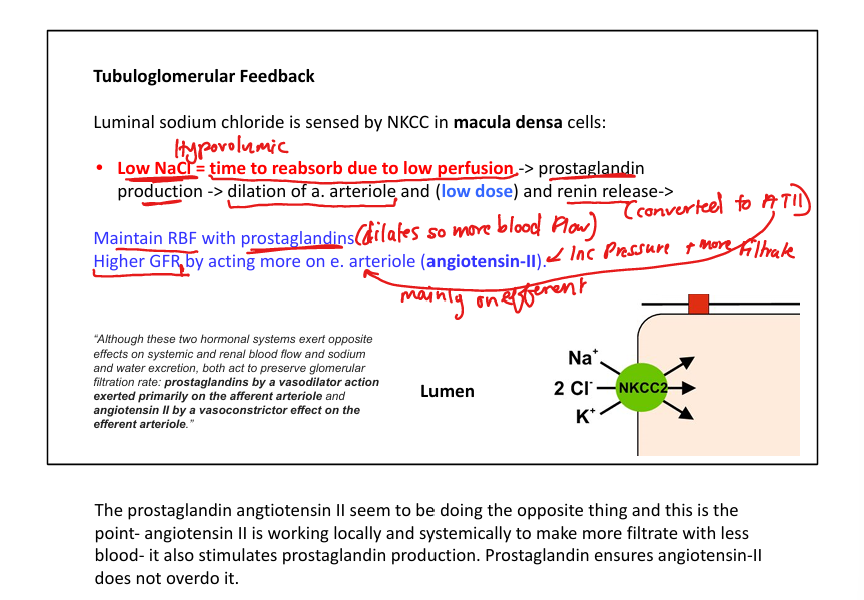
Macula‑densa–mediated(they sense NaCl)→ Increased NaCl(high BP, GFR is increased and moving fluid too fast for salt to be reabsorbed)→ release adenosine/ATP to vasoconstrict→afferent constriction to cut down blood flow in glomeruli)→glomerular capillary pressure drops→GFR drops back down to normal;
Decreased NaCl(low BP, GFR drops making fluid move slowly so more salt is reabsorbed before reaching end of loop)→low NaCl is sensed and release prostaglandins and renin→ afferent arteriole dilation to let more blood enter→capillary pressure increases→ increase GFR. It helps prevent excessive salt and water loses!
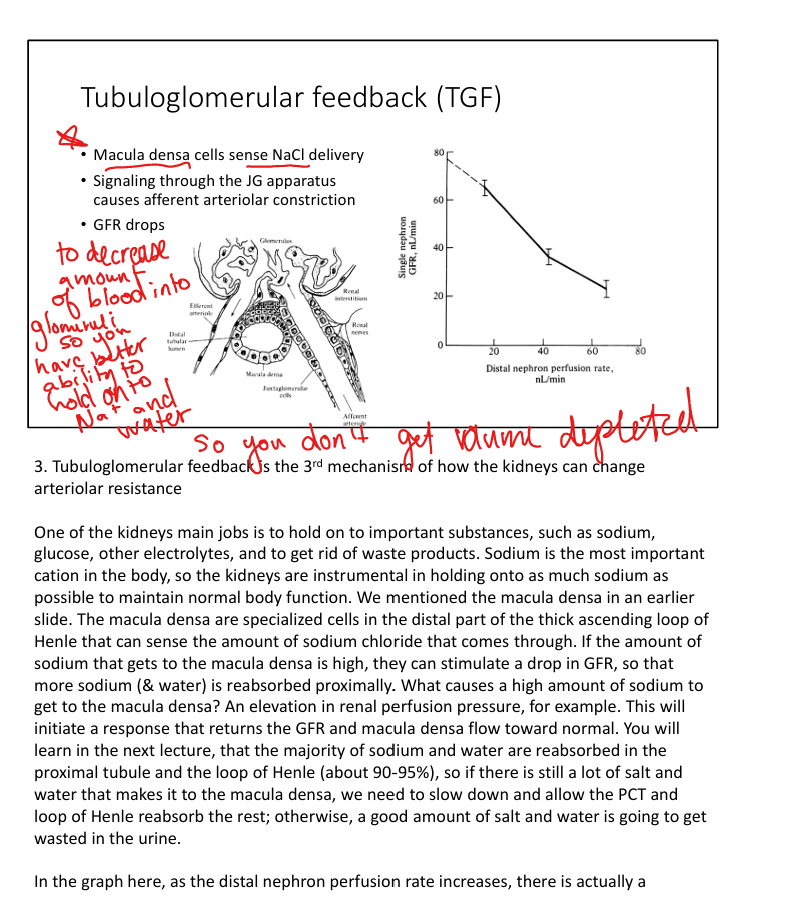

Explain macula densa sensing.
They detect tubular NaCl concentration. High NaCl → ATP/adenosine → afferent constriction to decrease GFR

Describe autoregulation of RBF and GFR. What MAP level has increased risk of AKI?
Autoregulation maintains stable RBF/GFR across wide arterial pressures. Uses myogenic afferent stretch receptors that contrict/dilate to keep glomerular pressure stable. MAP< 70 → AKI risk.
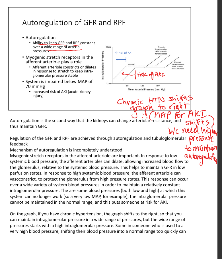
Explain the myogenic mechanism in autoregulation
Afferent arteriole smooth muscle constricts when stretched (high BP) and dilates when under‑stretched (low BP), stabilizing intraglomerular pressure.
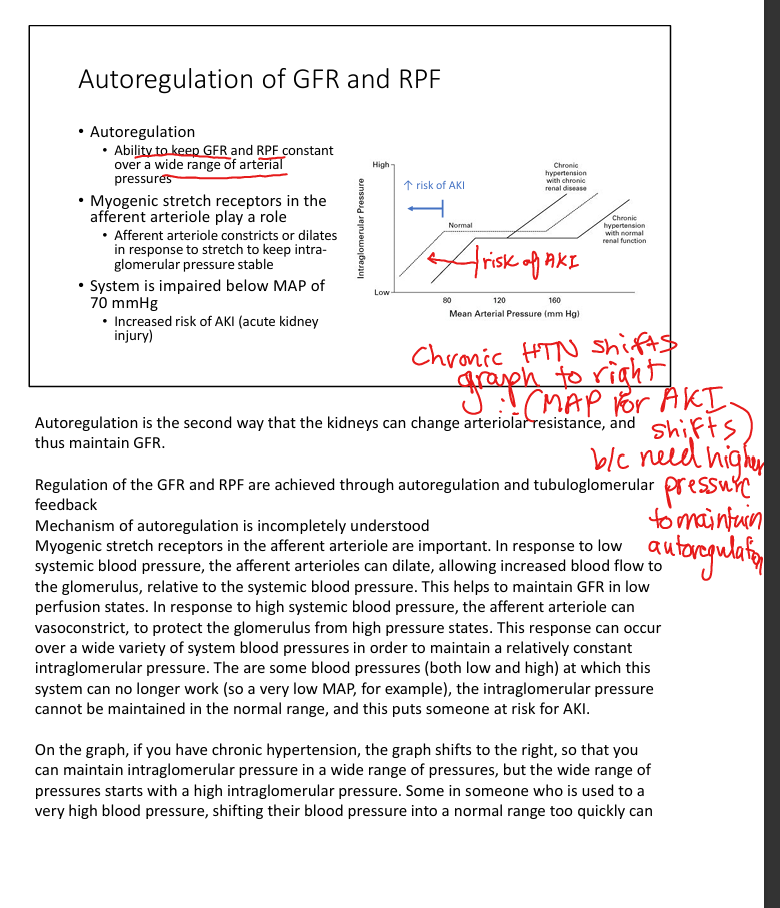
Explain how chronic hypertension shifts autoregulation
Chronic HTN shifts the autoregulatory curve rightward, requiring higher pressures to maintain GFR and increasing AKI risk when BP is rapidly lowered.

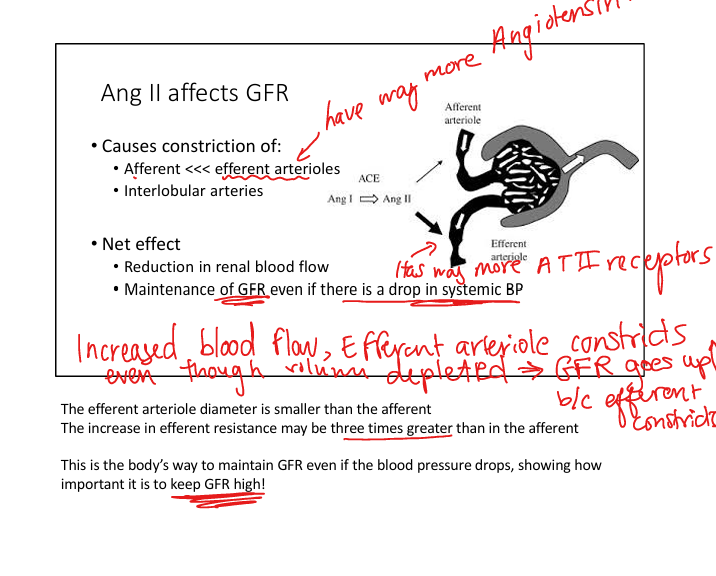
Describe RAAS influence on GFR.
RAAS activation causes Efferent >>> Afferent constriction via Ang II(inc GFR), raising Pgc to preserve GFR during hypoperfusion, also increases Na+ retention and systemic BP.


Explain prostaglandin protection of GFR. What medication impacts this in hypovolemic pts?
Prostaglandins preferentially dilate the afferent arteriole(to prevent ischemia)→ counterbalancing Ang II
NSAIDs block PGs → excessive afferent constriction → ↓GFR, especially in hypovolemia.


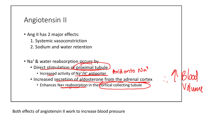
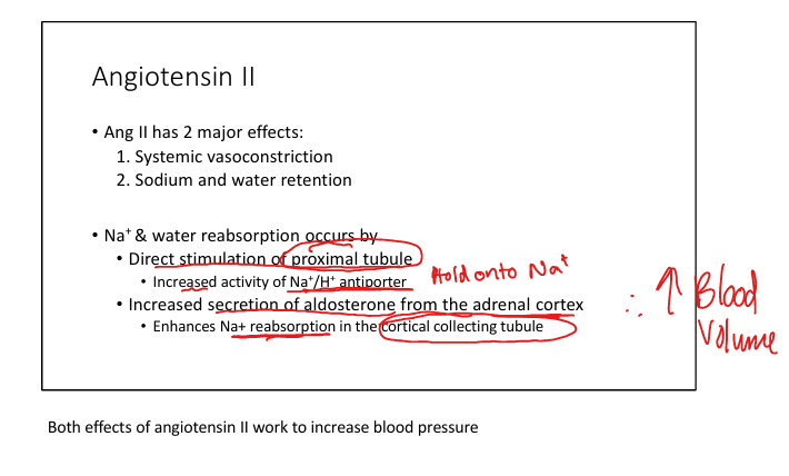
Angiotensin II major effects to increase BP
Systemic vasoconstriction and Na+/water reabsorption(via aldosterone stimulation)
GFR equation
GFR= Kf (Pgc - Pbs -ngc) where Kf= filtration coefficient, Pgc=hydraulic pressure in glomerular capillary, Pbs=hydraulic pressure in bowmans space, and ngc=oncotic pressure in plasma


Explain why creatinine is used for eGFR. Limitations?
Creatinine is produced at a steady rate, freely filtered, and only minimally secreted. Serum levels rise as GFR falls. Limitations: loss of nephrons leads to compensatory hyperfiltration, and when GFR begins to fall, tubular secretion of creatinine increase → results in creatinine remaining stable despite drop in GFR


At what stage and GFR do patients need to consider dialysis
Stage 4 (GFR is 15-29) start planning. Stage 5 (GFR <10-15) begin dialysis


Urine creatinine clearance vs urine urea clearance estimation tendencies"
Urine creatinine clearance tends to OVERestimate GFR
Urine Urea clearance tends to UNDERestimate GFR

Describe limitations of eGFR.
eGFR is inaccurate in extremes of muscle mass, acute kidney injury, pregnancy, and rapidly changing renal function, requires stable creatinine.
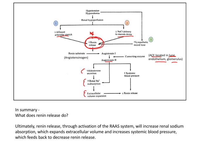
Stimuli for renin release
Renal hypoperfusion (hypotension/volume depletion), Decreased NaCl delivery to macula densa, or increased sympathetic activity

What cells store and release renin and where are they located?
Juxtaglomerular cells located on afferent arterioles

Why does Angiotensin II constrict efferent arterioles way more than afferent?
Efferent arterioles have more angiotensin II receptors on them

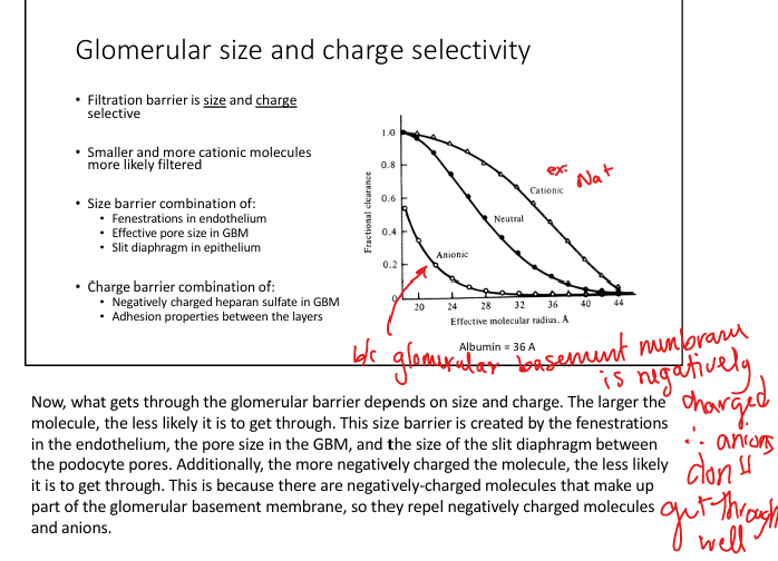
What does normal glomeruli filter let through?
Allows small molecules (Na, urea), less myoglobin, very little albumin (therefore albumin inurine is indicator of kidney damage)


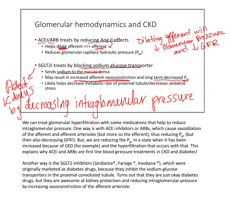
What is intraglomerular HTN(hyperinflation)?
Compensatory rise in filtration in other nephrons driven by afferent dilation to compensate for nephron loss (ex donated a kidney). Eventually this mechanism will lead to/resemble chronic HTN effects

Treatment for glomerular hyperinflation
ACEi/ARBs to reduce angiotensin II effects (will help dilate efferent arterioles >afferent); and SGLT2 inhibitors to decrease Na, glucose and inc afferent vasoconstriction
Order of blood flow through Renal Vessels
Renal artery>Segmental>Interlobar>Arcuate>Cortical radiate>Afferent arteriole>Glomerulus(capillaries)>Efferent arterioles
Renal Science Is Actually Cool And Generally Exciting

Role of the vasa recta + location
Regulates production of concentrated urine, seen in juxtamedullary nephron

Differentiate cortical vs juxtamedullary nephrons
Cortical nephrons have short loops and peritubular capillaries. Juxtamedulary nephrons have long loops and vasa recta


Describe PCT histology and major functions
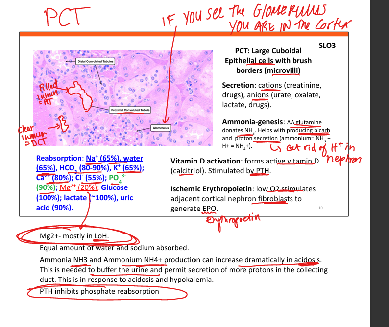
Large cuboidal cells with dense microvilli reabsorb ~65% Na/H2O, 90% HCO3, all glucose/AAs, some (20%) Mg2+, Site of ammoniagenesis, forms active vitamin D, generates EPO

Explain the transporters present in the PCT (think of whats filtered in PCT)
Na/K ATPase, Na/H+ exchanger, Na/Glucose exchanger, Na/lactate exchanger

How to tell if you are in the cortex of kidney on histology?
Presence of glomerulus= in the cortex
Transporters on Thick Ascending Loop of Henle
NaKCl transporter, Na/K+ ATPase, Renal Outer Medullary K+ Channel (ROMK)

Explain the countercurrent multiplier
Descending limb loses water, ascending limb pumps out NaCl→creating medullary gradient enabling concentrated urine formation.

Describe early DCT histology and transport
Small cuboidal cells, minimal microvilli. Has Na/K+ ATPase, NaCl cotransporters, Ca2+ ATPase(has receptor for PTH)


Describe late DCT and cortical collecting duct histology
Principal cells (ENaC, ROMK) regulate Na/K + Intercalated cells (regular H+ and HCO3)

Differentiate alpha vs beta intercalated cells
Alpha secrete H+ and reabsorb HCO3. Beta Cells secrete HCO3 and reabsorb H+

Explain aldosterone’s effects on principal cells
In principal cells increases ENaC and ROMK expression leading to salt reuptake without impacting K+ severely

Hyperosmolarity
High concentration of dissolved particles (like Na, glucose) making fluid more concentrated

Why does aldosterone increase pH?
Aldosterone activates H+ ATPase→Alpha cells(H+ secretion) outnumber Beta cells(HCO3) in medullary conducting duct + principal cell activity is increased(inc Na+ into cell making lumen more negative) →net negative charge in lumen increases pull force to pull protons out→alkalosis and inc pH

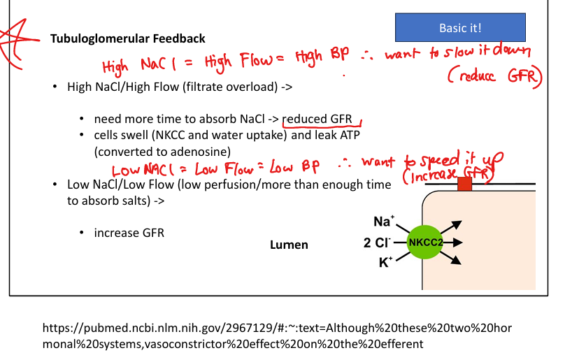
Explain how histology supports tubuloglomerular feedback
Macula densa cells sense high NaCl(high BP) via NKCC2 → release ATP/adenosine → afferent constriction
Low NaCl(dec BP)→release prostaglandins→afferent dilation

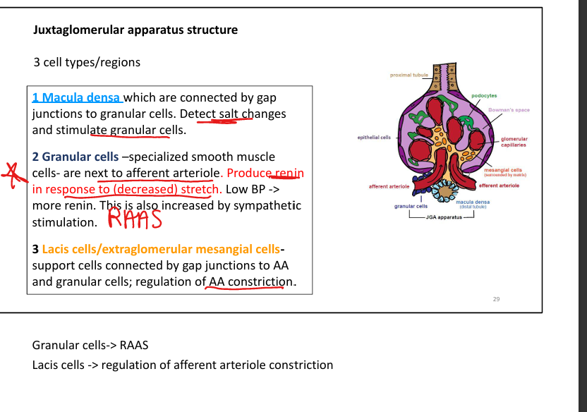
Describe juxtaglomerular apparatus 3 cell types and functions
Macula densa (DCT), granular cells (renin-producing smooth muscle), lacis cells (mesangial support) regulate GFR and BP.
Major site of Mg2+ absorption (70%)
Thick ascending loop of henle

Differentiating DCT from PCT on histology
DCT has clear lumen, while PCT has filled lumen
Tuboglomerular Feedback High NaCL
High NaCl=High Flow= High BP→need more time to absorb NaCl→release adenosine→afferent vasoconstriction→reduce GFR + inhibit renin release


Tuboglomerular Feedback Low NaCl
Low NaCl=Low Flow= Low BP→have too much time to absorb NaCl→release prostaglandins→afferent vasodilation→increase GFR + and increase renin release


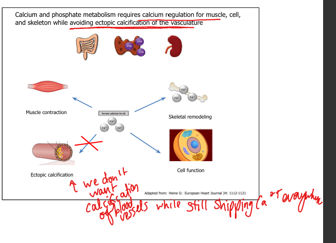
Why Ca/Phos regulation is complex?
Ca/Phos must support muscle/cell function + skeletal structure while preventing ectopic vascular calcification.
Major roles of calcium
Muscle contraction, neurotransmission, coagulation, intracellular signaling, structural bone matrix.

Major roles of phosphate
ATP production, phosphorylation reactions, nucleic acids, bone mineralization (hydroxyapatite).

Calcium distribution in body
99% bone, 1% intracellular, 0.1% extracellular.

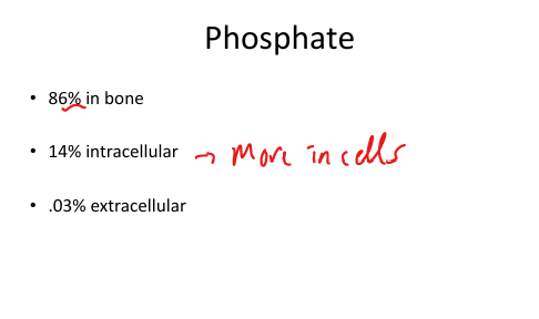
Phosphate distribution in body
86% bone, 14% intracellular, 0.03% extracellular.
Primary sources of Vitamin D
Sunlight (UVB → skin), fortified dairy, fish oils (salmon, trout), supplements (D2/D3).


Skin precursor to Vitamin D
7‑dehydrocholesterol → cholecalciferol (D3) via UV light.


Vitamin D3 vs D2
D3 = cholecalciferol (human + supplement), D2 = ergocalciferol (plant/supplement).

Where is 25‑hydroxy Vitamin D produced?
Liver (25‑hydroxylation) (not most active form)

Where is 1,25‑dihydroxy (calcitriol) Vitamin D produced?
Kidney (proximal tubule 1‑α hydroxylase). Therefore if you have kidney failure (even if you have 25 hydroxy D) you cannot make this (would have to supplement with this exactly)

What stimulates 1‑α hydroxylase(converts 25 hydroxyl D to 1,25 Dihydroxy-calcitriol)?
PTH, hypocalcemia, low phosphate.


Primary role of 1,25‑OH Vitamin D (active form)
↑ GI Ca absorption by increasing TRPV6, ↑ GI phosphate absorption by increasing NaPi‑IIb, ↑ bone Ca/Phos resorption.

Effect of Vitamin D on renal Ca handling
Slight ↓ Ca excretion.
Options for Vtiamin D supplementation (4 options)
Cholecalciferol (D3-in humans/supplemented in milk), Ergocalciferol (D2-plant based), Calcitriol(1,25 OH, most active form, used for kidney disease), Sunlight (D3)
Rickets mechanism + Signs
↓ 1,25‑OH Vitamin D → ↓ Ca absorption → decreased Ca for skeletal growth → osteoid accumulation. Sx: Leg bowing, Rosary sign, short stature


Rickets risk factors/causes
Breastfeeding w/o supplementation, dark skin + low sun, celiac disease, genetic defects.

Parathyroid Hormone Function generally to serum levels
Increase serum Ca2+ and normal to low Phosphate


PTH effects on bone
Stimulates osteoblast receptors → inc osteoclast activation → ↑ Ca + ↑ phosphate release.

PTH effects on kidney (calcium)
↑ Ca reabsorption in DCT by increasing TRPV5.

PTH effects on kidney (phosphate)
↓ Phosphate reabsorption in PT by downregulating NaPi‑2a/2b in the gut

Net serum effect of PTH
↑ Ca, ↓/normal phosphate.
Hyperparathyroidism
↑ Ca, ↓/normal phosphate, ↑ urinary Ca (hypercalciuria)→kidney stones and osteoporosis

Fiborblast Growth Factor‑23 (aka “Phosphaturin”)
Made by Osteocytes. Decreases PT phosphate reabsorption, decreases 1,25 OH D production, decreases PTH secretion


FGF‑23 nickname
“Phosphaturin.”

Calcitonin
Made from Parafollicular C‑cells of thyroid. Decreases Ca2+ and phosphate reabsorption(excretes them), inhibits osteoclast activity in bone, lowers Ca and phosphate. Opponent to PTH


Renal Ca reabsorption: PT
Passive, paracellular, linked to Na reabsorption (65–70%).
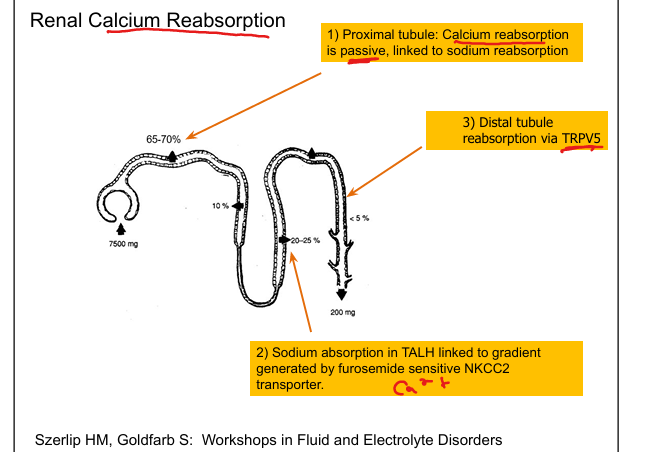

Renal Ca reabsorption: TALH
Paracellular via claudins, driven by NKCC2 gradient.


Renal Ca reabsorption: DCT
Active via TRPV5 (regulated by PTH, thiazides).


Effect of furosemide on Ca
Blocks NKCC2 → ↓ lumen‑positive gradient → ↓ Ca reabsorption → hypercalciuria.


Effect of thiazides on Ca
Blocks NaCl cotransporter → ↓ intracellular Na → ↑ TRPV5 → ↑ Ca reabsorption → hypocalciuria.


Where is Ca absorbed? What receptor? What is required?
Ca2+ is absorbed in duodenum+jejunum via TRPV6 and paraceullar transport requiring 1,25 OH D(most important part, allows for Ca to be absorbed).


Where is Phosphate absorbed? What receptor? What is helpful for absorption? What prevents absorption?
Duodenum/jejunum via NaPi‑IIb + paracellular (↑ with 1,25‑OH D). Calcium binding to phosphate prevents absorption

Phosphate renal handling
85% reabsorbed in PT via NaPi‑2a/2c (regulated by PTH + FGF‑23).

Total Calcium formula
=protein bound Ca + ionized Ca + complexed Ca

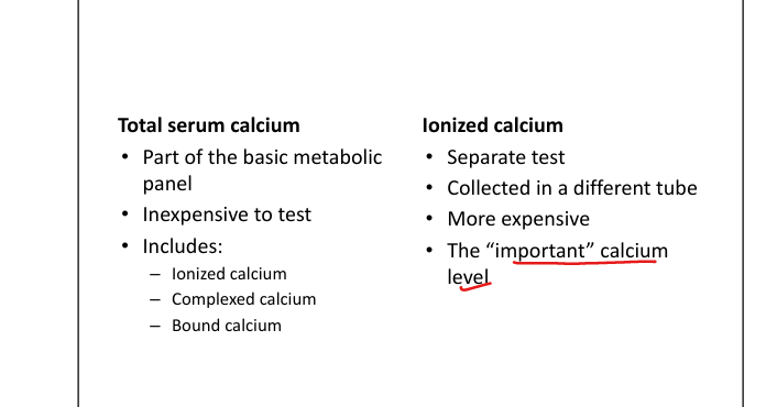
When to order ionized calcium
Whenever albumin abnormal or Ca result inconsistent with symptoms.

Hypocalcemia symptoms
Chvostek sign(tap on facial nerve causes contraction of ipsilateral facial muscles), Trousseau sign(carpal-pedal spasm with BP cuff inflation), tetany(involuntary muscle contractions), seizures, prolonged QT.

Hypercalcemia symptoms
Stones, bones, groans, moans (nephrolithiasis, bone pain, constipation, lethargy).

Causes of hypocalcemia with normal phosphate
Pancreatitis, hypomagnesemia, ↓ Vitamin D.

Causes of hypocalcemia with high phosphate
CKD, hypoparathyroidism.

Hyperphosphatemia causes
CKD, hemolysis, rhabdomyolysis, hypoparathyroidism.

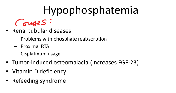
Hypophosphatemia causes
Proximal RTA, cisplatin, tumor‑induced osteomalacia (↑ FGF‑23), Vitamin D deficiency, refeeding syndrome.
Familial Hypocalciuric Hypercalcemia (FHH)
Inactivating mutation of Ca sensing receptor of parathyroid gland→leads to higher Ca levels needed to decrease PTH secretion(which increases Ca level)→hypercalcemia in blood, low Ca in urine

Magnesium GI absorption
Distal small intestine + colon (TRPM6/7).


Renal Mg reabsorption locations
PT (10–25%), Majority in TALH (50–75%), DCT (TRPM6).

Hypermagnesemia symptoms
↓ Deep Tendon Reflexes, respiratory depression, hypotension; Frequently seen in CKD or IV Mg.

Hypomagnesemia symptoms
Hyperreflexia, carpopedal spasm, prolonged QT, torsades.

Hypomagnesemia causes
Diuretics, malabsorption, cisplatin, Bartter/Gitelman→impaired reabsorption in Thick Ascending LoH

Effect of hypomagnesemia on calcium
Causes hypocalcemia (bc Mg is required for PTH secretion).

Renal disease effect on Ca/Phos
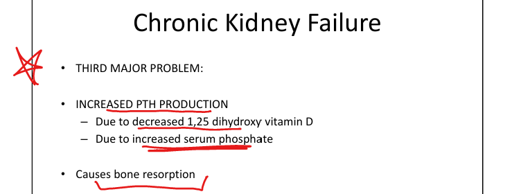
1. Decreased GFR→decreased phosphate excretion (increased serum Phosphate) →Results in increased FGF-23 to clear phosphate→2. Results in Decreased 1,25 OH D (calcitriol) →Calcitriol acts as inhibitor of PTH but dec Calcitriol results in →3. Increased PTH(due to dec 1,25 OH D, and inc serum P)→4.This Causes: Increased vascular calcification (dt inc serum P) and Decreased bone mineralization (dt increased PTH)




Treating Hyperphosphatemia in Chronic Kidney Failure
Decrease Phosphate in diet, use phosphate binders, remove phosphate with dialysis
